Amplified sensitization of Er3+ luminescence in silica by AuN quantum clusters upon annealing in a reducing atmosphere

Tiziana Cesca*, Boris Kalinic, Chiara Maurizio, Niccolò Michieli, Carlo Scian and Giovanni Mattei
University of Padova, Department of Physics and Astronomy, Nanostructures Group, via Marzolo 8, I-35131 Padova, Italy. E-mail: tiziana.cesca@unipd.it

Received 1st July 2016 , Accepted 8th October 2016

First published on 10th October 2016


Abstract

Finding effective strategies for improving the luminescence emission performance of Er3+ ions in silica is of paramount importance for the realization of efficient Er-based nanophotonic devices. To this aim, in the present work the sensitization mechanism of room temperature Er3+ luminescence by AuN quantum clusters, upon annealing in a reducing atmosphere, was investigated by photoluminescence characterization of Er–Au co-implanted samples in which Au ions were implanted at a low fluence of 3.4 × 10¹⁴ Au+ per cm2. The role of the annealing atmosphere was unveiled by comparing the emission properties of samples thermally treated in reducing and inert atmospheres. A significant amplification of the Er3+ sensitization efficiency (up to a 6× enhancement of the photoluminescent intensity) was revealed upon annealing in a reducing atmosphere and the possibility to get efficient room temperature Er3+ luminescence from Er–Au co-implanted samples with only a limited amount of sensitizing agents was demonstrated and discussed.
The research for effective strategies to improve the luminescence emission efficiency of Er3+ ions in silica has strongly motivated the scientific community in the last few decades. Owing to its spectroscopically sharp and thermally stable emission at 1.54 μm (i.e., matching the window of minimum losses in silica), Er represents the element of election in the field of optoelectronic and optical telecommunication,1 but severe limitations to its efficient employment in optoelectronic devices are posed by the low intrinsic Er3+ excitation cross-section (of the order of 10−21 cm2).2–4 Among the different strategies adopted during recent years to overcome such limitations, which include the interaction with fluorescence sensitizers as other rare-earth ions,5,6 silicon nanocrystals,7–10 or metallic species,11–13 it was recently demonstrated by our group that a particularly effective approach is the incorporation, in Er-doped silica matrices, of sub-nanometric AuN quantum clusters, made of N = 5–20 atoms, obtained by Au implantation and proper thermal treatment.14,15 The sensitization mechanism of the Er3+ luminescence in the Er–Au co-implanted systems was proved to be a non-resonant broad-band energy transfer process occurring at a very short range between the AuN quantum clusters and Er3+ ions located in close proximity. An enhancement of the effective Er excitation cross-section of more than 3 orders of magnitude was obtained in this way.14,16

Gold nanoparticles have been widely investigated during recent years for their plasmonic properties, with applications in many different fields such as photovoltaics,17,18 nonlinear optics,19,20 or sensing.21,22 Nonetheless, in the present case, due to their sub-nanometric size the AuN clusters incorporated in the samples do not exhibit any plasmonic behavior. No surface plasmon resonance peak can be detected in the absorbance spectra of the samples. Instead, these ultra-small AuN clusters have a “pre-plasmonic” or molecule-like character which definitely triggers their efficiency as nanoantennae for Er3+ luminescence. Indeed, it was demonstrated that such ultra-small AuN clusters exhibit discrete electronic energy levels which are resonant with corresponding absorption levels of the Er3+ ions and this represents the preferential path for the transfer of electromagnetic energy from the AuN clusters to the Er3+ ions.23 The process was proved to occur non-radiatively16 via short-range coupling with interaction distances in the range 0.4–0.8 nm.14 Moreover, since it is mediated by the formation of discrete energy levels related to the AuN clusters, the energy transfer process is strongly controlled by the clusters’ size in the sub-nanometer range. Furthermore, recent studies on the sensitization efficiency of Er–Au co-doped silica systems implanted with Au+ ions at different fluences and thermally treated in inert (N2) atmosphere have shown the beneficial effect of incorporating gold at low concentrations in the samples.14,24 In this way the kinetics of the cluster growth is slowed down and the annealing temperature to get AuN clusters in the optimal size range (of about N = 10–20 atoms) for the Er sensitization shifts to higher values, allowing also for a better recovery of the silica matrix from implantation damage, which is further advantageous for efficient Er3+ emission.14 Nonetheless, for annealing treatments in an inert atmosphere a lower limit for the Au concentration needed for the efficient Er3+ sensitization is observed, below which the efficiency of the energy transfer process drops drastically.14 The present work is therefore aimed at addressing the role of the annealing atmosphere (inert or reducing) on the sensitization efficiency of the Er3+ luminescence in Er–Au co-implanted silica samples in which Au is incorporated at low fluence (3.4 × 10¹⁴ Au+ per cm2). In particular, we demonstrated that a significant boost of the Er3+ emission properties can be obtained by thermal treatments in a reducing atmosphere with respect to analogous samples heated in an inert atmosphere, and a very efficient Er3+ sensitization is achieved in spite of the low Au content. These results are extremely valuable since the capability to get a strong amplification of the Er3+ luminescence sensitization and the understanding of the mechanisms that control this process have important technological implications for the effective employment of AuN quantum clusters as sensitizers of the Er3+ luminescence in photonic devices.

1 Experimental section

Sequential Au implantations were performed on Er-doped silica slabs (Infrasil by Haereus) at three energies (60, 110, 190 keV) with a total Au fluence of 3.4 × 10¹⁴ Au+ per cm2. The Er-doped silica slabs used as substrates for the Au implantations were obtained by an analogous three-energy implantation scheme (50, 100, 190 keV) at a total Er fluence of 6.7 × 10¹⁴ Er+ per cm2, and then thermally annealed at 800 °C in N2 atmosphere for 1 h to recover the implantation damage. Both Au and Er implantation schemes were chosen to get almost flat (about 70 nm thick) concentration profiles of the two elements and their complete overlap in the co-implanted samples. The Au and Er concentration ratio in the samples was [Au]/[Er] = 0.5. The concentration profiles were obtained by SRIM simulations,25 while the total fluence was experimentally determined by Rutherford Backscattering (RBS) measurements performed with a 2 MeV 4He+ beam. After Au implantation, isochronal annealing of the samples was performed for 1 h at different temperatures in the range 300–800 °C both in reducing (Ar + H2) and in inert (N2) atmospheres. The thermal treatments were necessary to induce the implantation damage recovery and consequently the Er3+ luminescence activation, and to promote the formation of AuN clusters in the silica matrix. An Er-implanted silica sample (annealed at 800 °C in N2 atmosphere), without Au incorporation, was used as a reference for the optical characterization. This sample will be referred to as the Er800 sample.

X-Ray Absorption Spectroscopy (XAS) measurements were performed to get structural information on the samples. The experiment was performed at the Au L3-edge at ID26 beamline of the European Synchrotron ESRF, Grenoble (F). A High-Energy Resolution Fluorescence Detection (HERFD) setup was used, with an energy resolution of the crystal analyzer of about 2 eV. In this way, a full insight on the Au site was achieved by combining the analysis of the high-resolution near-edge part of the X-ray absorption spectrum with the analysis of the full EXAFS (Extended X-ray Absorption Spectroscopy) spectrum. Details on the measurements and data analysis are reported in ref. 24.

Photoluminescence (PL) measurements (both integrated and time-resolved) of all the samples were performed at room temperature using a mechanically chopped cw multi-line Ar laser as the excitation source. The different laser lines are selected by interference filters. The laser line at 488 nm was used for the measurements to get resonant Er excitation conditions (matching the Er3+ absorption transition 4I15/24F7/2). The spectral PL emission was detected by a single-grating monochromator coupled to a N2-cooled photomultiplier tube. The photon flux was changed by varying the pumping power and the area of the beam spot at the sample position. For all the measurement configurations the spot-size was determined by the knife-edge method.26 The spectra were recorded with a lock-in amplifier using the chopper frequency as a reference. Time-resolved PL measurements were carried out by fixing the detected wavelength and collecting the PL intensity evolution as a function of time with a transient digitizer.

2 Results and discussion

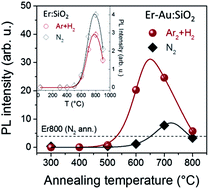
In Fig. 1 (main panel) the integrated Er3+ PL emission measured at 1540 nm under resonant excitation at 488 nm is reported as a function of the annealing temperature for the two Er–Au co-implanted sample sets annealed in reducing (Ar + H2) atmosphere (red dots) and in inert (N2) atmosphere (gray diamonds), respectively. The horizontal dashed line indicates the PL signal detected from the Er800 reference sample (i.e., the Er-doped silica sample, annealed at 800 °C in N2 atmosphere, not further implanted with Au). The inset shows the evolution as a function of the temperature, for thermal treatments in Ar + H2 and N2 atmosphere, of the PL intensity of Er-implanted silica samples in which only Er implantations were performed, at the same conditions as those used for the Er and Au co-implantations. In both cases the maximum Er3+ luminescence signal was obtained for the sample annealed at 800 °C, but the maximum PL intensity was 40% higher for the sample annealed in an inert atmosphere compared to the one treated in a reducing atmosphere. Concerning the Er–Au co-implanted samples, the PL data in Fig. 1 (main panel) show that for high temperature annealing treatments (above 600 °C) the Er3+ luminescence intensity is significantly increased with respect to the emitted intensity of the Er800 reference sample and the annealing atmosphere has a dramatic impact on such an enhancement. Upon annealing at 700 °C, a 2× amplification of the PL emission is obtained for thermal treatments in the N2 atmosphere, while a 6× enhancement is produced in the Ar + H2 atmosphere. At present, it is well observed that the sensitization of the Er3+ luminescence in Er–Au co-implanted silica samples is induced by the formation, upon thermal annealing, of sub-nanometric AuN quantum clusters that act very efficiently as energy-transfer agents to the rare-earth ions.14,15,27 Conversely, the role of the annealing atmosphere for Er–Au co-implanted samples in which Au is incorporated at a very low concentration, as in the present case, and particularly the demonstrated beneficial effect of thermal treatments in a reducing (Ar + H2) atmosphere is still to be clarified and needs to be deeply investigated.
image file: c6ra16931g-f1.tif
Fig. 1 Main panel: integrated PL intensity at 1540 nm as a function of the annealing temperature of Er–Au co-implanted samples annealed in reducing (Ar + H2, red dots) and in inert (N2, gray diamonds) atmospheres. The samples have been resonantly excited at 488 nm with a photon flux of 3 × 1018 cm−2 s−1. The horizontal dashed line indicates the PL intensity of the Er800 reference sample. Inset: PL emission at 1540 nm as function of the annealing temperature in N2 (open diamonds) and Ar + H2 (open dots) atmospheres of Er-implanted silica (the same Er implantation scheme as for the Er–Au co-implanted samples was used). In both panels the solid lines are best-fits to the experimental data according to the model described in the text.

A simple phenomenological model can be introduced to describe the observed evolution of the PL emission as a function of the annealing temperature.28 The basic idea is that the Er luminescent emission is proportional to the population of Er3+ ions that are optically active in the samples and competitive mechanisms occurring during the thermal treatments (as recovery of matrix defects or diffusion of quenching species) give rise to the net active Er3+ population, NEr,A. Assuming an initial population of Er ions, NEr, and defining as NEr,D the population of Er ions that can be eventually de-activated upon the annealing process due to quenching phenomena, and thus do not contribute to the PL emission, the evolution of NEr,A and NEr,D during the annealing duration (1 h in the present case) can be described by the following coupled rate-equations:

 
image file: c6ra16931g-t1.tif(1a)
 
image file: c6ra16931g-t2.tif(1b)
with initial conditions: NEr,A(0) = NEr,D(0) = 0 (no Er3+ PL signal was detected from the as-implanted samples).

RA and RD are, respectively, the rates of optical activation and de-activation of Er ions in the samples and they can be expressed by Arrhenius equations as:

 
RA(T) = R0,Ae(−EA/kBT) (2a)
 
RD(T) = R0,De(−ED/kBT) (2b)
where EA and ED are the corresponding Arrhenius activation energies; kB is the Boltzmann constant. By solving eqn (1) and taking into account the temperature dependence of the activation and de-activation rates in (2), an analytical expression for the optically active Er population versus annealing temperature, NEr,A(T), can be determined. Considering that IPLNEr,A, this expression can be used to fit the trend of the PL emission intensity as a function of annealing temperature, obtaining an estimation of the Arrhenius activation energies, EA and ED. The continuous lines in Fig. 1 are best-fits to the experimental data performed in this way. An important comment has to be done at this point. In the regime of low photon flux, as it is for the PL measurements reported in Fig. 1, the luminescent emission intensity is given more precisely by the expression IPLNEr,AσexcQ, in which σexc is the Er3+ excitation cross-section and Q = τ/τr is the quantum yield of the Er emission transition at 1540 nm (τ is the emission lifetime and τr is the radiative decay time of the Er3+ excited state in the Er–Au co-implanted samples).14 Even if the dominant behavior is expected to be related to the evolution upon annealing of NEr,A, nonetheless in principle all these parameters may depend on the annealing temperature and it is not easy to decouple the contribution of each term to the detected PL intensity. Moreover, several mechanisms can contribute at the same time to both the optical activation of Er ions as well as to their possible de-activation during the thermal annealing of the samples. Within this framework, the activation energies EA and ED that can be estimated by fitting the PL emitted intensity as a function of the annealing temperature have to be interpreted as effective energies to represent the dominant mechanisms.

As a first step, this approach has been used to fit the evolution of the PL intensity versus annealing temperature of the Er-implanted silica samples (see inset in Fig. 1). For both sets of samples (treated in inert and reducing atmosphere) we get EA = (1.37 ± 0.01) eV and ED = (2.63 ± 0.01) eV, with pre-factors R0,A ≈ 4 × 10³ s−1 and R0,D ≈ 4 × 10⁷ s−1. For Er-implanted silica systems, it is known that the progressive recovery of the SiO2 matrix from implantation damage (and the removal of irradiation defects that may act as nonradiative recombination centers),29 as well as the restoration of the full octahedral coordination of oxygen atoms around the Er3+ ions, is beneficial for the Er luminescence giving rise to an increase of the emitted intensity.30 The activation energy for molecular oxygen diffusion in silica was found to be in the range of 1.1–1.3 eV,31,32 in agreement with the values of EA determined by the fits. Concerning the de-activation mechanisms, instead, the PL intensity drop observed upon annealing at T > 900 °C can be attributed to possible Er clustering.30 An activation energy of 5.3 eV was recently determined for Er diffusion in silica33 that was significantly higher than the estimated ED. Nonetheless, possible structural rearrangements of the implanted silica matrix have to be considered that could speed up the clustering process. Moreover, upon annealing treatment, hydroxyl groups (OH) can penetrate the Er-containing layer (from the external environment or the silica substrate itself). OH groups are known to be efficient quenching species for the Er3+ luminescence34 and an activation energy of about 0.82 eV was found for their diffusion in amorphous silica upon annealing in the temperature range explored in the present work,35 thus giving rise to a lower effective activation energy for the de-activation process, as estimated in the present case.

Concerning the Er–Au co-implanted samples (main panel in Fig. 1) the fitting procedure yielded EA = (1.7 ± 0.2) eV and ED = (2.2 ± 0.3) eV for the samples annealed in N2 atmosphere, and very similar values, EA = (1.71 ± 0.02) eV and ED = (1.96 ± 0.01) eV, for those thermally treated in Ar + H2; in both cases the pre-factors were R0,A ≈ 4 × 10⁴ s−1 and R0,D ≈ 4 × 10⁷ s−1. For this class of systems, the Er3+ luminescence is controlled by the energy-transfer from the AuN quantum clusters, formed upon annealing, to the Er3+ ions and the most effective sensitization is obtained when the AuN cluster size is of the order of N = 10–20 atoms per cluster.14,15 The growth of AuN clusters with larger size produces instead a decrease of the sensitization efficiency and thus of the Er3+ PL emission. Within this framework, the activation energies EA and ED determined by the fits can be related to processes involving Au diffusion and nucleation. An activation energy of 2.14 eV is reported in the literature for Au diffusion in silica upon thermal annealing,36 but a much lower value was demonstrated for radiation damage assisted diffusion processes. As an example, for Au clustering in Au-implanted silica samples (at a fluence of 3 × 10¹⁶ Au+ per cm2) an activation energy of 1.17 eV was measured, which was interpreted taking into account the role of oxygen in promoting gold diffusivity and assuming a thermodynamic interaction between gold and oxygen.37,38 Furthermore, it is interesting to note that in the present case the same values of EA and ED have been obtained for the two sets of samples treated in inert or reducing atmospheres. This result is consistent with the findings of a structural study on the AuN cluster formation performed on these samples by HERFD-XAS analyses,24 as discussed below.

Structural characterization of the Er–Au co-implanted samples was performed by HERFD-XAS measurements at the Au L3 absorption edge. Due to its unique capability of revealing the aggregation of Au atoms in few-atom clusters since the very early stages of nucleation, the HERFD-XAS technique allowed us to follow the Au cluster growth upon annealing even in the ultra-low size range demonstrated for the samples investigated in the present work. All the details of the experimental conditions of the HERFD-XAS measurements and the data analysis are reported in ref. 24. In Table 1 we reported the main HERFD-XAS results obtained for the samples of the two series investigated in the present work. Ultra-small AuN clusters with the same size (number of atoms per cluster, N) and size evolution upon annealing are formed in both sample series, independently of the annealing atmosphere. Moreover, the HERFD-XANES (X-ray Absorption Near-Edge Structure) analysis reported in ref. 24 indicates that the Au clusters can be sketched as an effective core–shell system in which the Au atoms in the ‘core’ develop a metallic character, whereas the Au atoms in the ‘shell’ retain a partially covalent bond with O atoms of the matrix. Particularly, the Au–O coordination is favored for smaller cluster size and lower annealing temperature. Interestingly, a net difference between the two sample sets investigated in the present work is observed in the Debye–Waller (DW) factor (see Table 1) that is lower in the samples annealed in reducing atmosphere with respect to those treated in an inert atmosphere, especially for the samples annealed at 600 °C. The higher DW factor was related to a higher degree of configurational disorder of Au clusters induced by the defective matrix upon annealing in inert atmosphere.24 Indeed, while nitrogen permeation and diffusion in silica is very poor in this range of temperatures, H atoms from the reducing atmosphere can enter into the matrix and contribute to passivate Si dangling bonds. This favors the most stable Au cluster configurations, thus resulting in a lower structural disorder of Au sites and a smaller DW factor upon annealing in a reducing atmosphere than in the inert one.24

Table 1 HERFD-XAS results on the AuN cluster size evolution as a function of annealing temperature of Er–Au co-implanted samples annealed in N2 and in Ar + H2 atmospheres: dAu–Au is the Au–Au interatomic distance, N is the average number of Au atoms per cluster and DW is the Debye–Waller factor of the Au–Au correlation
Samples Er–Au Ann. T (°C) dAu–Au (Å) N (atoms) DW (×10−4 Å2)
N2 400 2.61 ± 0.03 2.2 ± 0.7 70 ± 30
600 2.69 ± 0.02 5.7 ± 1.2 131 ± 6
800 2.74 ± 0.01 12.7 ± 2.4 70 ± 2
Ar + H2 400 2.62 ± 0.02 2.5 ± 0.8 49 ± 6
600 2.68 ± 0.03 5.0 ± 1.9 61 ± 4
800 2.74 ± 0.01 12.7 ± 2.4 62 ± 3


Structural information on the samples was also obtained by investigation of their PL emission spectra. In Fig. 2, we report the room temperature PL spectra in the wavelength range 600–1700 nm of the Er–Au co-implanted samples annealed at the different indicated temperatures. Panel (a) refers to the samples annealed in a reducing atmosphere (Ar + H2), while panel (b) to those treated in an inert atmosphere (N2). The spectrum of the Er800 reference sample is also reported for comparison in both panels (black curve). The measurements have been taken under cw resonant Er3+ excitation at 488 nm. To increase the readability of the plots, different vertical scales have been used for the wavelengths shorter (left-hand scale) and longer (right-hand scale) than 1400 nm, and the spectra have been vertically shifted of a constant value. The peak visible in the wavelength region 1400–1700 nm is the characteristic luminescence band at 1540 nm of Er3+ ions in silica whose temperature evolution upon annealing is shown in Fig. 1. About the spectral features visible in the wavelength range 600–1400 nm (named as bands A, B and C in Fig. 2), it has been recently demonstrated on similar Er–Au co-implanted silica samples that such features are unambiguously related to the presence of ultra-small AuN quantum clusters in the samples.23 Owing to their sub-nanometric size, indeed, these clusters behave as quantum-dots and efficient radiative relaxation can be obtained even at room temperature.39,40 Particularly, bands A and C were attributed respectively to interband (d-sp) and intraband (sp) electronic transitions of the AuN quantum clusters, while band B was related to the formation of electronic surface states at the clusters. Specifically, the formation of this band, which is resonant with a corresponding Er3+ absorption band at 980 nm, was proved to be the channel for the transfer of electromagnetic energy from the Au clusters to the Er3+ ions, giving rise to the Er3+ luminescence sensitization.23 In order to highlight how these bands evolve as a function of the annealing temperature and the role of the annealing atmosphere, we have performed multiple Gaussian peak deconvolution of the spectra in the energy domain. As an example in Fig. 3a we have plotted the spectra and the fitting results of the Er–Au co-implanted samples annealed at 600 °C in the two atmospheres. Three main bands have been resolved, with peaks at about 1.7 eV (band A), 1.3 eV (band B) and 1 eV (band C). In Fig. 3b the amplitude of each Gaussian peak, as resulting from the fits, has been reported as a function of the annealing temperature. A similar trend with the temperature is observed for the three bands. Particularly, the amplitude of each band decreases for annealing at T > 600 °C in agreement with the growth of the AuN clusters that progressively loose their “quantum-dot” character. Furthermore, it is interesting to note that above 600 °C the amplitude of each band is higher for thermal treatment in a reducing atmosphere with respect to annealing in an inert atmosphere. This behavior is consistent with the higher degree of configurational disorder revealed by HERFD-XAS (higher DW factor) in the samples treated in N2 than in Ar + H2, which may induce the damping of the luminescence bands due to inhomogeneous broadening.


image file: c6ra16931g-f2.tif
Fig. 2 Room temperature PL emission spectra of the Er–Au co-implanted samples annealed (a) in a reducing (Ar + H2) atmosphere and (b) in an inert (N2) atmosphere at the different indicated temperatures. The measurements have been performed with resonant cw excitation at 488 nm. The PL spectrum of the Er800 reference sample is also reported for comparison (black line). In both panels, different vertical scales have been used for the wavelength range 600–1400 nm (left-hand scale) and 1400–1700 nm (right-hand scale), as marked by the vertical dashed line. The spectra are vertically shifted to increase the readability.

image file: c6ra16931g-f3.tif
Fig. 3 PL intensity versus emission energy of the Er–Au samples annealed at 600 °C in reducing (Ar + H2) and inert (N2) atmospheres. The continuous lines are the result of multiple Gaussian peak fittings. (b) Amplitude of the three main bands resulting from the Gaussian fits as a function of the annealing temperature for the two sample sets annealed in Ar + H2 (red symbols) and in N2 (open symbols) atmospheres.

From a structural point of view, all the experimental findings revealed strong similarities in the behavior of the two classes of samples, independent of the annealing atmosphere. In an attempt to explain the amplified sensitization demonstrated in the samples for thermal treatment in a reducing atmosphere (see Fig. 1), thus we addressed the issue from a different perspective: not only from the point of view of the sensitizing agents (AuN clusters), but also from that of the emitters (Er3+ ions). To do so, time resolved PL characterization of the samples has been performed to determine the lifetime of the Er3+ emission, and its evolution upon annealing in the different environments. As an example, in Fig. 4a we report the temporal decay curves of the PL emission at 1540 nm of the two Er–Au samples annealed at 700 °C. For both sample series, they represent the samples at which the measured Er3+ PL intensity is the highest (Fig. 1). The PL decay of the Er800 reference sample is also reported for comparison. Stretched exponential fits to the experimental decays have been performed, with the lifetime, τ, and the stretching parameter, β, as free parameters. Then the effective lifetime (τeff) of the PL emission has been calculated through eqn (3), as described in ref. 15:

 
image file: c6ra16931g-t3.tif(3)
Γ(x) is the Eulerian gamma function.


image file: c6ra16931g-f4.tif
Fig. 4 (a) Temporal decay curves of the PL emission at 1540 nm, resonantly excited at 488 nm, of the Er–Au samples annealed at 700 °C in the two atmospheres. The continuous curves are best-fits obtained with a stretched exponential decay function. The PL decay of the Er800 reference sample is also reported for comparison. (b) Effective lifetimes (τeff) and (c) stretching parameters (β) as a function of the annealing temperature of the two sample sets annealed in reducing (red closed symbols) and inert (open symbols) atmospheres. The horizontal dashed line in both panels indicates the values obtained from the PL decay of the Er800 reference sample.

In Fig. 4b and c the values of τeff and of the stretching parameters β estimated from the PL decays of the different samples have been reported as a function of the annealing temperature. In both panels the horizontal dashed line indicates the results of the Er800 reference sample: a single exponential decay (β = 1) was measured in this case (see Fig. 4a) and the lifetime is τ* = (10.7 ± 0.1) ms. A progressive increase of τeff by increasing the annealing temperature is observed for both samples sets, in agreement with the progressive recovery upon annealing of implantation-induced defects that may act as non-radiative recombination centers.30 Nonetheless, the emission lifetime of the Er800 reference sample cannot be recovered even for thermal treatments at the highest temperatures. This effect has been ascribed to the presence of the AuN clusters in the samples and the opening up of the energy-transfer paths to the Er ions,14 while the formation of non-radiative de-excitation channels as a consequence of the implantation process has been instead ruled out.30 The change of the local dielectric environment around the Er ions due to the incorporation of the AuN quantum clusters has been also taken into account as a possible further effect for the observed lifetime shortening in the Er–Au co-implanted samples.41,42 However, due to the structural characteristics of the samples (ultra-small Au cluster size and size distribution) a quantitative estimation of this effect was not possible and its role can only be guessed. By comparing the data of the two sets of samples in Fig. 4b and c, we observe that slightly longer lifetimes and larger values of the stretching parameter are obtained for the samples annealed in Ar + H2 with respect to those thermally treated in a N2 atmosphere. This suggests that a better local environment is created around the Er3+ ions if the annealing treatments are performed in a reducing atmosphere, with a reduction of the non-radiative decay rate due to the possible passivation of non-radiative de-excitation paths.

To investigate more deeply the effect of the annealing atmosphere on the Er3+ luminescence emission in the Er–Au co-implanted samples, we measured the evolution of the PL intensity emitted at 1540 nm as a function of the photon flux. This study has been done on the samples of both sets that showed the highest PL emission upon low pumping flux (in both cases such “best-performing” samples are those annealed at 700 °C, see Fig. 1). The data have been analyzed according to the phenomenological model reported in ref. 14, which allowed us to determine quantitatively the main photo-physical parameters that control the Er3+ emission properties of the samples, as the effective sensitization cross-section (σeff) and the fraction of Er3+ ions sensitized by the AuN clusters (fs), and the luminescence quantum yield of the Er emission transition at 1540 nm (Q). Fig. 5 shows the trend of the PL intensity at 1540 nm as a function of the photon flux of the two “best-performing” samples resonantly excited at 488 nm. According to the proposed model, for resonant Er3+ excitation the PL emitted intensity at 1540 nm of the Er–Au co-implanted samples can be written as a function of the photon flux by the following expression:

 
image file: c6ra16931g-t4.tif(4)


image file: c6ra16931g-f5.tif
Fig. 5 PL intensity at 1540 nm as a function of the photon flux for the Er–Au co-implanted samples annealed at 700 °C in a reducing (Ar + H2) atmosphere (red dots) and in an inert (N2) atmosphere (gray diamonds). The continuous lines are best-fits to the experimental data obtained according to the model described in the text. The measurements have been taken under resonant Er excitation at 488 nm.

The first term in the right-hand side of eqn (4) represents the contribution to the PL signal given by the fraction fs of optically active Er ions (NEr,A) indirectly sensitized by the AuN clusters, while the second term accounts for the direct excitation by the laser beam of the residual fraction fd (fs + fd = 1). The constant K is used to take into account the collection efficiency of the PL setup. σ represents the intrinsic emission cross-section of the 4I13/24I15/2 Er3+ transition and it was estimated by applying the Fuchtbauer–Ladenberg approach43 to the analysis of the Er3+ emission spectrum, obtaining σ = 4.4 × 10⁻²¹ cm2,14 in agreement with the values determined with different methods for various Er-implanted photonic materials.2 A summary of the main results determined by the analysis of the two “best-performing” samples according to this model is reported in Table 2. A comment is needed concerning the parameter reported in the last column, [Q with combining tilde]. As discussed in detail in ref. 14, by analyzing the data with the proposed model it is not possible to decouple the quantum yield Q and the parameter ξ = NEr,A/NEr, and the product of these two terms [Q with combining tilde] = ξQ is indeed obtained by the fits. ξ represents the fraction of optically active Er ions in the Er–Au co-implanted samples with respect to the population in the reference sample Er800, and it is introduced in the model to account for all the possible effects that may reduce such population (thus giving ξ ≤ 1), as for example residual damage due to Au implantation not completely recovered by the thermal treatment. By comparing the results in Table 2 we can observe that both samples are characterized by a similar sensitization cross-section (about 3 orders of magnitude larger than the intrinsic emission cross-section, σ) and sensitized fraction (fs < 1%). Conversely, very different values of the effective quantum yield [Q with combining tilde] have been determined, whose ratio is: ([Q with combining tilde])Ar+H2/([Q with combining tilde])N2 = 2.8 ± 0.2. With the definition of [Q with combining tilde] given above we can write:

 
image file: c6ra16931g-t5.tif(5)

Table 2 Effective lifetime, τeff, effective sensitization cross-section (at 488 nm), σeff, sensitized fraction, fs, and effective quantum yield, [Q with combining tilde] = ξQ of the Er–Au co-implanted samples annealed at 700 °C in reducing (Ar + H2) and inert (N2) atmosphere. The parameters have been determined by the analysis of the PL emitted intensity versus photon flux under resonant Er excitation at 488 nm, as described in the text
Samples τeff (ms) σeff (×10−18 cm2) fs (%) [Q with combining tilde] (%)
Er1Au0.5–H700 5.76 ± 0.04 2.2 ± 0.2 0.50 ± 0.06 73 ± 5
Er1Au0.5–N700 5.04 ± 0.04 2.9 ± 0.8 0.39 ± 0.08 26 ± 1


The second equality in eqn (5) is obtained by writing the luminescence quantum yield in the form Q = τeff/τr and assuming the same radiative lifetime (τr) for the two samples, i.e., independently of the annealing atmosphere. Considering the very strong structural similarities demonstrated for the two classes of samples, this hypothesis is quite reasonable. Therefore, by inserting in eqn (5) the values of τeff obtained for the two samples annealed at 700 °C (see Fig. 4b) we can provide an estimation of the ratio between the population of optically active Er ions in the two samples, that results: (NEr,A)Ar+H2/(NEr,A)N2 = 2.5 ± 0.2. Thus, upon annealing in an Ar + H2 atmosphere a much larger population of Er3+ ions (by a factor of 2.5) are optically active in the Er–Au co-implanted sample with respect to the sample thermally treated in N2 atmosphere. This is indeed the dominant effect, responsible for the improved performance demonstrated for the Er–Au co-implanted samples annealed in a reducing atmosphere with respect to those heated in an inert atmosphere, that allowed us to get a very efficient Er3+ luminescence emission in spite of the very limited amount of sensitizing agents (AuN clusters) incorporated in the samples.

By taking into account all the experimental findings, the following scenario can be tentatively depicted. When the Er–Au co-implanted samples are annealed in a reducing (Ar + H2) atmosphere, hydrogen passivation of Si dangling bonds around the AuN clusters may occur24 which reduces the degree of configurational disorder of the gold clusters, as it is testified by the smaller Debye–Waller factor estimated by XAS analysis and the higher amplitude of Au-related PL bands. At the same time, such a passivation limits the possibility of formation of Si–O bonds. In this way, a higher amount of oxygen atoms becomes available for coordinating with Er ions in the octahedral configuration, which is optimal for their optical activation, thus increasing the population of optically active Er ions (NEr,A) in close proximity with the Au clusters, which is, in turn, the population of Er3+ ions that can be effectively sensitized by short-range energy transfer from the Au clusters. Moreover, it is worth noting that, upon passivation, a certain amount of H atoms are immobilized, thus reducing also the probability of formation of quenching species for the Er emission which is further beneficial for the Er3+ luminescence. Indeed, as previously discussed, hydrogen is known to act as a quenching species for the Er3+ luminescence in silica and different quenching mechanisms can be considered: H2 molecules can penetrate the silica network forming hydroxyl (OH) groups, which act as quenchers of the Er3+ emission2 or, as it has been recently demonstrated,44 the deactivation of excited Er3+ ions can occur through the energy transfer to H2 molecular vibrations. Such quenching mechanisms can explain instead the slightly lower PL emission which was detected from the samples implanted with Er only (without gold) and annealed in a reducing atmosphere, with respect to those thermally treated in an inert atmosphere (as shown in the inset in Fig. 1).

3 Conclusions

The role of the annealing atmosphere (reducing or inert) on the sensitization process of the Er3+ luminescence by AuN quantum clusters in silica has been investigated by PL characterizations of Er–Au co-implanted samples in which Au+ ions were incorporated at low fluence. In spite of the structural similarities observed between the two sample sets, the Er3+ luminescence was demonstrated to be significantly boosted in the samples annealed in reducing (Ar + H2) atmosphere with respect to those thermally treated in an inert (N2) atmosphere. By decoupling all the different contributions to the PL emission, such amplified luminescence was proved to be due to the increase (by a factor of 2.5) of the population of Er3+ ions that are optically active in the Er–Au co-implanted samples, owing to the H passivation of Si dangling bonds around the AuN clusters. Thus, an intense Er3+ luminescence emission can be obtained from the samples heated in Ar + H2 despite their very low Au content. These results are extremely valuable for the employment of AuN quantum clusters as rare-earth sensitizers in photonic devices since they show that by properly selecting the annealing atmosphere (reducing) it is possible to strongly enhance the luminescence emission performance of Er-doped silica samples using, at the same time, only a very limited amount of sensitizing agents.

Acknowledgements

The support of the Italian-Mexican Project of Major Importance (Progetto Grande Rilevanza Italia-Messico, MX14MO09) of the Ministero degli Affari Esteri e della Cooperazione Internazionale (MAECI) of Italy is gratefully acknowledged.

References

  1. K. Binnemans, Chem. Rev., 2009, 109, 4283–4374 CrossRef CAS PubMed.
  2. A. Polman, J. Appl. Phys., 1997, 82, 1–39 CrossRef CAS.
  3. W. J. Miniscalco, J. Lightwave Technol., 1991, 9, 234–250 CrossRef CAS.
  4. Y. Molard, C. Labbé, J. Cardin and S. Cordier, Adv. Funct. Mater., 2013, 23, 4821–4825 CAS.
  5. C. Strohhofer and A. Polman, Opt. Mater., 2003, 21, 705–712 CrossRef.
  6. M. P. Hehlen, N. J. Cockroft, T. R. Gosnell and A. J. Bruce, Phys. Rev. B: Condens. Matter Mater. Phys., 1997, 56, 9302–9318 CrossRef CAS.
  7. M. Fuji, M. Yoshida, Y. Kanzawa, S. Hayashi and K. Yamamoto, Appl. Phys. Lett., 1997, 71, 1198–1200 CrossRef.
  8. C. E. Chryssou, A. J. Kenyon, T. S. Iwayama, C. W. Pitt and D. E. Hole, Appl. Phys. Lett., 1999, 75, 2011–2013 CrossRef CAS.
  9. D. Pacifici, G. Franzò, F. Priolo, F. Iacona and L. Dal Negro, Phys. Rev. B: Condens. Matter Mater. Phys., 2003, 67, 245301 CrossRef.
  10. P. G. Kik, M. L. Brongersma and A. Polman, Appl. Phys. Lett., 2000, 76, 2325–2327 CrossRef CAS.
  11. C. Strohhofer and A. Polman, Appl. Phys. Lett., 2002, 81, 1414–1416 CrossRef CAS.
  12. M. Eichelbaum and K. Rademann, Adv. Funct. Mater., 2009, 19, 2045–2052 CrossRef CAS.
  13. M. Mattarelli, M. Montagna, K. Vishnubhatla, A. Chiasera, M. Ferrari and G. C. Righini, Phys. Rev. B: Condens. Matter Mater. Phys., 2007, 75, 125102 CrossRef.
  14. T. Cesca, B. Kalinic, C. Maurizio, C. Scian, G. Battaglin, P. Mazzoldi and G. Mattei, ACS Photonics, 2015, 2, 96–104 CrossRef CAS.
  15. C. Maurizio, E. Trave, G. Perotto, V. Bello, D. Pasqualini, P. Mazzoldi, G. Battaglin, T. Cesca, C. Scian and G. Mattei, Phys. Rev. B: Condens. Matter Mater. Phys., 2011, 83, 195430 CrossRef.
  16. T. Cesca, B. Kalinic, N. Michieli, C. Maurizio, C. Scian, G. Devaraju, G. Battaglin, P. Mazzoldi and G. Mattei, Phys. Chem. Chem. Phys., 2014, 16, 15158–15163 RSC.
  17. H. A. Atwater and A. Polman, Nat. Mater., 2010, 9, 205–213 CrossRef CAS PubMed.
  18. K. R. Catchpole and A. Polman, Appl. Phys. Lett., 2008, 93, 191113 CrossRef.
  19. R. A. Ganeev, Nonlinear Optical Properties of Materials, Springer, New York, 2013 Search PubMed.
  20. T. Cesca, P. Calvelli, G. Battaglin, P. Mazzoldi and G. Mattei, Opt. Express, 2012, 20, 4537–4547 CrossRef CAS PubMed.
  21. J. N. Anker, W. P. Hall, O. Lyandres, N. C. Shah, J. Zhao and R. P. Van Duyne, Nat. Mater., 2008, 7, 442–453 CrossRef CAS PubMed.
  22. G. Mattei, P. Mazzoldi, M. L. Post, D. Buso, M. Guglielmi and A. Martucci, Adv. Mater., 2007, 19, 561–564 CrossRef CAS.
  23. T. Cesca, B. Kalinic, C. Maurizio, C. Scian, G. Battaglin, P. Mazzoldi and G. Mattei, Nanoscale, 2013, 6, 1716–1724 RSC.
  24. C. Maurizio, T. Cesca, G. Perotto, B. Kalinic, N. Michieli, C. Scian, Y. Joly, G. Battaglin, P. Mazzoldi and G. Mattei, Nanoscale, 2015, 7, 8968–8977 RSC.
  25. J. F. Ziegler, J. P. Biersack and M. D. Ziegler, SRIM – The Stopping Range of Ions in Matter, Ion Implantation Press, 2008 Search PubMed.
  26. J. M. Khosrofian and B. A. Garetz, Appl. Opt., 1983, 22, 3406–3410 CrossRef CAS PubMed.
  27. T. Cesca, C. Maurizio, B. Kalinic, C. Scian, E. Trave, G. Battaglin, P. Mazzoldi and G. Mattei, Nucl. Instrum. Methods Phys. Res., Sect. B, 2014, 326, 7–10 CrossRef CAS.
  28. S. Abedrabbo, B. Lahlouh and A. T. Fiory, J. Phys. D: Appl. Phys., 2011, 44, 315401 CrossRef.
  29. G. W. Arnold and P. Mazzoldi, in Ion beam modification of insulators, ed. P. Mazzoldi and G. W. Arnold, Elsevier, Amsterdam, 1987, ch. 5 Search PubMed.
  30. T. Cesca, C. Maurizio, B. Kalinic, G. Perotto, P. Mazzoldi, E. Trave, G. Battaglin and G. Mattei, Opt. Express, 2012, 20, 16639–16649 CrossRef CAS.
  31. F. J. Norton, Nature, 1961, 191, 701 CrossRef CAS.
  32. M. A. Lamkin, F. L. Riley and R. J. Fordham, J. Eur. Ceram. Soc., 1992, 10, 347–367 CrossRef CAS.
  33. Y.-W. Lu, B. Julsgaard, M. C. Petersen, R. V. S. Jensen, T. G. Pedersen, K. Pedersen and A. N. Larsen, Appl. Phys. Lett., 2010, 97, 141903 CrossRef.
  34. L. Slooff, M. de Dood, A. van Blaaderen and A. Polman, J. Non-Cryst. Solids, 2001, 296, 158–164 CrossRef CAS.
  35. R. Doremus, Diffusion of reactive molecules in solids and melts, John Wiley and Sons, New York, 2002 Search PubMed.
  36. D. R. Collins, D. K. Schroder and C. T. Sah, Appl. Phys. Lett., 1966, 8, 323–325 CrossRef CAS.
  37. A. Miotello, G. De Marchi, G. Mattei, P. Mazzoldi and C. Sada, Phys. Rev. B: Condens. Matter Mater. Phys., 2001, 63, 075409 CrossRef.
  38. P. Mazzoldi and G. Mattei, in Metal Nanoclusters in Catalysis and Materials Science, ed. B. C. S. Toshima, Elsevier, Amsterdam, 2008, pp. 269–291 Search PubMed.
  39. J. Zheng, C. Zhang and R. M. Dickson, Phys. Rev. Lett., 2004, 93, 077402 CrossRef PubMed.
  40. S. Link, A. Beeby, S. FitzGerald, M. A. El-Sayed, T. G. Schaaff and R. L. Whetten, J. Phys. Chem. B, 2002, 106, 3410–3415 CrossRef CAS.
  41. B. Kalinic, T. Cesca, N. Michieli, C. Scian, G. Battaglin, P. Mazzoldi and G. Mattei, J. Phys. Chem. C, 2015, 119, 6728–6736 CAS.
  42. J. Bao, N. Yu, F. Capasso, T. Mates, M. Troccoli and A. Belyanin, Appl. Phys. Lett., 2007, 91, 131103 CrossRef.
  43. W. Barnes, R. Laming, E. Tarbox and P. Morkel, IEEE J. Quantum Electron., 1991, 27, 1004–1010 CrossRef CAS.
  44. A. P. Bazakutsa and K. M. Golant, J. Non-Cryst. Solids, 2015, 411, 68–75 CrossRef CAS.

This journal is © The Royal Society of Chemistry 2016
Click here to see how this site uses Cookies. View our privacy policy here.