DOI:
10.1039/C6RA16847G
(Paper)
RSC Adv., 2016,
6, 75365-75375
Formation initiation and structural changes of phosphate conversion coating on titanium induced by galvanic coupling and Fe2+ ions
Received
30th June 2016
, Accepted 3rd August 2016
First published on 3rd August 2016
Abstract
The methodology of deposition of a phosphate chemical conversion coating on titanium (Ti) was modified by a galvanically coupled approach and addition of iron ions. The influences of the coupled Fe clip and Fe2+ ions in the coating bath on the formation, microstructure, phase composition and adhesion of the conversion coatings were studied by scanning electron microscopy, X-ray diffraction and scratch testing. Electrochemical measurements were also investigated to explore the formation of the coating. The results show that the coating formed on Ti was composed of scholzite (CaZn2(PO4)2·2H2O) and minor hopeite (Zn3(PO4)2·4H2O) crystals. During the coating deposition, the cathodic Ti promoted the dissolution of anodic Fe, which initiated the coating formation in the acidic bath. With increasing Fe2+ concentration, not only did the coating formation rate increase, but also the coating became fine and uniform with a good adhesion above 50 N. The deposition of the conversion coating was investigated in terms of the curves of pH–time and potential–time, which supported a better understanding of the formation mechanism of the conversion coating on Ti.
1. Introduction
Titanium (Ti) and its alloys are materials of choice for implantation due to their excellent chemical and physical characteristics such as high specific strength, the match of elastic modulus with human hard tissue and corrosion resistance.1–4 As one of the ideal implant candidates, research has concerned both its osteogenesis in the early period of implantation and long-term stability in an aggressive body environment.5–8 However, Ti is a biologically inert metal, which often somewhat influences its clinical applications.9 In spite of a passive oxide film on the surface, the poor tribological properties of Ti lead to release of wear debris from implants into surrounding tissue, which ultimately results in adverse pathophysiological effects, even failure of the implantation.10–12 A number of attempts have been made to improve the surface properties of Ti, such as alkali and heat treatment,7,13,14 pulsed laser deposition,15 plasma spraying,16,17 hydrothermal treatment,18 sol–gel,19,20 anodic oxidation21,22 and biomimetic methods.4,23,24 Nevertheless, limitations still exist such as high-cost, long coating duration, instability of the coating and interfacial separation under repeated loading conditions.1,25–27
Phosphate chemical conversion (PCC) technology is one of the effective surface treatments for metal materials in industry.28 Due to its advantages of low-cost, easy operation, suitable for treatment of irregular surfaces and ability to afford excellent adhesion and corrosion resistance of the matrix, it has been widely applied on steel,29,30 iron,31 magnesium (Mg) and its alloys32–34 for biomedical applications. However, there are few reports on coatings prepared on Ti using the method of PCC. It is difficult to convert a coating on Ti owing to the presence of the passive oxide layer on its surface using the traditional PCC technology.35 Therefore, some researchers have used an ultrasonic irradiation36 and electric field,37 or a hydrothermal treatment at 250 °C for 8 h (ref. 38) for forming a conversion coating on Ti. All these treatments above for Ti are feasible, nevertheless, either assistant external field or high temperature or long operating time is needed. In industry, besides these basic chemical, mechanical and electrochemical methods for accelerating low temperature zinc phosphating process,28,30,39–41 the utility of galvanic coupling on mild steel with other cathodic materials such as Ti, copper, brass, nickel, and stainless steel was established.42–44 In those researches, the galvanic coupling accelerated the dissolution of mild steel and enabled an earlier coating formation.
In this work, we took advantage of the anodic material of iron coupled with Ti to initiate the formation of conversion coatings on the surface of pure Ti in a PCC bath. Furthermore, we introduced pure iron powders into the PCC bath to increase the concentration of Fe2+ ions at the metal/solution interface. The microstructure and phase composition of the conversion coatings, the adhesive strength of coatings against Ti substrate were studied. Though the mechanism of coating formation on Fe or Mg by the PCC technology has been investigated in many researches,45–48 a more detailed coating deposition process is not completely understood. Moreover, unlike Fe or Mg substrates, Ti ions would not incorporate in the chemical conversion reactions under moderate conditions.38 It is suggested that the mechanism of coating deposition on Ti is different from that of on Fe and Mg. Therefore, we proposed a detailed deposition process of conversion coatings on Ti coupled with Fe in this study to help understand the mechanism of coating formation.
2. Materials and experimental methods
2.1. Specimens and coating
Ti disks with the dimension of Ø 10 × 3 mm were ground to a grit of 1000 and used as substrates. Prior to the PCC process, all Ti specimens were ultrasonically rinsed in acetone, alcohol and deionised water, sequentially. Then the Ti disk was connected with a pure iron clip to create a coupled combination noted as Ti/Fe. A point contact was used for the connection in order to increase the area ratio of Ti to Fe, as depicted in Fig. 1a. The uncoupled Ti disk was adopted as a control sample noted as Ti/UC. All the sample data are listed in Table 1. The Ti/Fe combination was pickled in 2% hydrofluoric acid (HF) at room temperature for 15 s to get a fresh surface of Ti. Afterwards, the combination underwent surface activation in an aqueous dispersion of 3 g L−1 colloidal titanium phosphate49–51 at room temperature for 30 s to reduce the surface potential of Ti and enhance the nucleation points on the surface. Same operations were carried out on the control samples for comparison.
 |
| | Fig. 1 Schematic illustration for (a) the sample Ti/Fe, (b) PCC processing steps. | |
Table 1 The information of the samples investigated in the work
| Sample ID |
Information |
| Ti substrate |
Commercially pure Ti disk with the composition in wt%: Fe: 0.09; C: 0.02; N: 0.01; Ti: balance |
| Ti/Fe |
A coupled combination of Ti disk connected with a pure iron clip |
| Ti/UC |
A control sample of Ti disk without coupling |
Then, the as-prepared samples were immediately immersed into a PCC bath contained 0.1 mol L−1 Zn(H2PO4)2·2H2O, 0.05 mol L−1 Ca(NO3)2·4H2O and 0.02 mol L−1 NaNO2. In order to increase the concentration of Fe2+ ions, as well as not to introduce other foreign ions into the baths, the PCC baths were cured with pure iron powders (AR, 98%) at room temperature for 24 h before immersion process. The total amount of added iron powders was 0 g L−1, 2.5 g L−1, 5.0 g L−1, 7.5 g L−1 and 10.0 g L−1, respectively. After curing treatment, the remaining iron powders in the baths were removed by filtration. Then the pH value of the PCC bath after curing was adjusted by H3PO4 or NaOH to 3.75. The PCC process was conducted at 55 °C for 30–50 min. Finally, coated Ti samples were washed with deionised water and dried for further characterization. The schematic diagram of the experimental procedures is presented in Fig. 1b. The obtained products and their experimental conditions are listed in Table 2.
Table 2 The products obtained from various experimental conditions
| Products |
Experimental conditions |
Coupled conditions |
| Solution |
Temperature |
Time |
| Pre-treated Ti |
Colloidal titanium phosphate |
Room temperature |
30 s |
Coupled with Fe |
| Ti/UC |
PCC bath with 5 g L−1 iron powders |
55 °C |
50 min |
Uncoupled |
| Ti/Fe |
PCC bath with 5 g L−1 iron powders |
55 °C |
50 min |
Coupled with Fe clip |
| a |
PCC bath without curing treatment |
55 °C |
30 min |
Coupled with Fe clip |
| b |
PCC bath with 2.5 g L−1 iron powders |
55 °C |
30 min |
Coupled with Fe clip |
| c |
PCC bath with 5.0 g L−1 iron powders |
55 °C |
30 min |
Coupled with Fe clip |
| d |
PCC bath with 7.5 g L−1 iron powders |
55 °C |
30 min |
Coupled with Fe clip |
| e |
PCC bath with 10 g L−1 iron powders |
55 °C |
30 min |
Coupled with Fe clip |
2.2. Characterization
The iron concentration of the PCC bath before and after coating process was determined by inductively coupled plasma-atomic emission spectrometry (ICP-AES) (iCAP 6300, Thermo Jarrell Ash Co.). The morphologies of the conversion coatings were observed using an SU-70 field emission scanning electron microscope (FE-SEM). The phase composition of the coating was examined by a Rigaku D/max-γB X-ray diffractometer (XRD) using a Cu-Kα radiation (λ = 1.5418 Å). The XRD profiles were recorded in step-scan intervals of 0.02° 2θ at a scanning speed of 4° per min between 5° to 60°, operated at 40 kV and 100 mA. The adhesive strength between the conversion coating and Ti substrate was investigated by a scratch tester (WS-2005, Lanzhou Institute of Chemical Physics, CAS, China) with a diamond stylus (120° cone with a 200 μm radius tip) under a continuously increasing load. Scratches were made to a maximum load 100 N at a loading rate of 100 N min−1 and the scratch length was 5 mm. The load at which the coating was totally peeled off from the Ti substrate was designated as the critical load (Lc) and the moment was recorded by an acoustic emission (AE) signal. The scratch tests were measured in a quiet environment and each specimen was scratched twice to ensure the reliability of the results.
2.3. Electrochemical measurements
Electrochemical measurements of the conversion coatings were carried out on a PARSTAT 2273 automatic laboratory corrosion measurement system. A classical three-electrode cell was used with a saturated calomel electrode (SCE) as the reference electrode, platinum as the counter electrode and the samples with 1 cm2 exposed area as the working electrode. A potential versus time curve was recorded to investigate the deposition of conversion coating by monitoring the open circuit potential (OCP) of the specimen in the PCC bath at 55 °C as a function of immersion time. The OCP was recorded from the beginning of immersion to 3000 s.
3. Results and discussion
3.1. Effect of galvanic coupling
Fig. 2a and b shows the FE-SEM images of the bare Ti specimen pretreated in colloidal titanium phosphate for 30 s. Fig. 2c–f shows the surface morphologies of Ti obtained from Ti/UC and Ti/Fe, which were treated for 50 min in the PCC bath cured with 5 g L−1 pure iron powders for 24 h. White irregular sites are observed on the surface of bare Ti, which are mainly Na4TiO(PO4)2·(0–7)H2O crystal nuclei performing as surface activated sites as a result of treatment in Ti colloids,49–51 as shown in Fig. 2a and b. Similar irregular sites are also observed for Ti/UC (Fig. 2c and d). The difference is that both the amount and area of Na4TiO(PO4)2·(0–7)H2O crystals on Ti/UC are declining, which can be attributed to the acid attack during PCC process. However, no phosphate crystals are observed except for some corrosion defects on the surface of Ti/UC. In contrast, the surface of Ti/Fe presents loose but uniform surface coating containing a number of laminar crystals with a thickness of less than 100 nm, as shown in Fig. 2e and f. These laminas accumulate to 3-dimensional radial clusters with a diameter of about 10 μm. It is suggested that the 3-dimensional matrix is not only relevant to cell expansion, but also conductive to regulating cell proliferation and differentiation.52,53
 |
| | Fig. 2 Surface morphologies of Ti obtained from (a) pre-treated Ti, (c) Ti/UC and (e) Ti/Fe. (b), (d) and (f) are their corresponding high magnification images, respectively. | |
Fig. 3 shows the surface morphologies of Fe obtained from Ti/Fe, which was also treated for 50 min in the PCC bath cured with 5 g L−1 pure iron powders for 24 h. It can be seen that the surface of Fe is covered with dense crystals with a size of about 1 μm. The coating is uniform but cracked, as shown by arrows in Fig. 3a and b. This indicates that the surface of Fe may be over treated after immersion for 50 min in the PCC bath. From the observation of Fig. 2 and 3, it is also indicated that the formation of a conversion coating on Ti needs longer time than that of Fe.
 |
| | Fig. 3 Surface morphologies of Fe obtained from Ti/Fe. (a) Low and (b) high magnification image. | |
Fig. 4a and b reveals the XRD patterns of the conversion coatings formed on Ti and Fe, respectively. In Fig. 4a, besides the diffraction peaks from the Ti substrate, the diffraction peaks of typical scholzite (CaZn2(PO4)2·2H2O, JCPDS #35-0495) with minor hopeite (Zn3(PO4)2·4H2O, JCPDS #37-0456) are detected on the surface of Ti. Scholzite crystals mainly grow along (200), (![[3 with combining macron]](https://www.rsc.org/images/entities/char_0033_0304.gif) 11) and (420) planes at 2θ degree of 10.3°, 21.3° and 31.8°, respectively. Hopeite crystals are mainly detected at 2θ degree of 9.7° and 19.4°, indicating the planes of (020) and (220). It is reported that scholzite can act as nucleating agent for hydroxycarbonate apatite54,55 and has a stimulatory effect on bone formation.31,56–58 Hopeite is considered as a potential versatile biomaterial which can be transformed into hydroxyapatite in calcium nitrate solution59–61 and it has been widely used in dentistry.62 Therefore, it is suggested that such a conversion coating would improve the biocompatibility and osteogenic effects of Ti implant. In Fig. 4b, the diffraction peaks detected on the surface of Fe are indexed to phosphophyllite (Zn2Fe(PO4)2·4H2O, JCPDS #29-1427) besides scholzite. Phosphophyllite exhibits better corrosion resistance in sodium chloride solution than hopeite or scholzite, due to its chemical stability in aqueous media.28,63 So the coating on Fe can inhibit the further corrosion of Fe in the acidic PCC bath.
11) and (420) planes at 2θ degree of 10.3°, 21.3° and 31.8°, respectively. Hopeite crystals are mainly detected at 2θ degree of 9.7° and 19.4°, indicating the planes of (020) and (220). It is reported that scholzite can act as nucleating agent for hydroxycarbonate apatite54,55 and has a stimulatory effect on bone formation.31,56–58 Hopeite is considered as a potential versatile biomaterial which can be transformed into hydroxyapatite in calcium nitrate solution59–61 and it has been widely used in dentistry.62 Therefore, it is suggested that such a conversion coating would improve the biocompatibility and osteogenic effects of Ti implant. In Fig. 4b, the diffraction peaks detected on the surface of Fe are indexed to phosphophyllite (Zn2Fe(PO4)2·4H2O, JCPDS #29-1427) besides scholzite. Phosphophyllite exhibits better corrosion resistance in sodium chloride solution than hopeite or scholzite, due to its chemical stability in aqueous media.28,63 So the coating on Fe can inhibit the further corrosion of Fe in the acidic PCC bath.
 |
| | Fig. 4 XRD patterns of the conversion coatings formed on (a) Ti substrate and (b) Fe clip obtained from Ti/Fe. | |
From the analyses of SEM and XRD, it is evident that the galvanic coupling plays a significant role on the formation of conversion coatings. Although the surface of Ti was activated in advance, it still exhibits chemical stabilization in the PCC bath at pH 3.75, 55 °C.35,64 Therefore, when the Ti specimen is immersed into the PCC bath by itself, it is difficult to deposit a scholzite coating on it because the electrochemical reaction in the electrolyte cannot occur, as shown in Fig. 2c and d. When the Ti substrate is connected with Fe, a bimetal couple is established.65 Fe with a lower potential in the galvanic couple will be anodic and will corrode with respect to cathodic cell of Ti in the acidic PCC bath. Galvanic corrosion of the combination initiates the chemical reaction of PCC process, and then a series of chemical conversion reactions take place.42,43 The details of the formation of PCC coating on the surface of Ti will be discussed in the following sections.
3.2. Effect of concentration of Fe2+
In order to better understand the effect of iron concentration on the coating formation, Ti/Fe samples were immersed into the PCC baths with various levels of pure iron powders. The diagram in Fig. 5 depicts the changes in iron concentrations in the bath before and after the PCC process following the increased amounts of iron powders from 0 to 10.0 g L−1. The pictures under the diagram in Fig. 5 show the PCC baths with 0–10 g L−1 iron powders before the PCC process. It can be seen that the iron concentration in the bath before coating process increases with the increase of the amounts of iron powders, which is accompanied by the change in color of the baths from white to yellow-green, and darkens. This change in color reflects the increasing of concentration of Fe2+ in the bath, caused by the dissolution of iron powders in the acidic PCC bath. After the PCC process, it is interesting to find that the iron concentration in the bath with 0 g L−1 iron powders increases from 0 to almost 1 × 10⁻⁵ mol L−1. It is suggested that without the addition of iron powders, Fe also can be detected in the bath after PCC process. Thus, the detected Fe in the bath is attributed not only to the dissolution of added iron powders, but also to the migration of Fe from the anodic Fe clip. For the other baths with addition of iron powders, the iron concentrations decreases after the PCC process, indicating that some of the Fe ions in these baths have been consumed during the coating deposition. The result of Fe release further proves that both the galvanic coupling with Fe and the addition of pure iron powders play an important role in the process of coating deposition.
 |
| | Fig. 5 Iron concentrations in the PCC bath before and after the PCC process following the increased mass of iron powders. The pictures under the diagram show the PCC bath after curing treatment with 0–10 g L−1 iron powders at room temperature for 24 h. | |
Fig. 6 shows the XRD patterns of the coatings on Ti obtained from Ti/Fe after PCC process in the baths with various mass of pure iron powders. It is revealed that the coatings consist of scholzite (JCPDS #35-0495) crystals which mainly grow along (200) and (11) planes at 2θ degree of 10.3° and 21.4°, respectively. The peak intensities increase with an increase in the concentration of Fe2+, indicating that the growth of scholzite is promoted by Fe2+ ions. This may be attributed to the increase of coating formation rate and the effect of Fe2+ ions on the scholzite crystallization. In addition, minor hopeite (JCPDS #37-0456) crystals are also detected in the coatings, which mainly grow along (020) and (220) planes at 2θ degree of 9.7° and 19.4°, respectively. Amorphous humps are detected at 2θ degree of about 15–25° in the XRD patterns of samples treated at 2.5–10.0 g L−1 conditions (Fig. 6b–e), indicating that the amorphous precipitation may take place during the coating deposition, due to the addition of iron powders.
 |
| | Fig. 6 XRD patterns of the conversion coatings on Ti obtained from Ti/Fe after treatment in the PCC bath cured with various mass of pure iron powder for 24 h. (a) 0 g L−1, (b) 2.5 g L−1, (c) 5.0 g L−1, (d) 7.5 g L−1 and (e) 10.0 g L−1. | |
Fig. 7 shows the surface morphologies of Ti obtained from Ti/Fe after PCC process for 30 min in the baths cured with different amounts of iron powders. It is observed that only several discrete crystals are formed on the surface of Ti without curing treatment (Fig. 7a). In contrast, coatings with plenty of crystallites are present on the surfaces of the samples treated in the cured solution (Fig. 7b–e). In particular, the coating treated with 2.5 g L−1 iron powders consists of randomly arranged crystals with a size of 10–20 μm and occasionally, a fraction of the substrate is exposed, as shown in Fig. 7b. When the mass of iron powders increases to 5.0 g L−1, the coating becomes uniform and compact. At the same time, the crystals of the coating become fine and start to accumulate to radial clusters, which show the natural morphology of scholzite mineral with hopeite (Fig. 7c).66 Moreover, as the increase of Fe2+ ions, not only the size of the crystals decreases to less than 1 μm, but also the shape of the crystals changes from plate-like to needle-like (Fig. 7d and e). However, as the amounts of the iron powders continue to increase, the needle-like crystals grow more and more intensively, resulting in the gaps between adjacent crystal clusters. Instead of being compact, the coating appears to be porous, as shown in Fig. 7e. This may be attributed the excess of Fe2+ in the bath. Fe2+ ions are easy to be oxidized to Fe3+ ions due to the oxidation of nitrate, which leads to the precipitation of FePO4. Invalid consumption of PO43− will change the chemical equilibrium and result in a loose structure and poor performance of the coating.28 So the addition of iron powders should keep in an appropriate range. It can be observed from Fig. 7 that a fine and uniform coating can be prepared with 5.0–7.5 g L−1 pure iron powders.
 |
| | Fig. 7 Surface morphologies of Ti obtained from Ti/Fe after PCC process in the PCC bath subjected to curing treatment with various mass of pure iron powder for 24 h. (a) 0 g L−1, (b) 2.5 g L−1, (c) 5.0 g L−1, (d) 7.5 g L−1 and (e) 10.0 g L−1. The insets are high magnification images. | |
The results of XRD and SEM indicate that the Fe2+ ions in the PCC bath not only promote the coating formation but also refine the crystal size of coatings. It was reported that for the metal substrate of iron or steel, the rate of PCC process depended on the rate of diffusion of Fe2+ ions from the structural lattice to the coating/solution interface through the formed coating.28,39 For the sample of Ti/Fe in this work, the increased Fe2+ ions at the metal/solution interface coming from both anodic Fe clip and iron powders enable an earlier deposition of insoluble phosphates. Furthermore, the increased Fe2+ ions enhance the amounts of crystal nuclei on the metal surface. It is reported that the crystal nuclei are formed initially at the micro-cathodic sites of the metal, and the amounts of them will not increase with the increasing immersion time.28 Machu67 claimed that these crystals nuclei were composed of mainly iron phosphates and they played a role as active sites on metal surface. The following crystallization and coating growth were based upon these iron phosphates crystal nuclei. Therefore, the increased amounts of crystal nuclei lead to the crystal refinement with the help of Fe2+ ions.
3.3. Adhesive strength
Fig. 8 depicts the AE signal curves of the coatings formed on the surface of Ti obtained from Ti/Fe samples, which were treated with various mass of iron powders. It can be seen that the first huge flex points of the AE signal curves appear at the load of 50 N upwards, indicating the onsets of mechanical damage to the coatings under the critical load (Lc). The AE signals afterwards correspond to the cracks generated in the coatings.68 The values of Lc of the coatings treated with 2.5, 5, 7.5 and 10 g L−1 iron powders are 52.3(±0.08), 80.8(±10.4), 64.2(±11.9) and 62.9(±10.0) N, respectively. In many researches, the various coatings, such as hydroxyapatite (HA),13,69 HA/TiO2![[thin space (1/6-em)]](https://www.rsc.org/images/entities/char_2009.gif) 70 or TiN71 coatings formed on Ti or its alloys using different technologies present lower adhesive strengths than 40 N through scratch testing. Compared with those results, the adhesion of the conversion coatings formed on Ti in this work is strong enough. In addition, it shows the best coating adhesion under the condition of 5 g L−1 iron powder (Fig. 8), which is related to the fine and compact structure of the coating shown in Fig. 7c.
70 or TiN71 coatings formed on Ti or its alloys using different technologies present lower adhesive strengths than 40 N through scratch testing. Compared with those results, the adhesion of the conversion coatings formed on Ti in this work is strong enough. In addition, it shows the best coating adhesion under the condition of 5 g L−1 iron powder (Fig. 8), which is related to the fine and compact structure of the coating shown in Fig. 7c.
 |
| | Fig. 8 AE signal curves of the coatings formed on the surface of Ti obtained from Ti/Fe after treatment in the PCC bath with various mass of pure iron powder. (a) 2.5 g L−1, (b) 5.0 g L−1, (c) 7.5 g L−1 and (d) 10.0 g L−1. | |
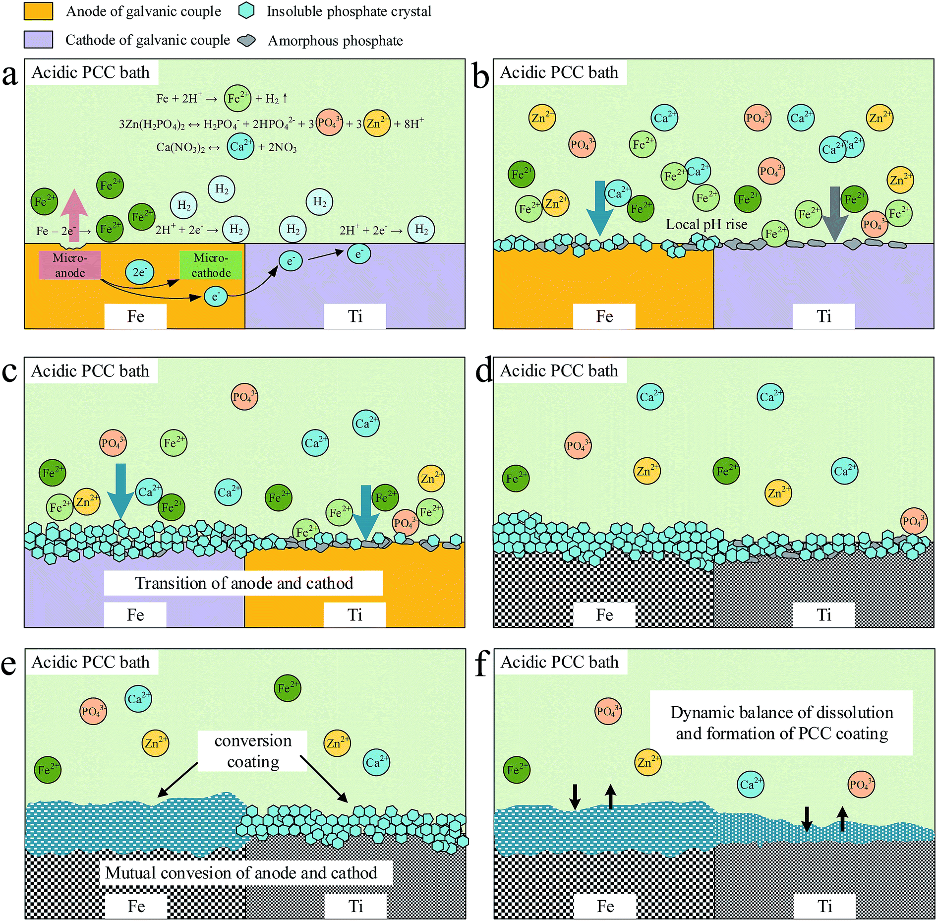
3.4. Formation mechanism of PCC coating on Ti
PCC can be regarded as complicated electrochemical and topochemical reactions, which is controlled by the change of pH at the metal/solution interface.58,72 Fig. 9 shows the pH–time curve by monitoring the pH value of the PCC bath as a function of immersion time during the process of PCC treatment of Ti/Fe. Before immersion of the sample, the PCC bath was adjusted to pH 3.75 as shown in the point of a in Fig. 9. The bath was in equilibrium due to the ionization of Zn(H2PO4)2 and Ca(NO3)2 under the test conditions (eqn (1) and (2)).| | |
3Zn(H2PO4)2 ↔ H2PO4− + 2HPO42− + 3PO43− + 3Zn2+ + 8H+
| (1) |
| | |
Ca(NO3)2 ↔ Ca2+ + 2NO3−
| (2) |
 |
| | Fig. 9 The pH–time curve of Ti/Fe during the coating deposition in the PCC bath at 55 °C. | |
When the sample is immersed into the PCC bath, the potential of the surface of the metal in the electrolyte changes with the growth of the coating. Therefore, the overall coating growth process can be followed by monitoring the open circuit potential (OCP) as a function of immersion time, which was first described by Machu67 and developed by several researchers.45,46,73,74 The potential–time curve obtained from OCP measurements indicates the various stages of coating deposition. Fig. 10a depicts the potential–time curve of the sample Ti/Fe which is monitored continuously from the beginning of immersion to 3000 s. However, since the coating is nonconductive, the potential measured in the present case is not that of the passive film as in the classical tests. The change of OCP is related to the nature and compactness of the coating and the stabilization of OCP indicates the completion of PCC treatment.46 It can be seen that the potential–time curve of Ti/Fe in Fig. 10a is very similar to the pH–time curve in Fig. 9. It is no coincidence that both the change of OCP and pH value of the bath is accompanied by a chain of chemical reactions during the PCC process. The detailed discussion of them is as follows.
 |
| | Fig. 10 (a) Potential–time curve of Ti/Fe during the coating deposition in the PCC bath at 55 °C. The inserted image shows local high magnification of the dashed box. (b) Potential–time curve of steel depicted by Ghali et al.46 | |
In addition, the typical model of the potential–time curve of steel reported by Ghali et al.46 is also shown in Fig. 10b for comparison. It is similar but not identical to the potential–time curve of Ti/Fe in Fig. 10a. According to Ghali et al.,46 the different periods of formation of conversion coatings on steel were defined in five steps: an electrochemical attack of steel in the acidic bath, an amorphous precipitation, a light attack of steel by the re-freed phosphoric acid, coarser crystallization and finally a crystalline reorganization. Learning from this, and taking into consideration the results of the evolution of chemical composition and microstructure of the coating, it is suggested that six stages are included in the deposition of coating on Ti/Fe. The schematic diagram is illustrated in Fig. 11.
 |
| | Fig. 11 Schematic diagram of the deposition of conversion coating on the surface of Ti/Fe. (a) Stage I, (b) Stage II, (c) Stage III, (d) Stage IV, (e) Stage V and (f) Stage VI. | |
3.4.1. Stage I. Electrochemical attack of anodic Fe in acid PCC bath (Fig. 11a). In Fig. 10a, the point of A is the initial potential indicating the nature of cathodic Ti undergoing corrosive attack by the free phosphoric acid present in the PCC bath.75 On account of the corrosion resistance, Ti exerts a nobler initial potential (about −30 mV) than steel (about −98 mV),42 shown in Fig. 10. The high potential difference between the two galvanic coupling materials promotes the dissolution of Fe, and the dissolution potential becomes less noble (A → B in Fig. 10a). Therefore, the topochemical reactions of Fe take place as eqn (3) and (4).At the micro-anodic sites on Fe, metal dissolution occurs:
At the micro-cathodic sites on Fe, hydrogen evolution occurs:
At the same time, the remaining electrons provided by the dissolution of Fe flow through Ti as a result of direct contact with Fe. Then the electrons are adopted by the hydrogen ions at the Ti/solution interface. Therefore, the hydrogen evolution also occurs on the surface of Ti. It is in agreement with M. Arthanareeswari et al.,42 who found that the hydrogen evolution occurred at the cathode of Ti coupled with mild steel.
3.4.2. Stage II. Crystallization and growth on anodic Fe and initiation of amorphous precipitation on cathodic Ti (Fig. 11b). The reactions of hydrogen evaluation at the Stage I cause the rise in pH of the PCC bath at the metal/solution interface as shown in Fig. 9, resulting in the conversion of primary soluble phosphates to insoluble tertiary phosphates (eqn (5)–(7)).28,31,46| | |
Fe2+ + 2Zn2+ + 2PO43− + 4H2O → Zn2Fe(PO4)2·4H2O↓
| (5) |
| | |
3Zn2+ + 2PO43− + 4H2O → Zn3(PO4)2·4H2O↓
| (6) |
| | |
Ca2+ + 2Zn2+ + 2PO43− + 2H2O → CaZn2(PO4)2·2H2O↓
| (7) |
It is shown in Fig. 10a that the OCP shifts towards anodic direction from B to C. B is the point of incipient precipitation that represents the onset of coating conversion, as defined by M. Arthanareeswari et al.28,42 It is the most negative potential during the entire coating process. During this period, there is an increase in the amount of scholzite, hopeite and phosphophyllite crystals formed on Fe. In fact, before the crystallization of insoluble phosphates, amorphous phases may be precipitated on the surface of Fe. Ghali et al.46 found that almost all the elements necessary for the formation of phosphates were precipitated principally in the amorphous state before crystallization and growth. However, due to the positive effect of galvanic coupling on coating formation, the deposition of amorphous phases on the surface of Fe is too fast to be observed.
With the growing of the coating on Fe, the surface active sites for new crystal nucleation decrease and the rate of coating formation reduces. Correspondingly, the acceleration of OCP goes down at the end of this stage.
On the other hand, at the interface of Ti/solution, the Fe2+ ions are enriched by the dissolution of both Fe and iron powders. Likewise, various amorphous ferrous phosphates may be formed on the surface of Ti (eqn (8)–(10)).28,76
| | |
Fe2+ + 2H2PO4− → Fe(H2PO4)2
| (8) |
| | |
Fe2+ + HPO42− → FeHPO4
| (9) |
| | |
3Fe2+ + 2PO43− → Fe3(PO4)2
| (10) |
This result can be proved by the detection of amorphous humps in the XRD patterns of the coatings treated with iron powders (Fig. 6b–e). These amorphous phases may be preferentially deposited on Fe–Ti phase of the Ti substrate. According to Cupr et al.,77 the development of the amorphous layer is based on the reaction between the ions of Fe2+ and ZnPO4− resulting from the dissociation of Zn(H2PO4)2 (eqn (11)).78
| | |
Fe2+ + 2ZnPO4− → Zn2Fe(PO4)2 + 2e−
| (11) |
3.4.3. Stage III. The re-dissolution of Fe and transition of anode and cathode of the galvanic couple (Fig. 11c). The consumption of PO43− ions at the Stage II results in the further ionization of Zn(H2PO4)2 (eqn (12)).| | |
H2PO4− ↔ HPO42− + H+ ↔ PO43− + 2H+
| (12) |
The slight cathodic shifting of OCP (C → D in Fig. 10a) indicates that the dissolution of Fe is promoted by the increasing free phosphoric acid. When the surface of Fe is fully covered with the non-conductive coating, the potential of the surface of Fe becomes higher than that of Ti. Therefore, it can assume that the transition between anode and cathode of the galvanic couple occurs. Ti substrate will take part in the following reactions as anode.
3.4.4. Stage IV. Crystallization and growth on Ti (Fig. 11d). Due to the lower potential of anodic Ti at this stage, the crystal nuclei of scholzite and hopeite are developed on Ti on the basis of the amorphous layer formed at early stages (eqn (13) and (14)). Therefore, the OCP is promoted to anodic direction (D → E in Fig. 10a). The conversion coating on Ti at this stage is thin and grows continuously.| | |
3Zn2+ + 2H2PO4− + 2H+ + 4H2O + 6e− → Zn3(PO4)2·4H2O↓ + 3H2↑
| (13) |
| | |
Ca2+ + 2Zn2+ + 2H2PO4− + 2H2O + 4e− → CaZn2(PO4)2·2H2O + 2H2↑
| (14) |
3.4.5. Stage V. Mutual conversion of cathode and anode and coating improvement (Fig. 11e). As the coating grows, the potential difference between Ti and Fe changes constantly, which probably result in the mutual conversion of cathode and anode of the bio-metal couple. Then, scholzite and hopeite crystals are further developed and the coating formed on Ti/Fe is being perfected, which is depicted in the plateau of period E → F in Fig. 10a. It is also reflected in the plateau of pH–time curve from about 500–1500 s in Fig. 9. Meanwhile, the new crystals exhibit epitaxial growth,28 as shown in eqn (15).| | |
Zn3(PO4)2·4H2O + Ca2+ → CaZn2(PO4)2·2H2O + 2H2O + Zn2+
| (15) |
3.4.6. Stage VI. Dynamic balance of dissolution and formation of the coating (Fig. 11f). In fact, the coating crystals are undergoing acidic corrosion throughout the entire growth, which is reflected in the small range of fluctuation of pH–time curve (Fig. 9). After immersing 1500 s, the OCP reaches a steady state (F → G in Fig. 10a), indicating the completion of PCC treatment.46 The reactions between the crystal reorganization and dissolution come to a chemical equilibrium. Finally, the dynamic balance of the coating dissolution and formation is established, the conversion coating is formed completely.
4. Conclusions
Flower-like scholzite and hopeite conversion coatings were formed on the surface Ti coupled with Fe through immersion in the PCC bath, where various mass of iron powders were applied. The dissolution of anodic Fe initiated the formation of the coating, and the introduction of iron powders increased the Fe2+ concentration in the bath. With increasing Fe2+ concentration, the coating formation rate increased and the coating became finer and denser. The coating with the highest critical load of 80.8 N was obtained from the bath with 5.0 g L−1 iron powders under galvanically coupled condition. The mechanism of formation of the conversion coatings on Ti/Fe was discussed with the help of pH–time curve and potential–time curve. The crystallization occurred on the surface of Fe first by the electrochemical attack of anodic Fe in the acidic bath. As the coating grew, the anode and cathode of the galvanic couple changed and the scholzite crystals precipitated on Ti. The coating was progressing until the dynamic balance of dissolution and formation of the coating was built.
Acknowledgements
This work was supported by Shandong Provincial Natural Science Foundation of China (ZR2013EMM013), Jiangsu Province Science Foundation for Youths (BK20140412), Shandong Provincial Science and Technology Development Plan (2014GGX102031), Fundamental Research Funds of Shandong University (2015JC018 and 2016JC024).
References
- M. Geetha, A. K. Singh, R. Asokamani and A. K. Gogia, Prog. Mater. Sci., 2009, 54, 397–425 CrossRef CAS.
- M. Niinomi, Sci. Technol. Adv. Mater., 2003, 4, 445–454 CrossRef CAS.
- M. Niinomi, Mater. Sci. Eng., A, 1998, 243, 231–236 CrossRef.
- I.-K. Yoon, J.-Y. Hwang, W.-C. Jang, H.-W. Kim and U. S. Shin, Appl. Surf. Sci., 2014, 301, 401–409 CrossRef CAS.
- M. Abdel-Hady Gepreel and M. Niinomi, J. Mech. Behav. Biomed. Mater., 2013, 20, 407–415 CrossRef CAS PubMed.
- S. Shadanbaz and G. J. Dias, Acta Biomater., 2012, 8, 20–30 CrossRef CAS PubMed.
- S. Tanaka, H. Tobimatsu, Y. Maruyama, T. Tanaki and G. Jerkiewicz, ACS Appl. Mater. Interfaces, 2009, 1, 2312–2319 CAS.
- J. E. Park, I.-S. Park, M. P. Neupane, T.-S. Bae and M.-H. Lee, Appl. Surf. Sci., 2014, 292, 828–836 CrossRef CAS.
- A. G. Gristina, Science, 1987, 237, 1588–1595 CAS.
- O. Addison, A. J. Davenport, R. J. Newport, S. Kalra, M. Monir, J. F. Mosselmans, D. Proops and R. A. Martin, J. R. Soc., Interface, 2012, 9, 3161–3164 CrossRef CAS PubMed.
- J. C. Oh and S. Lee, Metall. Mater. Trans. A, 2004, 35, 139–151 CrossRef.
- I. Gotman, J. Endourol., 1997, 11, 383–389 CrossRef CAS PubMed.
- N.-B. Li, G.-Y. Xiao, B. Liu, Z. Wang, R.-F. Zhu and Y.-P. Lu, Surf. Coat. Technol., 2016, 301, 121–125 CrossRef CAS.
- S. Nishiguchi, T. Nakamura, M. Kobayashi, H. M. Kim, F. Miyaji and T. Kokubo, Biomaterials, 1999, 20, 491–500 CrossRef CAS PubMed.
- V. Nelea, C. Morosanu, M. Iliescu and I. N. Mihailescu, Appl. Surf. Sci., 2004, 228, 346–356 CrossRef CAS.
- M. Cheng, Y. Qiao, Q. Wang, G. Jin, H. Qin, Y. Zhao, X. Peng, X. Zhang and X. Liu, ACS Appl. Mater. Interfaces, 2015, 7, 13053–13061 CAS.
- S. W. K. Kweh, K. A. Khor and P. Cheang, Biomaterials, 2002, 23, 775–785 CrossRef CAS PubMed.
- M. Lorenzetti, I. Dogša, T. A. Stošicki, D. Stopar, M. Kalin, S. Kobe and S. A. Novak, ACS Appl. Mater. Interfaces, 2015, 7, 1644–1651 CAS.
- H. W. Kim, Y. H. Koh, L. H. Li, S. Lee and H. E. Kim, Biomaterials, 2004, 25, 2533–2538 CrossRef CAS PubMed.
- A. Montenero, G. Gnappi, F. Ferrari, M. Cesari, E. Salvioli, L. Mattogno, S. Kaciulis and M. Fini, J. Mater. Sci., 2000, 35, 2791–2797 CrossRef CAS.
- A. M. Peterson, C. Pilz-Allen, T. Kolesnikova, H. Möhwald and D. Shchukin, ACS Appl. Mater. Interfaces, 2013, 6, 1866–1871 Search PubMed.
- B. Yang, M. Uchida, H.-M. Kim, X. Zhang and T. Kokubo, Biomaterials, 2004, 25, 1003–1010 CrossRef CAS PubMed.
- X. Chen, Y. Li, P. D. Hodgson and C. E. Wen, Mater. Sci. Eng., C, 2009, 29, 165–171 CrossRef CAS.
- X. Liu, P. Chu and C. Ding, Mater. Sci. Eng., R, 2004, 47, 49–121 CrossRef.
- X. X. Wang, S. Hayakawa, K. Tsuru and A. Osaka, J. Biomed. Mater. Res., 2001, 54, 172–178 CrossRef CAS PubMed.
- B. Wang, T. Lee, E. Chang and C. Yang, J. Biomed. Mater. Res., 1993, 27, 1315–1327 CrossRef CAS PubMed.
- S. H. Maxian, J. P. Zawadsky and M. G. Dunn, J. Biomed. Mater. Res., 1993, 27, 717–728 CrossRef CAS PubMed.
- T. Narayanan, Rev. Adv. Mater. Sci., 2005, 9, 130–177 CAS.
- X. Zhang, G.-Y. Xiao, Y. Jiao, X.-C. Zhao and Y.-P. Lu, Surf. Coat. Technol., 2014, 240, 361–364 CrossRef CAS.
- H.-Y. Su and C.-S. Lin, Corros. Sci., 2014, 83, 137–146 CrossRef CAS.
- H. Chen, E. Zhang and K. Yang, Mater. Sci. Eng., C, 2014, 34, 201–206 CrossRef CAS PubMed.
- H. Zhao, S. Cai, Z. Ding, M. Zhang, Y. Li and G. Xu, RSC Adv., 2015, 5, 24586–24590 RSC.
- X. B. Chen, D. R. Nisbet, R. W. Li, P. N. Smith, T. B. Abbott, M. A. Easton, D. H. Zhang and N. Birbilis, Acta Biomater., 2014, 10, 1463–1474 CrossRef CAS PubMed.
- N. Van Phuong and S. Moon, Mater. Lett., 2014, 122, 341–344 CrossRef CAS.
- C. E. Marino, E. M. de Oliveira, R. C. Rocha-Filho and S. R. Biaggio, Corros. Sci., 2001, 43, 1465–1476 CrossRef CAS.
- X.-C. Zhao, G.-Y. Xiao, X. Zhang, H.-Y. Wang and Y.-P. Lu, J. Phys. Chem. C, 2014, 118, 1910–1918 CAS.
- G.-Y. Xiao, X.-C. Zhao, X. Zhang, W.-H. Xu and Y.-P. Lu, Mater. Lett., 2015, 144, 30–32 CrossRef CAS.
- A. Valanezhad, K. Tsuru, M. Maruta, G. Kawachi, S. Matsuya and K. Ishikawa, Surf. Coat. Technol., 2012, 206, 2207–2212 CrossRef CAS.
- K. Abdalla, A. Rahmat and A. Azizan, J. Coat. Technol. Res., 2012, 10, 133–139 CrossRef.
- K. Abdalla, A. Rahmat and A. Azizan, Mater. Corros., 2014, 65, 977–981 CrossRef CAS.
- F. Fang, J.-H. Jiang, S.-Y. Tan, A.-B. Ma and J.-Q. Jiang, Surf. Coat. Technol., 2010, 204, 2381–2385 CrossRef CAS.
- M. Arthanareeswari, T. S. N. S. Narayanan, P. Kamaraj and M. Tamilselvi, Indian J. Chem. Technol., 2010, 17, 167–175 CAS.
- M. Arthanareeswari, T. S. N. Sankara Narayanan, P. Kamaraj and M. Tamilselvi, J. Coat. Technol. Res., 2011, 9, 39–46 CrossRef.
- K. Ravichandran, H. Sivanandh, S. Ganesh, T. Hariharasudan and T. S. Narayanan, Met. Finish., 2000, 98, 48–54 CrossRef CAS.
- Y. Song, D. Shan, R. Chen, F. Zhang and E.-H. Han, Corros. Sci., 2009, 51, 62–69 CrossRef CAS.
- E. I. Ghali and R. Potvin, Corros. Sci., 1972, 12, 583–594 CrossRef CAS.
- R.-C. Zeng, F. Zhang, Z.-D. Lan, H.-Z. Cui and E.-H. Han, Corros. Sci., 2014, 88, 452–459 CrossRef CAS.
- R. K. Gupta, K. Mensah-Darkwa and D. Kumar, J. Mater. Sci. Technol., 2014, 30, 47–53 CAS.
- M. Wolpers and J. Angeli, Appl. Surf. Sci., 2001, 179, 281–291 CrossRef CAS.
- P.-E. Tegehall, Colloids Surf., 1990, 49, 373–383 CrossRef CAS.
- P.-E. Tegehall, Colloids Surf., 1989, 42, 155–164 CrossRef CAS.
- Q. Feng, C. Chai, X. S. Jiang, K. W. Leong and H. Q. Mao, J. Biomed. Mater. Res., Part A, 2006, 78, 781–791 CrossRef PubMed.
- Y. Chen, A. F. T. Mak, M. Wang, J. Li and M. S. Wong, Surf. Coat. Technol., 2006, 201, 575–580 CrossRef CAS.
- S. Shruti, A. J. Salinas, G. Malavasi, G. Lusvardi, L. Menabue, C. Ferrara, P. Mustarelli and M. Vallet-Regì, J. Mater. Chem., 2012, 22, 13698 RSC.
- S. Horiuchi, M. Hiasa, A. Yasue, K. Sekine, K. Hamada, K. Asaoka and E. Tanaka, J. Mech. Behav. Biomed. Mater., 2014, 29, 151–160 CrossRef CAS PubMed.
- H. Kawamura, A. Ito, S. Miyakawa, P. Layrolle, K. Ojima, N. Ichinose and T. Tateishi, J. Biomed. Mater. Res., 2000, 50, 184–190 CrossRef CAS PubMed.
- R. Osorio, E. Osorio, I. Cabello and M. Toledano, Caries Res., 2014, 48, 276–290 CrossRef CAS PubMed.
- B. Liu, X. Zhang, G.-Y. Xiao and Y.-P. Lu, Mater. Sci. Eng., C, 2015, 47, 97–104 CrossRef CAS PubMed.
- S. M. A. Shibli and A. C. Jayalekshmi, Appl. Surf. Sci., 2008, 254, 4103–4110 CrossRef CAS.
- L. Herschke, J. Rottstegge, I. Lieberwirth and G. Wegner, J. Mater. Sci.: Mater. Med., 2006, 17, 81–94 CrossRef CAS PubMed.
- L. Herschke, I. Lieberwirth and G. Wegner, J. Mater. Sci.: Mater. Med., 2006, 17, 95–104 CrossRef CAS PubMed.
- A. Gerlach, B. Vincent, M. Lissac, X. Esnouf and G. Thollet, Biomaterials, 1993, 14, 770–774 CrossRef CAS PubMed.
- X. Zhang, G.-Y. Xiao, C.-C. Jiang, B. Liu, N.-B. Li, R.-F. Zhu and Y.-P. Lu, Corros. Sci., 2015, 94, 428–437 CrossRef CAS.
- L. B. Golden, I. R. Lane and W. L. Acherman, Ind. Eng. Chem., 1952, 44, 1930–1939 CrossRef CAS.
- P. Gordienko, T. Skorobogatov, O. Khrisanfova, A. Zavidnaya and M. Kandinskij, Zashch. Met., 1992, 28, 117–121 CAS.
- M. C. F. Magali-I, Mineral. Mag., 1986, 50, 33–39 Search PubMed.
- W. Machu, Die Phosphatierung, Verlag Chemie GmbH, 1950 Search PubMed.
- X. Ma, Y. He and D. Wang, Appl. Surf. Sci., 2011, 257, 10273–10281 CrossRef CAS.
- M. C. Kuo and S. K. Yen, Mater. Sci. Eng., C, 2002, 20, 153–160 CrossRef.
- X. Nie, A. Leyland and A. Matthews, Surf. Coat. Technol., 2000, 125, 407–414 CrossRef CAS.
- J. Stallard, S. Poulat and D. G. Teer, Tribol. Int., 2006, 39, 159–166 CrossRef CAS.
- T. Narayanan, Corros. Rev., 1994, 12, 201–238 CAS.
- T. S. Narayanan and M. Subbaiyan, Trans. Met. Finish. Assoc. India, 1992, 1, 9–12 CAS.
- T. Sankara Narayanan and M. Subbaiyan, Surf. Coat. Int., 1992, 75, 184–191 Search PubMed.
- J. Lakeman, D. Gabe and M. Richardson, Trans. Inst. Met. Finish., 1977, 55(2), 47–53 Search PubMed.
- N. V. Phuong, K. Lee, D. Chang, M. Kim, S. Lee and S. Moon, Met. Mater. Int., 2013, 19, 273–281 CrossRef CAS.
- V. Cupr and J. Pelikan, Metalloberflaeche, 1965, 19, 187–191 CAS.
- L. Kouisni, M. Azzi, M. Zertoubi, F. Dalard and S. Maximovitch, Surf. Coat. Technol., 2004, 185, 58–67 CrossRef CAS.
|
| This journal is © The Royal Society of Chemistry 2016 |
Click here to see how this site uses Cookies. View our privacy policy here.