
Genipin crosslinked soy-whey based bioactive material for atorvastatin loaded nanoparticles: preparation, characterization and in vivo antihyperlipidemic study†
Jovita Kanoujia,
Mahendra Singh,
Pooja Singh,
Poonam Parashar,
Chandra Bhusan Tripathi,
Malti Arya and
Shubhini A. Saraf*
Department of Pharmaceutical Sciences, Babasaheb Bhimrao Ambedkar University, (A Central University), Vidya Vihar, Raebareli Road, Lucknow, U.P., India-226025. E-mail: shubhini.saraf@gmail.com; Tel: +91-9415488410
First published on 26th September 2016
Abstract
The aim of the present study was to explore the lipid lowering potential of soy protein isolate (SPI) and whey protein concentrate (WPC) for the development of novel bioactive material as a drug delivery system for encapsulation of atorvastatin (ATR) in nanoparticles (NPs) and to enhance the antihyperlipidemic potential. SPI-WPC crosslinks were synthesized through amine–amine crosslinking reaction using genipin and characterized by FTIR, mass analysis and degree of crosslinking. ATR loaded NPs were prepared by desolvation process and the optimized formulation (ATR/SPI-WPC NPs-10) was selected on the basis of least particle size (158.1 ± 3.8 nm), zeta potential (−33.5 ± 2.6 mV), maximum EE (81.23 ± 1.1%) and DL (31.5 ± 2.9%). X-ray diffractometry spectra confirmed the encapsulation of ATR into NPs, whereas Transmission electron microscopy revealed that NPs were spherical in shape. Additionally, the ATR/SPI-WPC NPs significantly reduced cholesterol level as compare to SPI and WPC, individually. The results of internalization and MTT assay revealed efficient cellular uptake and cytocompatibility of the formulation. The results confirmed that SPI and WPC based bioactive material can be considered as promising biomaterial for encapsulation and enhancement of the therapeutic efficiency of ATR.
1. Introduction
According to WHO reports in 2015, obesity and cardiovascular disease (CVD) are the major causes of premature death worldwide annually. An estimated 17.5 million people died from CVDs in 2012, representing 31% of all global deaths.1 The numbers of heart patients suffering from CVD worldwide is gradually increasing.2 The patients associated with irregular levels of lipid profile, e.g. serum cholesterol (SC), serum triglycerides (ST), low density lipoprotein (LDL), high density lipoprotein (HDL), very low density lipoprotein (VLDL), will have a high risk of experiencing cardiovascular disorders3 and also contribute to develop secondary ailment, such as diabetes mellitus, obesity, hypothyroidism and renal insufficiency. The CVD related complications currently present a huge challenge for clinical practitioners to treat.4 Thus, a new drug delivery approach is required for the better management of hyperlipidemia.Statins have been reported as cholesterol reducing drugs in several studies to reduce CVD mortality.5 Atorvastatin (ATR) is a well known drug which is frequently used to decrease the cholesterol level and prevent cardiovascular diseases. It is a potent competitive inhibitor of the HMG Co-A (3-hydroxy-3-methylglutaryl coenzyme A) enzyme involved in conversion of HMG Co-A to mevalonate in cholesterol biosynthesis in the liver.
Recent progress in protein based nanotechnology has shown significant potential for novel drug delivery systems.6–8 Protein based NPs have unique properties including controlled particulate diameter, high surface to volume ratio, high loading capability, sustained release action and drug targeting potential.9 Proteins obtained from animal source (whey proteins, gelatin, casein) and from plant (soy proteins, pea proteins, cereal proteins) are widely used for encapsulation of active substances.10 These natural polymers present several advantages such as biodegradability, non-antigenicity, high nutritional value, easily modifiable surfaces, abundant renewable sources and astonishing binding capacity for various drugs as well as ease to scale up during manufacturing.11–13 Moreover, they have excellent functional properties and also possess therapeutic properties, which make them interesting to process as drug carriers for various drugs.
Among the diverse range of protein based materials used for preparing NPs, soy protein isolate (SPI) and whey protein concentrate (WPC) have gained increasing popularity because of their unique ability of forming intra and intermolecular bonds, whereas the degree of crosslinking depends on the amine groups of the proteins.14 SPI is a commercial form of soy protein that contains more than 90% protein and 18 diverse amino acids.15–17 It has been established that soy protein contributes to reduction of serum concentration of total cholesterol, low-density lipoproteins (LDLs), and triglycerides.18,19 The presence of amino and carboxyl groups in SPI creates prominent site for crosslinking reaction. Several components associated with soy protein have been implicated in lowering cholesterol: trypsin inhibitors, saponins, isoflavons. Small amounts of the heat-stable Bowman–Birk inhibitor (trypsin inhibitors) may exert a hypo cholestremic effect by increasing the secretion of cholecystokinin. Saponins may contribute to cholesterol lowering by increasing bile excretion. Soy protein containing isoflavones are known to lower cholesterol significantly and also to inhibit formation of atherosclerotic lesions in primates. On the other hand, whey proteins work by hampering the absorption of cholesterol by way of reduced micellar cholesterol solubility in intestine.20 Whey protein is a valuable by-product of the dairy industry21,22 and has also been reported to hold cholesterol reducing activity,23 which could be favorable for cardio vascular diseases. WPC consists of protein content of <80% (ref. 24) and is rich in hydrophilic groups such as –OH, –CONH–, –CONH2, –COOH, and –SO3H.23,25 Genipin (Gn) is obtained from fruits of Gardenia jasminoides.26 It is particularly advantageous in terms of being natural and a non toxic crosslinker with efficient property for the crosslinking of amino groups. Crosslinking process is also believed to improve the cholesterol reducing property of proteins (SPI and WPC).
It has been reported that SPI and WPC are promising biopolymers for the preparation of NPs by desolvation method.27,28 Desolvation is a beneficial method to obtain stable nanoparticles with narrow size distribution. The type of desolvating agent (DA), the pH of the protein solution, the rate of the organic solvent added, stirring rate of the protein solution during the desolvation process are all important parameters to be considered29 during optimization. Central composite design (CCD) was selected as the statistical based design of choice for the optimization of NPs. The novelty of the work is the use of Gn crosslinked SPI and WPC as an encapsulating material for the preparation of NPs with synergistic effect of biomaterial in lowering of cholesterol level in combination with ATR.30,31
The objective of our study was to design and develop a new delivery approach for ATR by synthesizing the cross/SPI-WPC using non toxic amine–amine crosslinker (Gn). Further, cross/SPI-WPC would be used as a bioactive material to encapsulate ATR by desolvation method. The in vivo antihyperlipidemic activity of the optimized formulation was evaluated in rat model. Cellular uptake and MTT assay was performed in J774 cell lines to evaluate the biocompatibility and internalization of NPs within the cells.
2. Material and methods
2.1. Materials
ATR was received as a gift sample from Sun Pharmaceutical Industries Ltd., (India). Soy protein isolate and whey protein concentrate was purchased from Adeesh Agro Foods, (Haryana, India). Trinitrobenzene sulfonic acid (TNBS) reagent and dialysis membrane (molecular weight cut off 12![[thin space (1/6-em)]](https://www.rsc.org/images/entities/char_2009.gif) 000–14000) were purchased from Sigma–Aldrich. Gn was purchased from Challenge Bioproducts (Taichung, Taiwan). Triton X-100 was purchased from Himedia Laboratories Pvt. Ltd, (Mumbai, India). Membrane filters (0.45 μm) were purchased from Pall Life Sciences, India. Diagnostic kit for determination of lipid profile was obtained from Span Diagnostics Ltd, Gujarat, India. All other chemicals were of analytical grade and were purchased from Himedia, Mumbai and used as obtained.
000–14000) were purchased from Sigma–Aldrich. Gn was purchased from Challenge Bioproducts (Taichung, Taiwan). Triton X-100 was purchased from Himedia Laboratories Pvt. Ltd, (Mumbai, India). Membrane filters (0.45 μm) were purchased from Pall Life Sciences, India. Diagnostic kit for determination of lipid profile was obtained from Span Diagnostics Ltd, Gujarat, India. All other chemicals were of analytical grade and were purchased from Himedia, Mumbai and used as obtained.
2.2. Synthesis of SPI-WPC conjugates
Cross/SPI-WPC was synthesized by Gn fixed crosslinking of –NH2 groups in proteins.30 SPI (5% w/v) and WPC (5% w/v) were dissolved in double distilled water (1 h). The resulting solution was immediately crosslinked by dropwise addition of aqueous Gn solutions (0.5, 1, 1.5, 2% w/w respectively) and stirred for 1 h. The excess of crosslinker was removed by frequent washing with distilled water and then purified by ultra centrifugation for 20 min at 18000 × g rpm (REMI CPR-24, Mumbai, India). The purified conjugates were lyophilized (Christ Alpha 1–4 lyophilizator, Osterode, Germany) and stored for further characterization.
2.3. Characterization of SPI-WPC conjugates
 | (1) |
| NPs code | Factors with levels | Responses | ||||
|---|---|---|---|---|---|---|
| X1 (w/v) | X2 (ml) | Particle sizeb (nm) | Zeta potential (mV) | EEb (%) | DLb (%) | |
| a X1 = cross/SPI-WPC w/v%, X2 = volume of DA (ml).b Average ± standard deviation. | ||||||
| ATR/SPI-WPC NPs-1 | 1 | 4 | 153 ± 1.24 | −24.1 | 82.4 ± 2.33 | 54 ± 2.6 |
| ATR/SPI-WPC NPs-2 | 3 | 4 | 157 ± 2.11 | −39.4 | 85 ± 1.32 | 28 ± 3.1 |
| ATR/SPI-WPC NPs-3 | 1 | 6 | 228 ± 0.89 | −30.5 | 73 ± 1.78 | 48.6 ± 2.2 |
| ATR/SPI-WPC NPs-4 | 3 | 6 | 272 ± 3.12 | −38.9 | 79 ± 2.54 | 22 ± 1.7 |
| ATR/SPI-WPC NPs-5 | 2 | 4 | 155 ± 1.76 | −26.4 | 84 ± 2.11 | 34 ± 1.4 |
| ATR/SPI-WPC NPs-6 | 2 | 6 | 248 ± 2.33 | −33.5 | 76 ± 1.43 | 30 ± 2.8 |
| ATR/SPI-WPC NPs-7 | 3 | 5 | 179 ± 1.82 | −26.3 | 79 ± 1.1 | 21 ± 2.5 |
| ATR/SPI-WPC NPs-8 | 1 | 5 | 169 ± 0.98 | −21.4 | 78 ± 1.55 | 52 ± 1.9 |
| ATR/SPI-WPC NPs-9 | 2 | 5 | 172 ± 2.12 | −24 | 80 ± 2.5 | 40 ± 2.6 |
| SPI-WPC NPs | 3 | 6 | 343 ± 2.18 | −11.1 | — | — |
For the MS analysis samples were prepared as per mentioned details. STD-Gn, pure form of Gn was dissolved in 5 ml HPLC grade methanol, R-Gn, cross/SPI-WPC was hydrolyzed in methanolic HCl at 60 °C for 1 h and supernatant obtained after centrifugation was evaporated to dryness. The residue was dissolved in 5 ml HPLC grade methanol and subjected to MS analyses, ME-Gn, cross/SPI-WPC was heated in HPLC grade methanol at 60 °C for 1 h and supernatant methanol was evaporated to dryness. The residue was redissolved in HPLC grade methanol and subjected to MS analyses.33
2.4. Fabrication of ATR/SPI-WPC nanoparticles
ATR/SPI-WPC nanoparticles were prepared by desolvation method with slight modification using ethanol as DA.26,27 In brief, cross/SPI-WPC was dissolved in double distilled water and stirred for 1 h. The pH of the solution was made alkaline (pH 9.0) using 2 M NaOH and kept overnight in a refrigerator. Desolvation of the solution was initiated by the addition of ethanol containing ATR at the rate of 1 ml min−1 with sonication (Labsonic®-M, Sartorious stedium) at 50% amplitude for 10 min. The turbidity in the solution revealed formation of nanoparticles. The nanoparticulate suspension was centrifuged at 18000 rpm (REMI CPR-24, Mumbai) for 10 min and pellets obtained were then washed, filtered (0.45 μm syringe filter), lyophilized (Christ Alpha 1–4 lyophilizator, Osterode, Germany) and stored at 4 °C for further studies.
2.5. Designs of experiment
There are several important factors which influence the physicochemical properties of the prepared NPs. Two factor, three level central composite design was developed to investigate the influence of independent variables such as amount of cross/SPI-WPC (%, w/v) and volume of DA (ml) at three different levels. Particle size (Y1), zeta potential (Y2), entrapment efficiency (EE, Y3) and drug loading (DL, Y4) were selected as response variables shown in Table 1. As per the CCD generated by software (Design Expert, Trial Version 8.0.7.1, Stat-Ease Inc., MN) nine experiments were conducted.The second order polynomial model (eqn (2)) was used to establish the mathematical relationship between the independent variables.
| Y = b0 + b1X1 + b2X2 + b3X1X2 + b4X12 + b5X22 | (2) |
2.6. Characterization of ATR/SPI-WPC nanoparticles
 | (3) |
The ultracentrifugation method was used to determine EE and DL of ATR/SPI-WPC NPs suspension. For this purpose, specified suspensions were centrifuged (REMI CPR-24, Mumbai, India) for 20 min at 15000 rpm and 4 °C to remove the free drug. The free drug in supernatant was diluted and absorbance taken using UV-VIS Spectrophotometer (Labtronics LT-2910) at 246 nm. All the experiments were performed in triplicate and mean values reported. The EE and DL was determined by the following equation.
 | (4) |
 | (5) |
MTT assay was carried out by seeding the cells (5000 cells per well) in 96 well plate and incubated at 37 °C for 24 h. After incubation, cells were exposed to different concentrations of ATR, SPI-WPC NPs and ATR/SPI-WPC NPs (6.25, 12.5, 25, 50 and 100 μg ml−1) dispersed in culture media for 24 h and 48 h followed by washing with HBSS (Hank's balanced salt solution). Thereafter, cells were incubated with 20 μl MTT for another 3 h. The purple formazan crystals were dissolved using DMSO. The absorbance of each well was measured at 570 nm through a microplate reader (Alere Microplate reader, AM, 2100).
Group 1 – control, carboxy methyl cellulose, (CMC: 0.5% w/v, p.o.).
Group 2 – hyperlipidemic control, triton X-100 (100 mg kg−1, p.o.).
Group 3 – positive control, ATR (10 mg per kg per day p.o.) + triton X-100.
Group 4 – SPI (10 mg per kg per day p.o.) + triton X-100.
Group 5 – WPC (10 mg per kg per day p.o.) + triton X-100.
Group 6 – SPI + WPC + ATR (10 mg per kg per day p.o.) + triton X-100.
Group 7 – SPI-WPC NPs (10 mg per kg per day p.o.) + triton X-100.
Group 8 – ATR/SPI-WPC NPs-10 (10 mg per kg per day p.o.) + triton X-100.
Blood samples were collected in tubes by retro orbital route, under mild ether anesthesia on 8th day and were centrifuged (2400 × g for 10 min) to separate serum. The extracted serum was assayed for ST, SC, HDL, LDL and VLDL by using commercial kits (Span Diagnostics Ltd, Gujarat, India).
HMG-CoA reductase activity was determined by method recommend by Venugopala Rao and Ramakrishnan.35 Fresh liver homogenate, 0.5 ml, (10% tissue homogenate was prepared by using arsenate solution 1g L−1) was mixed with 0.5 ml of perchloric acid (50 ml L−1). Samples were centrifuged (2000 rpm, 10 min) after 5 minutes. The obtained solution was filtered and 1 ml filtrate was added to 0.5 ml of freshly prepared hydroxyl amine reagent (2 mol L−1 reagent; pH 5.5 for HMG-CoA and 2 mol L−1 reagent; pH 2.1 for mevalonate). After 5 minutes, 1.5 ml of ferric chloride reagent was added and shaken well for 10 min. Absorbance was taken at 540 nm against blank (without homogenate) using double beam UV-Visible spectrophotometer (Labtronics LT-2910).
2.7. Statistical analysis
All experiments were conducted three times and results expressed as a mean ± standard deviation. The cell viability and in vivo study were analyzed by one-way analysis of variance (ANOVA). The difference between the means was assessed using Dunnett's test using GraphPad Prism 5. p < 0.05, p < 0.01 was considered as statistically significant.3. Result and discussion
3.1. Synthesis of SPI-WPC conjugate
SPI and WPC are obtained from natural sources and possess ample free amine groups representing ideal material for crosslinking and are also known to be cholesterol lowering proteins.36,37 On the other hand, Gn is a non toxic crosslinker which is involved either, inter or intra-molecularly in crosslinking between free amine groups of proteins. Furthermore, it was thought worthwhile to consider ATR/SPI-WPC NPs as appropriate drug delivery systems for enhanced therapeutic efficiency of ATR with negligible toxicity.3.2. Characterization of SPI-WPC conjugates
![[double bond, length as m-dash]](https://www.rsc.org/images/entities/char_e001.gif) O stretching) respectively in FTIR peaks (Fig. 1D and E) of NPs.26,40
O stretching) respectively in FTIR peaks (Fig. 1D and E) of NPs.26,40
 | ||
| Fig. 1 FTIR spectra of (A) SPI, (B) WPC, (C) cross/SPI-WPC, (D) ATR, (E) ATR/SPI-WPC nanoparticles. | ||
3.3. Fabrication of ATR/SPI-WPC nanoparticles
ATR/SPI-WPCNPs were prepared by desolvation process due to robustness and reproducibility for the preparation of stable and size controllable NPs and also to allow surface functionalization.42,43 The addition of DA to the protein solution resulted in folding of protein initiated by the expulsion of water from the hydrophobic core. Furthermore, continuous addition of DA leads to nanoprecipitation of cross/SPI-WPC attributed to loss of water from the hydrophobic core resulting in folding of proteins and encapsulation of drug. The yield of nanoparticles was found to be high ranging from 66 ± 1.84% to 87 ± 3.11%.3.4. Designs of experiment
ATR/SPI-WPC NPs preparation is based on desolvation method and the critical factors influencing the desolvation process were investigated by conducting nine experiments. The design of experiment with their observed response values is presented in Table 1. The model fitting was carried out using the values of investigated responses by applying one-way ANOVA (p < 0.05) for statistical evaluation and regression coefficient of the model equation was calculated (Table 2). Multiple regression analysis was used for model simplification through exclusion of non-significant terms (p > 0.05) from model equations.| Response | Source | Sum of squares | dfa | Mean square | F value | p-Value (Prob > F) | R2 |
|---|---|---|---|---|---|---|---|
| a Is degree of freedom, S is significant at p > 0.05. | |||||||
| Y1 | Model | 19.45 | 5 | 3.89 | 213.62 | 0.0005 (S) | 0.9972 |
| Cor total | 19.50 | 8 | |||||
| Y2 | Model | 2.62 | 4 | 0.65 | 10.86 | 0.0201 (S) | 0.9157 |
| Cor total | 2.86 | 8 | |||||
| Y3 | Model | 0.34 | 2 | 0.17 | 32.29 | 0.0006 (S) | 0.9150 |
| Cor total | 0.37 | 8 | |||||
| Y4 | Model | 8.46 | 5 | 1.69 | 12.78 | 0.0309 (S) | 0.9552 |
| Cor total | 8.85 | 8 | |||||
3.5. Optimization of conditions for preparation ATR/SPI-WPC NPs
The ATR/SPI-WPC NPs were characterized for particle size, zeta potential, EE and DL. The particles obtained were found in range from 153 ± 4.2 to 272 ± 1.5 nm. Amount of cross/SPI-WPC (% w/v) and volume of DA (ml) were the most important factors affecting particle size. The simplified model explains the relationship between the independent variables and the particle size presented in eqn (6).| Y1 = +16.37X1 + 3.13X2 + 1.54X1X2 + 4.08X12 + 0.24X22 | (6) |
Zeta potential is an important electrokinetic parameter which furnishes information regarding stability of NPs. The adjusted model for zeta potential is presented in eqn (7).
| Y2 = +5.12X1 − 1.75X2 + 0.82X1X2 + 0.19X22 | (7) |
According to eqn (7), it is clear that linear term of cross/SPI-WPC amount (X1) significantly affects the response variable. The positive coefficient of X1 indicated that increase in cross/SPI-WPC increases the zeta potential. The cross/SPI-WPC is an encapsulating material for the preparation of NPs and it displayed a negative charge when prepared at high pH (9.0).21 In contrast, increase in DA volume weakened the electrostatic repulsion between protein molecules which resulted in protein aggregate and prevented their dissociation.26
The simplified linear model which demonstrated the relationship between the independent variables and % EE is presented by eqn (8).
| Y3 = +0.90X1 − 0.11X2 | (8) |
It is observed from equation that positive coefficient of X1 revealed that an increasing amount of cross/SPI-WPC lead to increase in % EE. The eqn (9) presents the modified quadratic model which demonstrates the relationship between the independent variables and DL. The linear term of cross/SPI-WPC had a positive effect on the response variable (Y4, DL). High cross/SPI-WPC concentration resulted in small inter-particles spaces or crowded protein strands which could increase resistance to drug diffusion into the dispersing phase, leading to the incorporation of greater amount of ATR inside NPs. Another possible reason for high entrapment and drug loading is that ATR is a high molecular weight drug (1209.38 Da) and this attribute can be credited for slower diffusion of drug molecules from the polymeric matrix of particles.
| Y4 = +38.47X1 − 0.56X2 + 0.28X1X2 + 13.83X12 − 0.026X22 | (9) |
3.6. Validation
Optimization and validation of the experimental model was done in order to check the reliability of applied design. Response surface methodology (RSM) is a well established statistical approach which was applied to investigate the effect of independent variables on responses via 3D plots (plots shown in Appendix Fig. 2). The predicted model was generated by applying the criteria of minimum particle size and maximum zeta potential, EE and DL. The optimum level of cross/SPI-WPC concentration and volume of DA were determined to be 2.10% w/v and 4 ml respectively. At these levels, predicted responses of Y1 (particle size), Y2 (zeta potential), Y3 (EE) and Y4 (DL) were 154.36 nm, −30.26 mV, 83.67% and 35.46%, respectively. To validate the predicted model a new batch of NPs (ATR/SPI-WPC NPs-10) was prepared according to the optimized formula. The obtained response for the particle size, zeta potential, EE and DL were found to be 158.1 ± 3.8 nm, −33 ± 2.6 mV, 81.23 ± 1.1% and 31.5 ± 2.9% respectively. The comparison between particles and their predicted and obtained values indicated a good agreement with each other.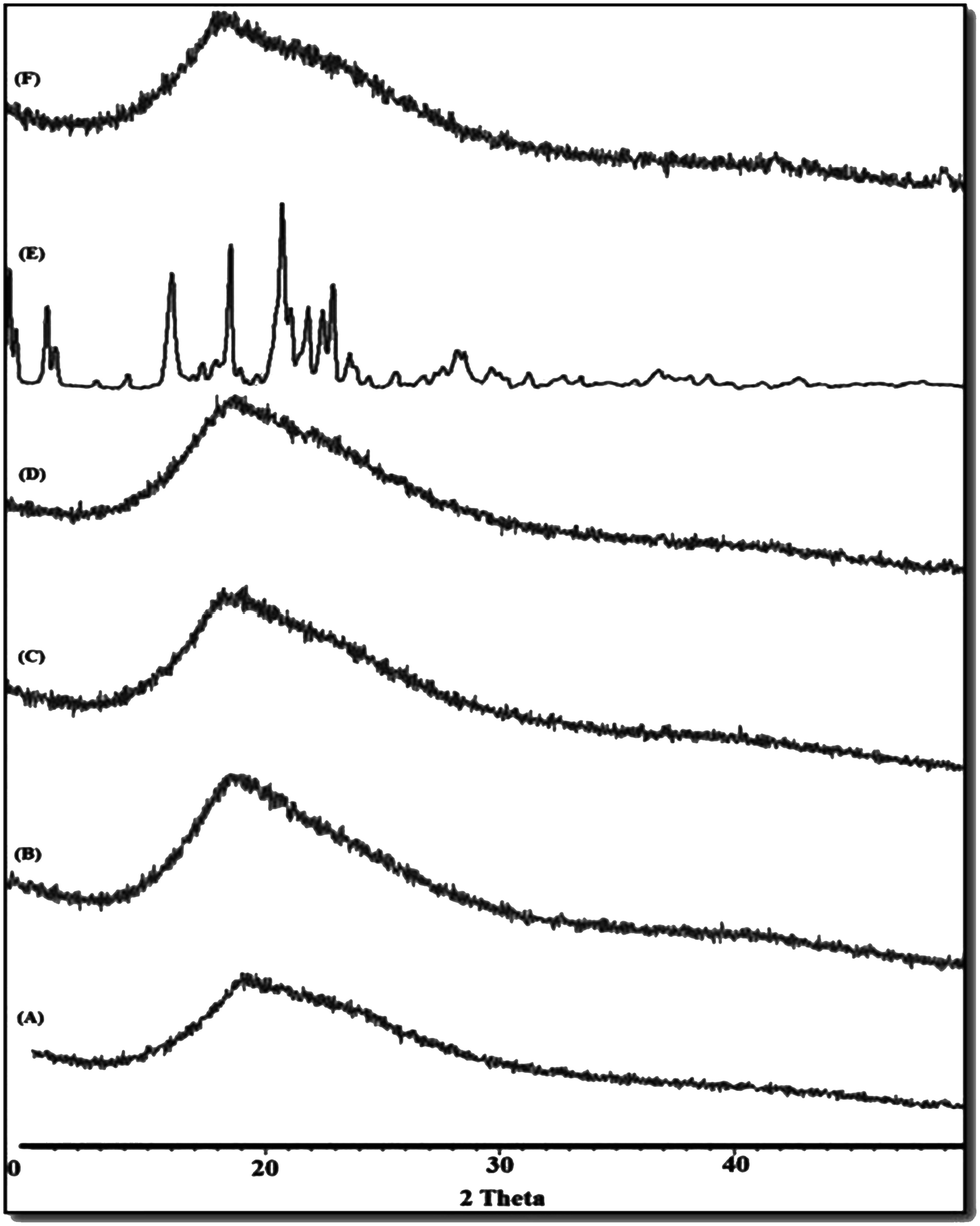 | ||
| Fig. 2 XRD Pattern of (A) SPI, (B) WPC, (C) pm/SPI-WPC, (D) cross/SPI-WPC, (E) ATR, (F) ATR/SPI-WPC NPs-10. | ||
 | ||
| Fig. 3 SEM images of (A) SPI, (B) WPC, (C) cross/SPI-WPC and TEM images of (D) SPI WPC NPs, (E) ATR/SPI WPC NPs-10. | ||
3.7. Characterization of the optimal ATR/SPI-WPC nanoparticles
 | ||
| Fig. 4 In vitro release profile of ATR in phosphate buffer pH 7.4 from ATR/SPI-WPC NPs-10 and ATR solution. | ||
| Time (days) | Particle size (nm) | PDI | Drug loading (%) |
|---|---|---|---|
| 0 | 158.1 ± 11.3 | 0.239 ± 2.0 | 31.5 ± 2.3 |
| 10 | 162 ± 14.1 | 0.265 ± 2.4 | 30.6 ± 1.6 |
| 90 | 177 ± 10.7 | 0.281 ± 2.2 | 27.3 ± 2.6 |
| 180 | 181.6 ± 15.2 | 0.283 ± 1.3 | 25.7 ± 3.9 |
 | ||
| Fig. 5 MTT assay for percentage cell viability of ATR, SPI-WPC NPs and ATR/SPI-WPC NPs after (A) 24 h and (B) 48 h incubation period with increasing concentration of NPs, data are expressed as mean ± SD of three independent experiments. * denotes a statistically significant (p < 0.05) difference from the control group. Confocal microscopic images of control (C–F) and FITC labeled ATR/SPI-WPC NPs (G–J) showing the cellular uptake of nanosized NPs in J774 cells. (C and G) Bright field, (D and H) green fluorescent channel, (E and I) blue channel, (F and J) overlay of channels. | ||
Conclusively, cell viability studies suggested that prepared NPs appeared to affect cell viability less than ATR. The NPs were non-toxic, probably due to the use of naturally occurring dietary proteins (SPI, WPC) as well as natural crosslinker (Gn). SPI, WPC and Gn proved to be non toxic47–49 and thus it could be assumed that prepared NPs would also be biocompatible. The combination of SPI and WPC is inert and acts as single component system. Furthermore, the NPs can be regarded as safe drug delivery carriers for future applications.
The majority of NPs co-localized in the cell cytoplasm and not in the nucleus of the cell, which was revealed by strong green fluorescent channel as shown in Fig. 5H. Nano-sized (below 250 nm) and negatively charged NPs are appropriate for cellular uptake and these features ensure better interaction with cationic site of cell surface. Furthermore, this can occur due to an increased retention time of NPs at the cell surface which increases the probability of particle internalization.50,51
 | ||
| Fig. 6 Serum lipids levels & H/M ratio in liver extract, Con: rats + CMC (0.5% w/v), triton: rats + triton, ATR: rats + triton + ATR (10 mg kg−1), SPI: rats + triton + SPI, WPC: rats + triton + WPC, SPI + WPC + ATR: rats + triton + SPI + WPC + ATR, ATR/SPI-WPC NPs: rats + triton + ATR/SPI-WPC NPs, SPI-WPC NPs: rats + triton + SPI-WPC NPs data are expressed as means ± SEM, n = 6. *p < 0.05 and **p < 0.01 represent significant differences when compared with the Control group. #p < 0.05 and ##p < 0.01 represent significant differences when compared with the Triton group. SC: serum cholesterol, ST: serum triglyceride, HDL: high density lipoproteins, LDL: low density lipoproteins, VLDL: very low density lipoproteins, AI: anthrogenic index = ST/HDL, H/M ratio: HMG-CoA/Mevalonate ratio. | ||
The highlight of this work is that ATR/SPI-WPC NPs group displayed lower level of lipids (SC = 62.5 ± 3.1, ST = 56.7 ± 3.8, LDL = 16.2 ± 4.6, VLDL = 12.8 ± 0.95) as compared to ATR group (SC = 89.67±, ST = 65.67 ± 4.10, LDL = 25.6 ± 3.1, VLDL = 13 ± 0.9) and triton treated group (SC = 125 ± 3.2, ST = 110 ± 3.2, LDL = 75 ± 2.9, VLDL = 17.6 ± 1.9). Therefore, it was concluded that ATR/SPI-WPC NPs showed potential hypolipidemic effect. A possible explanation for this might be the synergistic effect of SPI, WPC and ATR, as several studies reported that soy protein18–20 and whey protein23 holds cholesterol reducing activity which could be favorable for cardio vascular diseases. The mechanisms of the lipid-lowering effects of soy protein are due to the presence of several components although the whey protein works by hampering the absorption of cholesterol through reduced cholesterol solubility in lipid micelles.23 Antherogenic index (AI) can be calculated by total cholesterol (SC)/total high density lipoprotein (HDL), and is an indicator of cardiovascular diseases. A significant (p < 0.05 and p < 0.01) reduction in AI ratios of ATR/SPI-WPC NPs (1.46 ± 0.69) was found when compared with triton administered group (4.46 ± 0.76). Hence, it can be concluded that optimized ATR/SPI-WPC NPs would provide better protection against cardiovascular diseases.
The degree of cholesterol synthesis was determined by H/M assay based on quantifying the activity of HMG-CoA reductase enzyme. The H/M ratio for control, triton, ATR, SPI, WPC, SPI + WPC + ATR, ATR/SPI-WPC NPs and SPI-WPC NPs was found to be 3.41, 2.52, 3.30, 2.8, 2.5, 3.04, 3.7 and 2.5, respectively. The higher H/M ratio indicates a lower extent of cholesterol synthesis, which was observed in the ATR/SPI-WPC NPs treated group. In contrast triton administered group displayed a higher cholesterol synthesis (2.52) compared to control group (3.41). Thus, our results suggest that developed nanoparticulate system can be used to lower the risk of cardiovascular heart disease.
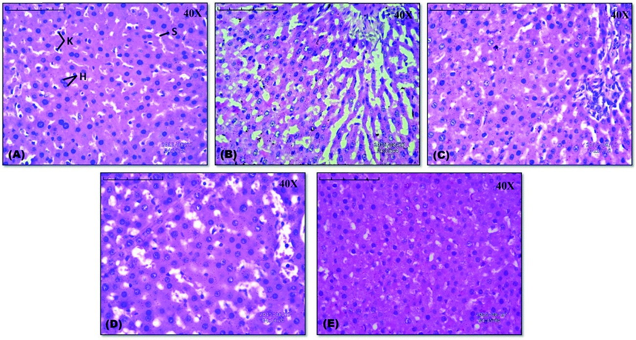 | ||
| Fig. 7 Histological evaluation of the liver stained by H & E stain (A) rats + CMC (0.5% w/v) (40×); (B) rats + Triton (40×); (C) rats + Triton + ATR (40×); (D) rats + Triton + SPI-WPC NPs (40×); (E) rats + Triton + ATR/SPI-WPC NPs-10 (40×). | ||
| Histopathology scores of observation for each group | Control | Toxic | ATR | SPI-WPC NPs | ATR/SPI-WPC NPs-10 |
|---|---|---|---|---|---|
| a Where (0) indicates no changes and (1), (2) and (3) indicate mild, moderate and severe changes respectively. | |||||
| Activated Kupffer cells | 0 | 3 | 2 | 2 | 1 |
| Sinusoidal dilatation | 0 | 3 | 2 | 2 | 1 |
| Cytoplasmic vacuolation | 0 | 2 | 1 | 2 | 1 |
| Hepatocytes | 0 | 2 | 1 | 2 | 0 |
| Mean score | 0 | 2.5 | 1.5 | 2 | 0.75 |
4. Conclusions
Recent reports show frequent use of biomaterial based excipients for encapsulation of various drugs. It is considered to be a green approach to utilize entities already present in nature, or their modified forms, than to load the environment with new chemical entities. In the present study, we synthesized a novel biomaterial by genipin induced crosslinking of SPI and WPC and reported the physicochemical characteristics by FTIR, degree of crosslinking, MS/MS spectroscopy. The NPs were successfully prepared using novel biomaterial and loaded with ATR by desolvation method. The characterization and optimization of NPs was done by central composite statistical design using particle size, zeta potential, EE and DL. The results indicated that optimized NPs (ATR/SPI-WPC NPs-10) showed sustained release of ATR over a period of 72 h. The findings of cell viability studies suggest that using SPI, WPC, Gn makes the delivery system non-toxic and biocompatible. From the in vivo and histological characterization, it may be concluded that ATR loaded in novel biomaterial is a promising delivery system which proved to have better anticholesterol activity as compared to that of pure drug. However, further evaluation of these carriers can be performed for their potential to treat hypercholesterolemia associated diseases such as diabetes, obesity, hypothyroidism and Gaucher disease, as future scope.Conflict of interest
The authors confirm that content of this article has no conflict of interest.Acknowledgements
The kind support of Sophisticated Analytical Instrument Facility (SAIF), Panjab University, Chandigarh, in XRD and TEM studies is acknowledged.References
- World Health Organization-WHO, Cardiovascular diseases (CVDs), Fact sheet N°, Geneva, 2015, p. 317 Search PubMed.
- D. Kumar, V. Parcha, A. Maithani and I. Dhulia, Effect and evaluation of antihyperlipidemic activity guided isolated fraction from total methanol extract of Salvador aoleoides (Decne.) in Triton WR-1339 Induced hyperlipidemic rat, Pharmacogn. Mag., 2012, 8, 314–318 CrossRef CAS PubMed.
- M. E. Shahira, A. S. Essam, M. H. Fathalla and A. G. Salah, Antihyperglycemic and antihyperlipidemic effects of the methanol extracts of Cleome ramosissima Parl., Barleriabispinosa (Forssk.) Vahl. and Tribulusmacropterus Boiss, Bull. Fac. Pharm., 2014, 52, 1–7 Search PubMed.
- P. K. Biswal, N. R. Pani and P. K. Dixit, Effects of carbohydrate polymers in self-microemulsified tablets on the bioavailability of atorvastatin: In vitro–in vivo study, Life Sci., 2015, 135, 92–100 CrossRef CAS PubMed.
- F. L. Pereira, V. B. Oliveira, C. T. R. Viana, P. P. Campos, M. A. N. Silva and M. G. L. Brandao, Antihyperlipidemic and antihyperglycemic effects of the Brazilian salsaparrilhas Smilax brasiliensisS preng. (Smilacaceae) and Herreriasal saparrilha Mart. (Agavaceae) in mice treated with a high-refined-carbohydrate containing diet, Food Res. Int., 2015, 76, 366–372 CrossRef.
- M. Kim, S. Jin, J. Kim, H. J. Park, H. S. Song, R. H. Neubert and S. J. Hwang, Preparation, characterization and in vivo evaluation of amorphous atorvastatin calcium nanoparticles using supercritical antisolvent (SAS) process, Eur. J. Pharm. Biopharm., 2008, 69, 454–465 CrossRef CAS PubMed.
- V. P. Torchilin, Nanotechnology in Drugs, Imperial College Press, London, 2nd edn, 2008 Search PubMed.
- S. K. Sahoo and V. Labhasetwar, Nanotech approaches to drug delivery and imaging, Drug Discovery Today, 2008, 8, 1112–1120 CrossRef.
- A. O. Elzoghby, W. M. Samy and N. A. Elgindy, Protein-based nanocarriers as promising drug and gene delivery systems, J. Controlled Release, 2012, 161, 38–49 CrossRef CAS PubMed.
- A. Nesterenkoa, I. Alric, F. Silvestre and V. Durrieu, Vegetable proteins in microencapsulation: A review of recent interventions and their effectiveness, Ind. Crops Prod., 2013, 42, 469–479 CrossRef.
- J. A. Jenkins, H. Breiteneder and E. N. Mills, Evolutionary distance from human homologs reflects allergenicity of animal food proteins, J. Allergy Clin. Immunol., 2007, 120, 1399–1405 CrossRef CAS PubMed.
- H. Li, K. Zhu, H. Zhou and W. Peng, Effects of high hydrostatic pressure treatment on allergenicity and structural properties of soybean protein isolate for infant formula, Food Chem., 2012, 132, 808–814 CrossRef CAS.
- L. Chen, G. E. Remondetto and M. Subirade, Food protein-based materials as nutraceutical delivery systems, Trends Food Sci. Technol., 2006, 17, 272–283 CrossRef CAS.
- F. Hammann and M. Schmid, Determination and Quantification of Molecular Interactions in Protein Films: A Review, Materials, 2014, 7, 7975–7996 CrossRef.
- P. Kumar, K. P. Sandeep, S. Alavi, V. D. Truong and R. E. Gorga, Preparation and characterization of bio-nanocomposite films based on soy protein isolate and montmorillonite using melt extrusion, J. Food Eng., 2010, 100, 480–489 CrossRef CAS.
- L. Xiang, C. Tang, J. Cao, C. Wang, K. Wang, Q. Zhang, Q. Fu and S. G. Zhao, Preparation and Characterization of Soy Protein Isolate (Spi)/Montmorillonite (mmt) Bionanocomposites, Chin. J. Polym. Sci., 2009, 27, 843–849 CrossRef CAS.
- R. Kumar, V. Choudhary, S. Mishra, I. K. Varma and B. Mattiason, Adhesives and plastics based on soy protein products, Ind. Crops Prod., 2001, 16, 155–172 CrossRef.
- W. W. Wong, E. O. B. Smith, J. E. Stuff, D. L. Hachey, W. C. Heird and H. J. Pownell, Cholesterol-lowering effect of soy protein in normocholesterolemic and hypercholesterolemic men, Am. J. Clin. Nutr., 1998, 68, 1385–1389 Search PubMed.
- R. M. Arliss and C. A. Biermann, Do soy isoflavones lower cholesterol, inhibit atherosclerosis and play a role in cancer prevention?, Holistic Nursing Practice, 2002, 16, 40–48 CrossRef PubMed.
- J. W. Erdman, Soy Protein and Cardiovascular Disease, A Statement for Healthcare, Professionals From the Nutrition Committee of the AHA, Circulation, 2000, 14, 2555–2559 CrossRef.
- S. Gunasekaran, S. Ko and L. Xiao, Use of whey proteins for encapsulation and controlled delivery applications, J. Food Eng., 2007, 83, 31–40 CrossRef CAS.
- A. Abbasi, Z. Emam-Djomeh, M. A. E. Mousavi and D. Davoodi, Stability of vitamin D3 encapsulated in nanoparticles of whey protein isolate, Food Chem., 2014, 143, 379–383 CrossRef CAS PubMed.
- K. Marshall, Therapeutic Applications of Whey Protein, Altern. Med. Rev., 2004, 9, 136–156 Search PubMed.
- H. Winkler, W. Vorwerg and M. Schmid, Synthesis of hydrophobic whey protein isolate by acylation with fatty acids, Eur. Polym. J., 2015, 62, 10–18 CrossRef CAS.
- P. Aramwit, T. Siritientong, S. Kanokpanont and T. Srichana, Formulation and characterization of silk sericin–PVA scaffold crosslinked with genipin, Int. J. Biol. Macromol., 2010, 47, 668–675 CrossRef CAS PubMed.
- Z. Teng, Y. Luo and Q. Wang, Nanoparticles Synthesized from Soy Protein: Preparation, Characterization and Application for Nutraceutical Encapsulation, J. Agric. Food Chem., 2012, 60, 2712–2720 CrossRef CAS PubMed.
- I. Gulseren, Y. Fang and M. Corredig, Zinc incorporation capacity of whey protein nanoparticles prepared with desolvation with ethanol, Food Chem., 2012, 135, 770–774 CrossRef CAS PubMed.
- S. R. Kaup, N. Arunkumar, L. K. Bernhardt, R. G. Vasavi, S. S. Shetty, S. R. Pai and B. Arun Kumar, Antihyperlipedemic activity of Cynodondactylon extract in high-cholesterol diet fed Wistar rats, Genomic Med., Biomarkers, Health Sci., 2011, 3, 98–102 CrossRef.
- M. R. Law, N. J. Wald and A. R. Rudnicka, Quantifying effect of statins on low density lipoprotein cholesterol, ischaemic heart disease, and stroke: systematic review and meta-analysis, BMJ, 2003, 326, 1423–1427 CrossRef CAS PubMed.
- R. You, Y. Xu, G. Liu, Y. Liu, X. Li and M. Li, Regulating the degradation rate of silk fibroin films through changing the genipin crosslinking degree, Polym. Degrad. Stab., 2014, 109, 226–232 CrossRef CAS.
- W. A. Bubnis and C. M. Ofner, Anal. Biochem., 1992, 207, 129–133 CrossRef CAS PubMed.
- R. N. Kale and A. N. Bajaj, Ultraviolet Spectrophotometric Method for Determination of Gelatin Crosslinking in the Presence of Amino Groups, J. Young Pharm., 2010, 2, 90–94 CrossRef CAS PubMed.
- R. Meena, K. Prasad and A. K. Siddhanta, Development of a stable hydrogel network based on agar–kappa-carrageenan blend cross-linked with genipin, Food Hydrocolloids, 2009, 23, 497–509 CrossRef CAS.
- J. Kanoujia, M. Singh, P. Singh and S. A. Saraf, Novel genipin crosslinked atorvastatin loaded sericin nanoparticles for their enhanced antihyperlipidemic activity, Mater. Sci. Eng., C, 2016, 69, 967–976 CrossRef CAS PubMed.
- A. Venugopala Rao and S. Ramakrishnan, Indirect Assessment Hydroxymethylglutaryl Co-A Reductase (NADPH) Activity in Liver Tissue, Clin. Chem., 1975, 21, 1523–1525 Search PubMed.
- C. E. West, C. J. K. Spaaij, W. M. Clous, H. P. Twisk, M. P. Goertz, R. W. Hunnard, M. W. Kuyvenhoven, R. Vander meer, W. F. Roszkowaski and A. Sanchez, Comparison of the hypocholesterolemic effects of dietary soybean protein with those of formaldehyde-treated casein in rabbits, J. Nutr., 1989, 119, 843–856 CAS.
- A. C. Beynen, C. E. West, C. J. K. Spaaij, J. Huisman, P. Van Leeuwen, J. B. Schutte and W. H. Hackeng, Cholesterol metabolism, digestion rates and postprandial changes in serum of swine fed purified diets containing either casein or soybean protein, J. Nutr., 1990, 120, 422–430 CAS.
- Q. Li, X. Wang, X. Lou, H. Yuan, H. Tu, B. Li and Y. Zhang, Genipin-crosslinked electrospun chitosan nanofibers: Determination of crosslinking conditions and evaluation of cytocompatibility, Carbohydr. Polym., 2015, 130, 166–174 CrossRef CAS PubMed.
- F. I. Mi, S. S. Shyu and C. K. Peng, Characterization of ring-opening polymerization of genipin and pH-dependent cross-linking reactions between chitosan and genipin, J. Polym. Sci., Part A: Polym. Chem., 2005, 43, 1985–2000 CrossRef CAS.
- Z. Teng, Y. Luo and Q. Wang, Carboxymethyl chitosan–soy protein complex nanoparticles for the encapsulation and controlled release of vitamin D3, Food Chem., 2013, 141, 524–532 CrossRef CAS PubMed.
- R. Meena, K. Prasad and A. K. Siddhanta, Effect of genipin, a naturally occurring crosslinker on the properties of kappa-carrageenan, Int. J. Biol. Macromol., 2007, 41, 94–101 CrossRef CAS PubMed.
- M. Wackera, A. Zensi, J. Kufleitner, A. Ruff, J. Schutz, T. Stockburger, T. Marstaller and V. Vogel, A toolbox for the upscaling of ethanolic human serum albumin (HSA) desolvation, Int. J. Pharm., 2011, 414, 225–232 CrossRef PubMed.
- X. H. Li, Y. Li, Y. F. Hua, A. Y. Qiu, C. Yang and S. Cui, Effect of concentration, ionic strength and freeze-drying on the heat-induced aggregation of soy proteins, Food Chem., 2007, 104, 1410–1417 CrossRef CAS.
- Y. W. Won and Y. H. Kim, Recombinant human gelatin nanoparticles as a protein drug carrier, J. Controlled Release, 2008, 127, 154–161 CrossRef CAS PubMed.
- G. M. Kavanagh, A. H. Clark and S. B. Ross-Murphy, Heat induced gelation of globular proteins: part 3. Molecular studies on low pH beta-lactoglobulin gels, Int. J. Biol. Macromol., 2000, 28, 41–50 CrossRef CAS PubMed.
- A. Bathool, G. D. Vishakante, M. S. Khan and H. G. Shiva kumar, Development and characterization of atorvastatin calcium loaded chitosan nanoparticles for sustain drug delivery, Adv. Mater. Lett., 2012, 3, 466–470 CAS.
- J. Zhang, L. Liang, Z. Tian, L. Chen and M. Subirade, Preparation and in vitro evaluation of calcium-induced soy protein isolate nanoparticles and their formation mechanism study, Food Chem., 2012, 133, 390–399 CrossRef CAS PubMed.
- M. Mekhaila, K. Jahan and M. Tabrizian, Genipin-crosslinked chitosan/poly-l-lysine gels promote fibroblast adhesion and proliferation, Carbohydr. Polym., 2014, 108, 91–98 CrossRef PubMed.
- M. L. Cross and H. S. Gill, Modulation of immune function by a modified bovine whey protein concentrate, Immunol. Cell Biol., 1999, 77, 345–350 CrossRef CAS PubMed.
- S. Patil, A. Sandberg, E. Heckert and W. Self, Protein adsorption and cellular uptake of cerium oxide nanoparticles as a function of zetapotential, Biomaterials, 2007, 28, 4600–4607 CrossRef CAS PubMed.
- M. G. Mekhail, A. O. Kamel, G. A. S. Awad and N. D. Mortada, Anticancer effect of atorvastatin nanostructured polymeric micelles based on stearyl-grafted chitosan, Int. J. Biol. Macromol., 2012, 51, 351–363 CrossRef PubMed.
Footnote |
| † Electronic supplementary information (ESI) available. See DOI: 10.1039/c6ra16830b |
| This journal is © The Royal Society of Chemistry 2016 |