A high energy output and low onset temperature nanothermite based on three-dimensional ordered macroporous nano-NiFe2O4†
Liming Shia,
Wenchao Zhang*a,
Jia Chenga,
Chunpei Yua,
Ruiqi Shena,
Jiahai Yea,
Zhichun Qina and
Yimin Chao *b
*b
aSchool of Chemical Engineering, Nanjing University of Science and Technology, Nanjing 210094, China. E-mail: zhangwenchao303@aliyun.com
bSchool of Chemistry, University of East Anglia, Norwich NR4 7TJ, UK. E-mail: Y.Chao@uea.ac.uk
First published on 19th September 2016
Abstract
Three-dimensional ordered macroporous (3DOM) Al/NiFe2O4 nanothermite has been obtained by a colloidal crystal templating method combined with magnetron sputtering processing. Owing to the superior material properties and unique 3DOM structural characteristics of composite metal oxides, the heat output of the Al/NiFe2O4 nanothermite is up to 2921.7 J g−1, which is more than the values of Al/NiO and Al/Fe2O3 nanothermites in literature. More importantly, by comparison with the other two nanothermites, the onset temperature of about 300 °C from Al/NiFe2O4 is remarkably low, which means it can be ignited more easily. Laser ignition experiments indicate that the synthesized Al/NiFe2O4 nanothermite can be easily ignited by a laser. In addition, the preparation process is highly compatible with the MEMS technology. These exciting achievements have great potential to expand the scope of nanothermite applications.
1. Introduction
Energetic thermites, a class of mixture or compound that contain fuel metal and matching oxide, have been used in a wide range of applications because of their high heat quantity.1,2 At present, the research of nanometer scale thermites has become mainstream owing to a higher-energy output and faster energy release rate compared with traditional thermite materials.3 Some processes to prepare nanocomposites have been reported in the literature: electrophoretic deposition,4 self-assembly,5 sol–gel,6 and multilayered foils.7 However, in the vast majority of reported nanothermites, the metal oxides are single metal oxide. High onset temperature is still a problem for the similar systems, which is leading to the difficulty of ignition with limited application in MEMS initiating explosive devices. For example, the onset temperature of Al/CuO, Al/Fe2O3, Al/NiO nanothermite reaction is 500 °C, 550 °C, 400 °C, respectively.8–10 Recent studies have indicated that the composite metal oxides (combination of two or more metals in an oxide matrix) possess unique properties, such as synergistic effect, catalytic property and electronic properties.11 This can bring a superior performance in energetic material applications.12 In addition, composite metal oxides can meet with the demand of the reaction of energetic materials that require oxygen in self-sustaining reaction. In literature, sol–gel13 and the hydrothermal method14 have been employed to create the nano-composite metal oxides. However, the poor dispersion is the common disadvantage of this kind. Three-dimensionally ordered macroporous (3DOM) structure with pores sized in the sub-micrometer range is a viable alternative to solve dispersion problem because of the stable porous structures and uniform pore size.15,16In the present work, Ni–Fe composite metal oxide has been chosen to explore the possibility of the utilization in nanothermites owing to the high theoretical calorific value. The synthesized nanothermite with compound metal oxide has shown significant performance compared with traditional thermite materials, such as lower onset temperature, higher energy output, and fully compatible with MEMS technology. It is expected that composite metal oxides are to be the focus of nanothermite studies because of their unique performance.
2. Experimental section
2.1 Sample preparation
All purchased chemicals were of analytical grade and used without further purification. The highly monodispersed polystyrene (PS) microspheres were successfully prepared via emulsifier-free polymerization method. Subsequently the colloidal crystal template was assembled onto as-cleaned microslide substrate by depositing the suspension. The certain amount of Fe(NO3)3·9H2O and Ni(NO3)2·6H2O were dissolved into mixed solution of methanol and ethylene glycol (1![[thin space (1/6-em)]](https://www.rsc.org/images/entities/char_2009.gif) :
:![[thin space (1/6-em)]](https://www.rsc.org/images/entities/char_2009.gif) 1 in volume) under stirring for 4 h. Afterwards, the colloidal crystal template was immersed vertically into the resulting precursor for 5 min, followed by drying at 50 °C for 2 h. This process was repeated once more to let the precursor completely fill into the close-packed PS colloidal crystal. The as-fabricated samples were then pyrolyzed at 500 °C for 5 h. The 3DOM NiFe2O4 skeleton was formed during calcination process. Finally, nano-Al was deposited onto the 3DOM NiFe2O4 by magnetron sputtering under a vacuum level of 5 × 10⁻³ Pa and a temperature at 30 °C in order to obtain 3DOM Al/NiFe2O4 nanothermite membrane on the substrate.
1 in volume) under stirring for 4 h. Afterwards, the colloidal crystal template was immersed vertically into the resulting precursor for 5 min, followed by drying at 50 °C for 2 h. This process was repeated once more to let the precursor completely fill into the close-packed PS colloidal crystal. The as-fabricated samples were then pyrolyzed at 500 °C for 5 h. The 3DOM NiFe2O4 skeleton was formed during calcination process. Finally, nano-Al was deposited onto the 3DOM NiFe2O4 by magnetron sputtering under a vacuum level of 5 × 10⁻³ Pa and a temperature at 30 °C in order to obtain 3DOM Al/NiFe2O4 nanothermite membrane on the substrate.
2.2 Characterization
The crystal structures of all the samples were analyzed by powder X-ray diffractometer (XRD, Bruker, D8 Advance). The morphology was characterized using a field emission scanning electron microscope (SEM, Hitachi, S4800). Elemental analyses of NiFe2O4 and Al/NiFe2O4 were carried out using energy dispersive spectrometry (EDS). And the differential scanning calorimetry (DSC, Mettler Toledo, DSC 1) measurement was carried out to obtain the thermodynamic performance. A pulsed laser (Beamtech, DAWA-350) was used to investigate the ignition performance of the 3DOM Al/NiFe2O4 nanothermite membrane. The combustion behavior of samples was recorded by a high-speed camera (HG-100K Motionxtra, Redlake MASD Inc., USA).3. Results and discussion
Fig. 1 shows the XRD patterns of NiFe2O4 membrane, Al/NiFe2O4 membrane and the final product of Al/NiFe2O4 membrane after a DSC test. Fig. 1(a) is completely indexed to NiFe2O4 (JCPDS No. 10-0325). Compared with Fig. 1(a), four extra peaks are observed in Fig. 1(b) and these peaks are completely indexed to Al (JCPDS No. 4-787), indicating the Al has been deposited onto NiFe2O4. The results are confirmed by EDS (in Fig. 2). The peaks of Ni, Fe and Ni, Fe, Al are observed in Fig. 2(a) and (b), respectively. EDS analysis also shows the atom ratio of Al, Fe and Ni is 6.85![[thin space (1/6-em)]](https://www.rsc.org/images/entities/char_2009.gif) :
:![[thin space (1/6-em)]](https://www.rsc.org/images/entities/char_2009.gif) 1.92
1.92![[thin space (1/6-em)]](https://www.rsc.org/images/entities/char_2009.gif) :
:![[thin space (1/6-em)]](https://www.rsc.org/images/entities/char_2009.gif) 0.85 (see Table S1 in the ESI†). The molar ratio of Al and NiFe2O4 is 6.85
0.85 (see Table S1 in the ESI†). The molar ratio of Al and NiFe2O4 is 6.85![[thin space (1/6-em)]](https://www.rsc.org/images/entities/char_2009.gif) :
:![[thin space (1/6-em)]](https://www.rsc.org/images/entities/char_2009.gif) 0.96. Fig. 1(c) is the XRD pattern from the product of Al/NiFe2O4 membrane after the DSC test. From the pattern, in the complex product, [Fe Ni] alloy (JCPD No. 18-0877) and Al2O3 (JCPD No. 10-0173) are the main components.
0.96. Fig. 1(c) is the XRD pattern from the product of Al/NiFe2O4 membrane after the DSC test. From the pattern, in the complex product, [Fe Ni] alloy (JCPD No. 18-0877) and Al2O3 (JCPD No. 10-0173) are the main components.
 | ||
| Fig. 1 XRD patterns of (a) NiFe2O4 membrane after calcination, (b) Al/NiFe2O4 membrane, (c) Al/NiFe2O4 membrane after a DSC test. | ||
The morphologies of all the samples were examined by SEM. All the results are given in Fig. 3. The surface view, in Fig. 3(a), of the PS template shows that the particle diameter of PS particle is about 300 nm and every sphere is surrounded by six others. High dimensional consistency and closely packed are its main characteristic. Cross-section view in Fig. 3(b) indicates that the thickness of PS film is about 2.8 μm. The NiFe2O4 membrane was prepared after calcination process. Fig. 3(c) shows that the NiFe2O4 membrane has a honeycomb structure. The size of the pore and the wall thickness are about 182 nm and 27 nm, respectively. From Fig. 3(d), it can be seen that the thickness of NiFe2O4 membrane is about 1.5 μm. The changing of aperture size and thickness is mainly caused by the deformation of the PS template in the process of calcination. After the Al deposition, as shown in Fig. 3(e) and (f), the NiFe2O4 have been obviously coated by Al to form dense coalition.
 | ||
| Fig. 3 SEM images of (a and b) PS template, (c and d) 3DOM NiFe2O4 membrane and (e and f) 3DOM Al/NiFe2O4 membrane after Al deposition, (a, c and e) surface view and (b, d and f) cross-section view. | ||
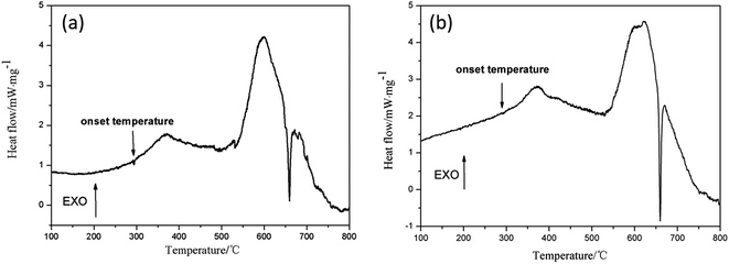
Heat release and onset temperature are the main indicators of evaluating thermite. The thermal property of the Al/NiFe2O4 nanothermite was evaluated by DSC in the temperature range from 100 to 800 °C. As shown in Fig. 4, two exothermic peaks and one small endothermic peak in the DSC curve can be observed. At 359.2 °C and 594.6 °C, the first and second exothermic peaks are observed, respectively. Through integral calculation of the two exothermic peak areas, from 298.2 to 464.3 °C and 510.1 to 655.7 °C, the outputs of heat are 279.4 J g−1 and 2642.3 J g−1, respectively. The total heat release is 2921.7 J g−1. The value is more than that from Al/NiO (2200 J g−1) and Al/Fe2O3 (2830 J g−1), which have been reported before.10,16 High heat release is due to two factors. First, unique three-dimensional reticular structure greatly increases the oxide surface area in contact with the fuel, conducive to the full response. Second, calcination treatment of oxide and magnetron sputtering aluminizing method is adopted to reduce the content of impurities such as alumina ratio. The melting of Al caused a small endothermic peak at 660 °C and the very small endothermic peak indicate that Al has almost completely reaction. In particular, the onset temperature of Al/NiFe2O4 (298.2 °C) thermite reaction is much low compared with that of Al/NiO (400 °C) and Al/Fe2O3 (550 °C).9,10 Gradient experiments were done to verify the reproducibility of the phenomenon, the results were shown in Fig. 5. It shows that the onset temperature is between 295–300 °C. In addition, the DSC curve of pure NiFe2O4 (see Fig. S1 in the ESI†) shows that there is no exothermic peak near 300 °C. This means that the first slow rising heat release curve was caused by the mild react of the nanoscale Al/NiFe2O4 membrane. It is noteworthy that the second exothermic peak occurs below the melting point of Al, 660 °C. This phenomenon is not common in the previous literature. The possible reason for the phenomenon is synergistic effect or catalytic property of composite metal oxide,11 but the mechanism is not fully understood.
 | ||
| Fig. 4 DSC curve of Al/NiFe2O4 membrane with an aluminizing duration of 15 min obtained in a temperature range from 100 to 800 °C with a heating rate of 20 °C min−1 under a 30.0 mL min−1 N2 flow. | ||
Laser ignition experiment has been employed to further verify the ignition performance of Al/NiFe2O4 membrane, see Fig. 6. Combustion is completed within ca. 99 μs and a bright flame has been observed. It is noteworthy that the incident energy is only 74 mJ per pulse. High burning rate, bright flame and low input energy indicate that the synthesized Al/NiFe2O4 nanothermite can be easily ignited by laser.
4. Conclusions
NiFe2O4 nanothermite has been successfully developed by unique PS template, with Al deposited onto it by magnetron sputtering processing. With this unique pathway, the following achievements have been verified: increased effective component, improved heat release and combustion performance of Al/NiFe2O4 nanothermite owing to reduced impurities and alumina. Furthermore, the self-catalysis of NiFe2O4 has significantly contributed to a decreased onset temperature of nanothermite reaction, leading to the easy ignition of thermite. These characteristics are important for the nanothermite application in initiating explosive devices, especially the use in MEMS pyrotechnics that require low ignition energy and high output power.Acknowledgements
This work was supported by National Natural Science Foundation of China (Grant 51576101), National Science Foundation of Jiangsu Province of China (Grant BK20141399), the Fundamental Research Funds for the Central Universities (Grant 30915012101).References
- C. Zhou, G. Li and Y. Luo, New Chem. Mater., 2010, 38, 4–7 CAS.
- L. L. Wang, Z. A. Munir and Y. M. Maximov, J. Mater. Sci., 1993, 28, 3693–3708 CrossRef CAS.
- L. Menon, S. Patibandla, K. B. Ram, S. I. Shkuratov, D. Aurongzeb, M. Holtz, J. Berg, J. Yun and H. Temkin, Appl. Phys. Lett., 2004, 84, 4735–4737 CrossRef CAS.
- D. Zhang, X. Li, B. Qin, C. Lai and X. Guo, Mater. Lett., 2014, 120, 224–227 CrossRef CAS.
- R. Thiruvengadathan, C. Staley, J. M. Geeson, S. Chung, K. E. Raymond, K. Gangopadhyay and S. Gangopadhyay, Propellants, Explos., Pyrotech., 2015, 40, 729–734 CrossRef CAS.
- V. A. Chaudhari and G. K. Bichile, Q. J. Speech, 2015, 75, 1–24 CrossRef.
- J. Y. Ahn, S. B. Kim, J. H. Kim, N. S. Jang, D. H. Kim, H. W. Lee, J. M. Kim and S. H. Kim, J. Micromech. Microeng., 2016, 26, 015002 CrossRef.
- K. Zhang, C. Rossi, G. A. Ardila Rodriguez, C. Tenailleau and P. Alphonse, Appl. Phys. Lett., 2007, 91, 113–117 Search PubMed.
- N. Zhao, C. He, J. Liu, H. Gong, T. An, H. Xu, F. Zhao, R. Hu, H. Ma and J. Zhang, J. Solid State Chem., 2014, 219, 67–73 CrossRef CAS.
- K. Zhang, C. Rossi, P. Alphonse, C. Tenailleau, S. Cayez and J.-Y. Chane-Ching, Appl. Phys. A: Mater. Sci. Process., 2008, 94, 957–962 CrossRef.
- Y. Wang, X. Xia, J. Zhu, Y. Li, X. Wang and X. Hu, Combust. Sci. Technol., 2011, 183, 154–162 CrossRef CAS.
- D. J. Stacchiola, S. D. Senanayake, P. Liu and J. A. Rodriguez, Chem. Rev., 2013, 113, 4373–4390 CrossRef CAS PubMed.
- N. Bayal, P. Jeevanandam, N. Bayal and P. Jeevanandam, Ceram. Int., 2014, 40, 15463–15477 CrossRef CAS.
- X. Xia, J. Tu, Y. Zhang, X. Wang, C. Gu, X. Zhao and H. J. Fan, ACS Nano, 2012, 6, 5531–5538 CrossRef CAS PubMed.
- T. Charoensuk, U. Boonyang, C. Sirisathitkul, P. Panchawirat and P. Senthongkaew, Mater. Sci., 2014, 20, 97–102 CrossRef.
- W. Zhang, B. Yin, R. Shen, J. Ye, J. A. Thomas and Y. Chao, ACS Appl. Mater. Interfaces, 2013, 5, 239–242 CAS.
Footnote |
| † Electronic supplementary information (ESI) available. See DOI: 10.1039/c6ra16429c |
| This journal is © The Royal Society of Chemistry 2016 |


