Altenusin derivatives from mangrove endophytic fungus Alternaria sp. SK6YW3L

Yayue Liuab, Yingnan Wua, Rui Zhaia, Zhaoming Liua, Xishan Huang*a and Zhigang She*ac
aSchool of Chemistry and Chemical Engineering, Sun Yat-sen University, Guangzhou 510275, China. E-mail: huangxishan13@foxmail.com; cesshzhg@mail.sysu.edu.cn; Fax: +86 020 84113356; Tel: +86 020 84113356
bCollege of Food Science and Technology, Guangdong Ocean University, Zhanjiang 524025, China
cSouth China Sea Bio-Resource Exploitation and Utilization Collaborative Innovation Center, Guangzhou 510006, China

Received 22nd June 2016 , Accepted 21st July 2016

First published on 22nd July 2016


Abstract

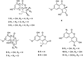
Five new altenusin derivatives, compounds 1–5, along with six known analogues 6–11, were isolated from a culture of the endophytic fungus Alternaria sp. SK6YW3L, which was isolated from a fresh fruit of the mangrove plant Sonneratia caseolaris, collected from the South China Sea. Their structures were elucidated by analysis of 1D and 2D NMR and high resolution mass spectroscopic data. The absolute configurations of compounds 1–3 and 5 were assigned by quantum chemical calculations of the electronic circular dichroic (ECD) spectra. Structures of compounds 1 and 4 were further confirmed by single-crystal X-ray diffraction experiments using Cu Kα radiation. All isolated compounds were evaluated for α-glucosidase inhibitory activity, and compounds 2, 3 and 9 exhibited moderate inhibitory activity. The plausible biosynthetic pathways for all the compounds were proposed.


Introduction

Alternaria is a cosmopolitan genus of ascomycetes that occurs on a large variety of substrates,1 and is usually known as a plant pathogen which almost produces phytotoxic secondary metabolites.2 However, not all Alternaria species are pests and pathogens; some have shown promise as biocontrol agents against invasive species.3 Some species have also been reported as endophytic microorganisms with a high variety of bioactive metabolites.4–7 Altenusins are a kind of important Alternaria metabolite, with a polyketide origin, that form various tricyclic, including 6/6/6, 6/6/5, 6/5/5 and 6/7/7 ring, skeletons. Their derivatives are reported to exhibit diverse and remarkable pharmacological activities, such as cytotoxic,8 antimicrobial,9 and anti-influenza10 activities.

In our ongoing search for new and potent α-glucosidase inhibitors from mangrove entophytic fungi,11–14 five new altenusin derivatives, compounds 1–5, together with six known analogues 6–11, were isolated from the culture of the endophytic fungus Alternaria sp. SK6YW3L, which was isolated from a fresh fruit of the mangrove plant Sonneratia caseolaris collected from the South China Sea. We reported the isolation, structure elucidation, and α-glucosidase inhibitory activity of these compounds. A possible biosynthetic pathway for 1–11 was also proposed in this paper.

Results and discussion

The fungus Alternaria sp. SK6YW3L was cultured on a rice-based medium and then extracted with CH2Cl2. The CH2Cl2 extract was found to show strong α-glucosidase inhibitory activity (IC50 < 10 μg mL−1). The extract was subjected to repeated column chromatography and sephadex LH-20 CC to afford compounds 1–11. The structures of talaroflavone (6),15 deoxyrubralactone (7),16 rubralactone (8),17 2-OH-AOH (9),18 alternariol (10)19 and alternariol methyl ether (11)20 were established by comparison of their NMR, MS and optical rotation data with literature values.

Compound 1 was obtained as a white crystal. Its molecular formula was assigned as C13H12O5 on the basis of HR-ESI-MS analysis at m/z 247.0608 [M − H] (calcd for 247.0612), and determined to possess 8 degrees of unsaturation. The presence of hydroxyl and carbonyl groups was revealed by the IR absorption bands at νmax 3331 and 1690 cm−1. Overall inspection of the 1H and 13C NMR spectra indicated that it contained one carbonyl (δC 167.1) and 8 sp2-hybridized carbons which resonated between δC 100.2 and 164.9, consuming altogether five degrees of unsaturation. The remaining degrees of unsaturation supported a tricyclic system in the molecular. In the 1H NMR spectrum, the signals for two meta-coupled aromatic protons at δH 6.44 (d, J = 2.1 Hz, H-4) and 6.33 (d, J = 2.1 Hz, H-6), one oxymethine (δH 5.21, t, J = 5.6 Hz, H-9), one methyne (δH 3.32, m, H-7), one methylene (δH 2.25 and 2.19, H-8), one methyl (δH 1.27, d, J = 6.9 Hz, H-10) and one chelated hydroxyl (δH 11.31) were observed (Table 1). The 1H–1H COSY correlations of H-7/H-8, H-7/H-10 and H-8/H-9 indicated the presence of spin fragment –CH(O)–CH2–CH–CH3 (Fig. 2). Combined with the HMBC correlations of the chelating hydroxyl (3-OH) to C-2a, C-3, and C-4; H-6 to C-2a, C-4, C-5, C-7a; H-10 to C-7a, together with H-9 to C-7a, C-9a, the planar structure of 1 was established as an altenusin derivative with 6/6/5 tricyclic ring skeleton. The relative configuration of H-9 and methyl was established as a syn relationship by a single-crystal X-ray diffraction experiment21 (Fig. 3).

Table 1 1H and 13C NMR data for compounds 1–3
Position 1 (CDCl3) 2 (methanol-d4) 3 (methanol-d4)
δC δH, mult (J in Hz) δC δH, mult (J in Hz) δC δH, mult (J in Hz)
2 167.1   164.1   168.5  
2a 100.2   99.0   100.1  
3 164.9   163.8   167.5  
4 102.7 6.44, d (2.1) 105.1 6.56, d (1.5) 102.1 6.36, d (2.1)
5 163.8   166.4   165.9  
6 101.5 6.33, d (2.1) 104.0 6.72, d (1.5) 103.0 6.37, d (2.1)
6a 137.6   134.9   138.9  
7 32.5 3.32, m 27.8 3.43, m 43.0 3.06, m
7a 121.0   144.7   119.6  
8 40.5 2.25, m, 2.19, m 42.4 2.91, dd (6.4, 18.9), 2.21, d (18.9) 77.9 4.02, dd (2.2, 5.5)
9 71.9 5.21, t (5.6) 195.7   72.4 4.89, d (5.5)
9a 154.0   147.5   154.0  
10 21.2 1.27, d (6.9) 20.3 1.34, d (7.0) 17.5 1.31, s
3-OH   11.31, s   11.07, s    


image file: c6ra16214b-f1.tif
Fig. 1 Structures of the isolated compounds 1–11.

image file: c6ra16214b-f2.tif
Fig. 2 Selected 1H–1H COSY (bold line) and HMBC (arrow) correlations of compounds 1–4.

image file: c6ra16214b-f3.tif
Fig. 3 X-ray crystallographic analysis of compounds 1 and 4.

To assign the absolute configuration of 1, the CD spectrum was measured in methanol and compared with its calculated ECD of 7S,9S-1 using quantum chemical method. After conformational analysis and geometry optimizations at the B3LYP/6-31G (d) level, three low-energy conformers with energies from 0–2.5 kcal mol−1 were obtained. The ECD were calculated at the B3LYP/6-311+G (d) level using the reported effective methods which Zhu has summarized.22 As illustrated in Fig. 4, the theoretical ECD curve for 7S,9S-1 showed an excellent fit with the experimental one. Therefore, the absolute configuration of 1 was determined to be 7S,9S.


image file: c6ra16214b-f4.tif
Fig. 4 Experimental and calculated ECD spectra of compounds 1–3 and 5 in methanol.

Compound 2 was obtained as a white powder. The molecular formula was assigned as C13H10O5 on the basis of HR-ESI-MS analysis at m/z 245.0454 [M − H]. The 1H and 13C NMR data of 2 were similar to those of 1, which indicated 2 has a structural similarity to 1, except for the lack of an oxymethine signal and the addition of α,β-conjugated keto carbonyl signal (δC 195.7). The carbonyl group was deduced to be located at C-9 in 2 for the HMBC correlation from H-8 to C-9. The predicted ECD curves of 2 were calculated by a quantum chemical method at the B3LYP/6-311+G (d)//B3LYP/6-31G (d) level, and the predicted ECD curve of 7S-2 was similar to the experimental one (Fig. 4). Therefore, the absolute configuration of 2 was identified as 7S.

A molecular formula of C13H12O6 (eight degrees of unsaturation) was determined for compound 3 on the basis of HR-ESI-MS analysis at 263.0554 [M − H]. The 1H and 13C NMR data indicated 3 was resembled to those of 1, except for the lack of methylene signal and the addition of an oxymethine signal at δC 77.9/δH 4.02. Combined with the 1H–1H COSY correlations of H-7/H-8 and H-8/H-9, together with the HMBC correlations of CH3-10 to C-7a, the planar structure of 3 was established as Fig. 1. The NOESY correlations of CH3-10 with H-8 and H-9 supported a syn relationship between them. Therefore, the relative configuration of 3 was designated as 7R*,8S*,9S*. The absolute configuration of 3 was established as 7R,8S,9S by comparing the experimental CD with the predicted ECD curves of 7R,8S,9S-3 isomers, calculated by the quantum chemical method at the B3LYP/6-311+G (d)//B3LYP/6-31G (d) level.

Compound 4 was isolated as a yellow crystal. Its molecular formula C14H12O6 (nine degrees of unsaturation) was established on the basis of HR-EI-MS analysis at 276.0625 [M]+. Analysis of 1H and 13C NMR spectroscopic data (Table 2) revealed that compound 4 possessed the same 6/6/5 skeleton,23 as in 2. The HMBC correlations of H-10 to C-7a, C-9, C-9a (δC 81.6), and H-9 to C-7, C-7a, C-8, C-9a indicated that the methyl group was attached at C-9a, and carbonyl group at C-8. The methyloxy group (δC 56.0/δH 3.87) was assigned at C-5 supported by the HMBC correlation of methyloxy protons to C-5. Thus, the planar structure of 4 was established as Fig. 1, which supported by a single-crystal X-ray diffraction experiment using Cu Kα radiation (Fig. 4). However, since the optical rotation value of 4 was zero and its CD spectrum showed no cotton effect, it was considered to be a racemate. And unfortunately, the separation of 4 on a chiralcel OD column (hexane/2-propanol or hexane/MeOH) was failed, with only one symmetrical peak in the chromatogram, as detected by HPLC.

Table 2 1H and 13C NMR data for compounds 4 and 5
Position 4 (DMSO) 5 (methanol-d4)
δC δH, mult (J in Hz) δC δH, mult (J in Hz)
2 167.9   171.2  
2a 99.3   107.0  
3 163.9   159.4  
4 101.7 6.60, d (2.4) 102.8 6.41, d (1.9)
5 165.7   168.4  
6 105.9 6.92, d (2.4) 100.9 6.21, d (1.9)
6a 132.8   152.3  
7 149.6   98.5  
7a 129.8      
8 196.6   86.8 4.39, d (5.6)
9 46.8 3.05, d (17.8), 2.80, d (17.8) 78.3 4.67, m
9a 81.6      
10 27.4 1.61, s 132.5 5.80, m
11     141.5  
11-CH3     11.8 1.44, t (1.8)
5-OCH3 56.0 3.87, s 56.4 3.83, s
3-OH 11.35, br s    
7-OH 11.32, br s    


Compound 5 was obtained as white amorphous powder. Its molecular formula was deduced to be C14H14O6 on the basis of HR-ESI-MS analysis at 277.0714 [M − H]. Overall inspections of the 1H and 13C NMR spectroscopic data (Table 2) indicated compound 5 and talaroflavone (6) shared the same skeleton, except for the lack of α,β-conjugated keto carbonyl signal and the addition of an oxymethine signal (δC 78.3/δH 4.67). Further HMBC correlations confirmed that 5 was the reduced product of talaroflavone.

In the NOESY spectrum, the NOE correlations between H-6 and H-9 instead of H-8 established the relative configuration of 5 was 7R*,8S*,9R* (Fig. 5). The absolute configuration of 5 was established as 7R,8S,9R by the quantum chemical method at the B3LYP/6-311+G (d)//B3LYP/6-31G (d) level.


image file: c6ra16214b-f5.tif
Fig. 5 Selected HMBC (arrow) and NOESY correlations of 5.

Based on literature reports, the biosynthetic routes of compounds 1–11 were proposed based on the polyketide pathway, originating from seven acetate units (Fig. 6).16,23,24


image file: c6ra16214b-f6.tif
Fig. 6 Proposed biosynthetic pathways for compounds 1–11.

All isolates were tested for their in vitro inhibitory activities against α-glucosidase.25 The results were given in Table 3. Compounds 2, 3 and 9 exhibited strongest inhibitory activities in comparison with other compounds and acarbose (used as a positive control, IC50 of 553.7 μM), with IC50 values of 78.2, 78.1, and 64.7 μM, respectively. The activity of compounds 1 and 8 were two-fold better than that of acarbose. While compounds 4, 6 and 10 showed moderate inhibitory activity against α-glucosidase with IC50 values of 334.4, 348.4, and 474.3 μM. Other altenusin derivatives with 6/5/5 ring skeleton such as (5 and 7) showed weak activities (IC50 > 500 μM). The chelated hydroxyl group at C-3 (9 and 10) enhanced the inhibitory activity compared with 11, bearing a methoxy group.

Table 3 α-Glucosidase inhibitory activitiesa
Compounds IC50 (μM) Compounds IC50 (μM)
a IC50 values are shown as mean from three independent experiments.b Positive control.
1 235.2 ± 1.6 6 348.4 ± 1.8
2 78.2 ± 1.2 7 >500
3 78.1 ± 0.9 8 194.4 ± 0.7
4 334.4 ± 2.2 9 64.7 ± 1.5
5 >500 10 474.3 ± 2.2
Acarboseb 553.7 ± 9.8 11 >500


Experimental section

General experimental procedures

The NMR spectra were performed on Bruker Advance 500 spectrometer (1H/500 MHz, 13C/125 MHz). All chemical shifts (δ) were given in ppm with reference to the solvent signal (δC 49.0/δH 3.31 for CD3OD, δC 39.5/δH 2.50 for DMSO and δC 77.1/δH 7.26 for CDCl3), and coupling constants (J) were given in Hz. Optical rotations were recorded with an MCP 300 (Anton Paar, Shanghai, China) polar meter at 25 °C. UV spectra were measured on a PERSEE TU-1900 spectrophotometer. IR spectra were carried out on a Nicolet Nexus 670 spectrophotometer, in KBr discs. CD spectra were measured on a Chira scan™ CD spectrometer (Applied Photo physics, London, UK). EI-MS on a DSQ EI-mass spectrometer (Thermo) and HR-EI-MS data were measured on a DMAT95XP high-resolution mass spectrometer. ESI-MS spectra were recorded on a Finnigan LCQ-DECA mass spectrometer and HR-ESI-MS spectra were recorded on a Thermo Fisher Scientific Q-TOF mass spectrometer. Single-crystal data were carried out on an Agilent Gemini Ultra diffractometer (Cu Kα radiation). Column chromatography (CC) was performed on silica gel (200–300 mesh, Qingdao Marine Chemical Factory) and Sephadex LH-20 (Amersham Pharmacia). Thin-layer chromatography (TLC) was performed on silica gel plates (Qingdao Huang Hai Chemical Group Co., G60, F-254).

Fungal material

The fungal strain SK6YW3L used in this study was isolated from a fresh fruit of Sonneratia caseolaris, which was collected from the Shankou Mangrove reserve, Guangxi Province, China, in April 2014. The strain was identified as Alternaria sp., according to morphologic traits and molecular identification. Its 444 base pair ITS sequence had 99% sequence identity to that of Alternaria longipes (KJ722535). The sequence data has been submitted to GenBank with accession number KP059101. A voucher specimen (registration number SK6YW3L) has been deposited at Sun Yat-sen University, China.

Fermentation, extraction and isolation

The fungus Alternaria sp. SK6YW3L was grown on a solid autoclaved rice substrate medium (thirty 500 mL Erlenmeyer flasks, each containing 50 g of rice and 50 mL 3‰ of saline water) for 28 days at 25 °C. The mycelia and solid rice medium were extracted with CH2Cl2 three times. The organic solvents were evaporated to dryness under reduced pressure to yield 1.6 g. And then was subjected to a silica gel column (30 × 6 cm), eluting with gradient of PE/CH2Cl2 and CH2Cl2/MeOH to afford six fractions (Fr. 1–Fr. 6), containing 0.2, 0.3, 0.5, 0.4, 0.1 and 0.1 g of material, respectively. Fr. 2 (0.3 g) was subjected to a silica gel column (10 × 3 cm) using gradient mixtures of petroleum ether and CH2Cl2 to yield five subfractions (Fr. 2-1–Fr. 2-5). Fr. 2-3 (103 mg) was purified by recrystallization with MeOH–H2O (100[thin space (1/6-em)]:[thin space (1/6-em)]1, v/v) to yield 1 (20.8 mg). Fr. 2-4 (115 mg) was re-chromatographed on silica gel column (10 × 2 cm) eluting with PE/CH2Cl2 (50[thin space (1/6-em)]:[thin space (1/6-em)]50) to give 6 (1.9 mg) and 4 (9.5 mg). Fr. 3 (0.5 g) was subjected to column chromatography (CC) on silica gel (10 × 4 cm column) eluting with a gradient of PE/CH2Cl2 from 50[thin space (1/6-em)]:[thin space (1/6-em)]50 to 0[thin space (1/6-em)]:[thin space (1/6-em)]100 v/v affording 7 (1.8 mg) and 5 (3.2 mg). Fr. 4 (0.4 g) was further fractioned on Sephadex LH-20 CC (110 × 4 cm) eluting with CHCl3–MeOH (1[thin space (1/6-em)]:[thin space (1/6-em)]1, v/v), to obtain seven subfractions (Fr. 4-1–Fr. 4-7). Fr. 4-1 (110 mg) was further fractioned by silica gel (10 × 2 cm column) eluting with a gradient of petroleum ether and ethyl acetate from 80[thin space (1/6-em)]:[thin space (1/6-em)]20 to 20[thin space (1/6-em)]:[thin space (1/6-em)]80 v/v to give 2 (2.1 mg) and 3 (2.0 mg). Fr. 4-4 (310 mg) was chromatographed on silica gel (20 × 4 cm column) eluting with CH2Cl2–MeOH from 1[thin space (1/6-em)]:[thin space (1/6-em)]0 to 10[thin space (1/6-em)]:[thin space (1/6-em)]1 v/v to obtain 8 (3.0 mg) and 11 (5.6 mg). Fr. 4-6 (201 mg) was chromatographed on silica gel (20 × 4 cm column) petroleum ether and ethyl acetate (60[thin space (1/6-em)]:[thin space (1/6-em)]40 to 40[thin space (1/6-em)]:[thin space (1/6-em)]60 v/v) to afford 9 (2.0 mg) and 10 (6.2 mg).
Compound 1. Colorless crystals; mp 108.1–108.2 °C, [α]25D +164.1 (c 0.005, MeOH); UV (MeOH) λmax (log[thin space (1/6-em)]ε): 332 (1.5), 247 (2.5) nm; CD (CH3OH) λmaxε) 290 (+2.9), 259 (0.7), 245 (−6.48), 275 (+9.6) nm; IR (KBr) νmax, 3641, 3331, 2971, 1690, 1626, 1563, 1507, 1401, 1345, 1239, 1190, 1063, 852 cm−1; 1H NMR (CDCl3, 500 MHz) and 13C NMR (CDCl3, 125 MHz), see Table 1; ESI-MS m/z 247 [M − H]; HR-ESI-MS m/z 247.0608 [M − H] (calcd for C13H12O5, 247.0612).
Compound 2. White powder; mp 281.5–281.6 °C, [α]25D +35.3 (c 0.002, MeOH); UV (MeOH) λmax (log[thin space (1/6-em)]ε): 347 (0.9), 318 (1.0), 276 (1.3), 258 (1.7) nm; CD (CH3OH) λmaxε) 359 (−0.7), 325 (+0.2), 257 (−1.4), 209 (+1.9) nm; IR (KBr) νmax 3436, 2922, 2845, 1675, 1612, 1570, 1401, 1176, 1113, 789 cm−1; 1H NMR (methanol-d4, 500 MHz) and 13C NMR (methanol-d4, 125 MHz), see Table 1; ESI-MS m/z 245 [M − H]; HR-ESI-MS m/z 245.0454 [M − H] (calcd for C13H10O5, 245.0456).
Compound 3. White powder; mp 182.8–182.9 °C, [α]25D +89.9 (c 0.004, MeOH); UV (MeOH) λmax (log[thin space (1/6-em)]ε): 332 (1.7), 248 (2.5) nm; CD (CH3OH) λmaxε) 294 (+3.1), 261 (+0.8), 245 (−6.4), 207 (+8.8) nm. IR (KBr) νmax 3655, 2127, 1683, 1612, 1443, 1352, 1302, 859 cm−1; 1H NMR (methanol-d4, 500 MHz) and 13C NMR (methanol-d4, 125 MHz), see Table 1; ESI-MS m/z 263 [M − H]; HR-ESI-MS m/z 263.0554 [M − H] (calcd for C13H12O6, 263.0561).
Compound 4. Yellow crystal; mp 217.3–217.4 °C, [α]25D 0 (c 0.04, MeOH); UV (MeOH) λmax (log[thin space (1/6-em)]ε): 341 (2.1), 317 (2.1), 265 (2.4), 203 (2.3) nm; IR (KBr) νmax 3259, 2931, 1712, 1659, 1616, 1572, 1359, 1335, 1160, 1058, 841 cm−1; 1H NMR (DMSO, 500 MHz) and 13C NMR (DMSO, 125 MHz), see Table 2; EI-MS m/z 276 [M]+; HR-EI-MS m/z 276.0625 [M]+ (calcd for C14H12O6, 276.0628).
Compound 5. White crystal; mp 104.5–104.6 °C, [α]25D −10.3 (c 0.002, MeOH); UV (MeOH) λmax (log[thin space (1/6-em)]ε): 294 (1.5), 258 (2.0), 220 (2.4) nm; IR (KBr) νmax 3493, 3345, 1725, 1612, 1443, 1373, 1330, 1232, 1197, 1134, 1035, 964 cm−1; 1H NMR (methanol-d4, 500 MHz) and 13C NMR (methanol-d4, 125 MHz), see Table 2; ESI-MS m/z 277 [M − H]; HR-ESI-MS m/z 277.0714 [M − H] (calcd for C14H14O6, 277.0718).

X-ray crystallographic analysis of compounds 1 and 4

Colorless crystals of 1 were obtained from MeOH–H2O. Colorless crystals of 4 were obtained from MeOH; all single-crystal X-ray diffraction data were collected at 123 K on an Oxford Gemini S Ultra diffractometer with Cu Kα radiation (λ = 1.54178 Å). The structures were solved by direct methods (SHELXL-2013) and refined using full-matrix least-squares difference Fourier techniques. All non-hydrogen atoms were refined anisotropically, and all hydrogen atoms were placed in idealized positions and refined as riding atoms with the relative isotropic parameters. The crystallographic data of 1 and 4 have been deposited at the Cambridge Crystallographic Data Centre with the deposition numbers CCDC 1435614 and CCDC 1435618, respectively.

Crystal data of 1: orthorhombic, C13H12O5, space group P2(1)2(1)2(1), a = 9.8293 (4) Å, b = 19.0054 (6) Å, c = 6.7783(4) Å, α = 90, β = 90, γ = 90, Z = 3, Dcalcd = 1.449 mg cm−3, μ = (Cu Kα) 0.976 mm−1, and F(000) = 576, Flack = 0(4). Crystal size: 0.40 × 0.20 × 0.20 mm3. Independent reflections: 2216 [Rint = 0.0286]. The final indices were R1 = 0.0544, wR2 = 0.1511 [I > 2σ(I)].

Crystal data of 4: monoclinic, C14H12O6, space group P21/n, a = 9.2401(3) Å, b = 12.8808(5) Å, c = 10.0633(3) Å, α = 90.00, β = 93.788(3), γ = 90°, Z = 4, Dcalcd = 1.535 mg cm−3, μ = (Cu Kα) 1.034 mm−1, and F(000) = 576. Crystal size: 0.36 × 0.27 × 0.14 mm3. Independent reflections: 2085 [Rint = 0.0263]. The final indices were R1 = 0.0421, wR2 = 0.1075 [I > 2σ(I)].

α-Glucosidase inhibitory activity

All the assays were performed using 0.01 M KH2PO4/K2HPO4 buffers, pH 7.0, and a microtiter plate reader. Enzyme solution was prepared to give 2.0 units per mL in 2 mL aliquots. The assay medium contained phosphate buffer, pH 7.0 (140 μL), 10 μL of enzyme solution, 20 μL DMSO or inhibitor (dissolved in DMSO) and 30 μL of 0.01 M substrate (p-nitrophenyl) (PNP) glycoside (3 mg mL−1). The substrate was immediately added to a 96-well microtiter plate containing enzyme and buffer with inhibitor after 15 min of incubation time. The activity was determined by measuring the increase in absorbance at 405 nm for 1 min interval at 37 °C. Calculations were performed according to the equation: η (%) = [(BS)/B] × 100% (B stands for the assay medium with DMSO; S stands for the assay medium with inhibitor). All measurements were done in triplicate from two independent experiments. The reported IC50 was the average value of two independent experiments.

Quantum mechanical calculation

In theoretical calculations, the preliminary conformational distribution search was performed by Spartan'10 software (Wavefunction, Inc., Irvine, CA, USA) using the MMFF94S force field. The corresponding minimum geometries were further fully optimized with the Gaussian 03 (Gaussian, Wallingford, CT, USA) program package at the B3LYP/6-31G (d) computational level as frequency calculations. The obtained stable conformers were submitted to CD calculation by the TDDFT B3LYP/6-311+G(d) method. ECD spectra were generated using the program SpecDis 1.6 (University of Würzburg, Würzburg, Germany) and OriginPro 8.5 (OriginLab, Ltd., Northampton, MA, USA) from dipole-length rotational strengths by applying Gaussian band shapes with sigma = 0.19 eV. All calculations were performed with the High-Performance Grid Computing Platform of Sun Yat-sen University.

Conclusions

In summary, a chemical investigation of mangrove endophytic fungus Alternaria sp. SK6YW3L led to the isolation and identification of eleven secondary metabolites, including five new altenusin derivatives 1–5 bearing 6/6/5 or 6/5/5 tricyclic ring skeletons. In bioactivity assays, compounds 2, 3 and 9 exhibited moderate inhibitory activity against α-glucosidase. To the best of our knowledge, it is the first time to report the α-glucosidase inhibitory activity of altenusins derivatives. Our findings would enrich chemical context of the genus Alternaria and expand the chemical and biological diversity of altenusins.

Conflicts of interest

The authors declare no conflict of interest.

Acknowledgements

We thank the National Natural Science Foundation of China (21472251, 41276146), the Key project of Natural Science Foundation of Guangdong Province (2016A040403091), the Science & Technology Plan Project of Guangdong Province of China (2013B021100011), Special Financial Fund of Innovative Development of Marine Economic Demonstration Project (GD2012-D01-001), China's Marine Commonwealth Research Project (201305017), the Fundamental Research Funds for the Central Universities (141gjc16). We also give thanks to the High-Performance Grid Computing Platform of Sun Yat-sen University for generous support of our computational chemistry research.

Notes and references

  1. J. D. Miller and S. N. Richardson, The Regulatory Governance Initiative, 2013 Search PubMed.
  2. M. J. Kelman, J. B. Renaud, K. A. Seifert, K. A. Seifert, J. Mack, K. Sivagnanam, K. K. C. Yeung and M. W. Sumarah, Rapid Commun. Mass Spectrom., 2015, 29, 1805–1810 CrossRef CAS PubMed.
  3. V. Ostry, World Mycotoxin J., 2008, 1, 175–188 CrossRef CAS.
  4. H. W. Zhang, Y. C. Song and R. X. Tan, Nat. Prod. Rep., 2006, 23, 753–771 RSC.
  5. G. A. Strobel, Crit. Rev. Biotechnol., 2002, 22, 315–333 CrossRef CAS PubMed.
  6. J. L. Azevedo, W. Maccheroni Jr, J. O. Pereira and W. L. deAraujo, Electron. J. Biotechnol., 2000, 3, 15–16 Search PubMed.
  7. R. X. Tan and W. X. Zhou, Nat. Prod. Rep., 2001, 18, 448–459 RSC.
  8. M. Zhou, K. Zhou, P. He, K. M. Wang, R. Z. Zhu, Y. D. Wang and G. Du, Planta Med., 2016, 82, 414–417 CrossRef CAS PubMed.
  9. J. F. Tian, P. J. Li, X. X. Li, P. H. Sun, H. Gao, X. Z. Liu and X. S. Yao, Bioorg. Med. Chem. Lett., 2016, 26, 1391–1396 CrossRef CAS PubMed.
  10. W. Gu, W. Wang, X. N. Li, Y. Zhang, L. P. Wang, C. M. Yuan and X. J. Hao, Fitoterapia, 2015, 107, 60–62 CrossRef CAS PubMed.
  11. Y. Liu, Q. Yang, G. Xia, H. Huang, H. Li, L. Ma, Y. Lu, L. He, X. Xia and Z. She, J. Nat. Prod., 2015, 78, 1816–1822 CrossRef CAS PubMed.
  12. S. Chen, Y. Liu, Z. Liu, R. Cai, Y. Lu, X. Huang and Z. She, RSC Adv., 2016, 6, 26412–26420 RSC.
  13. S. Chen, Z. Liu, Y. Lu, L. He and Z. She, Beilstein J. Org. Chem., 2015, 11, 1187–1193 CrossRef CAS PubMed.
  14. Y. Liu, S. Chen, Z. Liu, G. Xia, L. He and Z. She, Mar. Drugs, 2015, 13, 3091–3102 CrossRef CAS PubMed.
  15. M. Naganuma, M. Nishida, K. Kuramochi, F. Sugawara, H. Yoshida and Y. Mizushina, Bioorg. Med. Chem., 2008, 16, 2939–2944 CrossRef CAS PubMed.
  16. W. A. Ayer and J. S. Racok, Can. J. Chem., 1990, 68, 2085–2094 CrossRef CAS.
  17. Y. Kimura, T. Yoshinari, H. Koshino, S. Fujioka, K. Okada and A. Shimada, Biosci., Biotechnol., Biochem., 2007, 71, 1896–1901 CrossRef CAS PubMed.
  18. K. Koch, J. Podlech, E. Pfeiffer and M. Metzler, J. Org. Chem., 2005, 70, 3275–3276 CrossRef CAS PubMed.
  19. O. Mumm, Phys. Chem., 1907, 59, 459–491 CAS.
  20. G. G. Freeman, Phytochemistry, 1966, 5, 719–725 CrossRef CAS.
  21. H. D. Flack and G. Bernardinelli, Chirality, 2008, 20, 681–690 CrossRef CAS PubMed.
  22. J. Ren, J. Jiang, L. Li, R. Tian, X. Chen, S. Jiang, C. U. Pittman and H. Zhu, Eur. J. Org. Chem., 2009, 23, 3987–3991 CrossRef.
  23. W. A. Ayer and J. S. Racok, Can. J. Chem., 1990, 68, 2095–2101 CrossRef CAS.
  24. T. M. Harris and J. V. Hay, J. Am. Chem. Soc., 1977, 99, 1631–1637 CrossRef CAS.
  25. Z. Y. Du, R. R. Liu, W. Y. Shao, X. P. Mao, L. Ma, L. Q. Gu, Z. S. Huang and A. S. C. Chan, Eur. J. Med. Chem., 2006, 41, 213–218 CrossRef CAS PubMed.

Footnote

Electronic supplementary information (ESI) available: Spectra of all new compounds (1H NMR, 13C NMR, 2D NMR, HR-EI-MS and HR-ESI-MS). CCDC 1435614 and 1435618. For ESI and crystallographic data in CIF or other electronic format see DOI: 10.1039/c6ra16214b

This journal is © The Royal Society of Chemistry 2016
Click here to see how this site uses Cookies. View our privacy policy here.