
MnO2 nanosheet-based heparin and OSCS fluorescent biosensor with lowered background and amplified hybridization chain reaction†
Ruifen Tiana,
Hong Jianga and
Guangfeng Wang*ab
aKey Laboratory of Chem-Biosensing, Key Laboratory of Functional Molecular Solids, College of Chemistry and Materials Science, Center for Nano Science and Technology, Anhui Normal University, Wuhu 241000, Anhui Province, P. R. China. E-mail: wangyuz@mail.ahnu.edu.cn
bState Key Laboratory of Chemo/Biosensing and Chemometrics, Hunan University, Changsha 410082, P. R. China
First published on 1st September 2016
Abstract
We report a “turn-on” ultrasensitive method with ultralow background for heparin or OSCS detection in aqueous solutions. The signal amplification was realized by a hybridization chain reaction (HCR). The background was lowered by the quenching effect of MnO2 nanosheet. MnO2 nanosheet served as an efficient fluorescence quencher to reduce the background signal. It was used as a platform for absorbing the single-stranded DNA (ssDNA) while desorbing double-stranded DNA (dsDNA). In the presence of the target analytes, the HCR process would happen. The long chain dsDNA would detach from the surface of MnO2 nanosheet. The fluorescence of SYBR Green I (SG I) inserted in dsDNA cannot be quenched. Therefore, a signal-on mode was employed in the signal-amplified and background-lowered fluorescence sensor. This can increase the biosensing reliability and overcome the disadvantages of susceptibility to false positive response or inferior sensitivity. In the heparin biosensing system, the sensor exhibits high sensitivity with a linear range from 10−5 to 1 μM and a detection limit of 4 pM. In the OSCS detection process, the linear range is 10−10% to 10% w/w and the detection limit reaches 10−8% w/w. The sensor shows enhanced sensitivity, stability and reproducibility in sample analysis. In addition, the quenching effect of MnO2 nanosheet toward SG inserted into double-stranded DNA (dsDNA) was not studied in this work. The MnO2 nanosheet-SG holds great promise as a simple and background-lowered novel fluorescent biosensing platform that may be extended for other related analytes.
1. Introduction
Heparin overall, including trisulfated disaccharide repeating units, is reported to be a kind of effective anticoagulant drug.1 It can treat venous thromboembolism and prevent blood clots in the regulation of various normal physiological and pathological processes, such as blood coagulation and inflammatory response, cell growth, immune defense, lipid transport and metabolism.2 However, the overdose of heparin can induce adverse effects such as hemorrhages and thrombocytopenia.3 Therefore, the development of rapid and sensitive methods for quantification of heparin is highly desirable. Recently, various methods have been developed for the determination of heparin such as fluorescence,4–6 anion exchange chromatography, capillary electrophoresis7 and potentiometric methods.8–10 Furthermore, the main contaminant in heparin is over-sulfated chondroitin sulfate (OSCS), which can cause severe and acute adverse events within several minutes, for example, allergic or hypersensitivity-type reactions.11 Current methods for identification of OSCS include proton nuclear magnetic resonance spectrometry,12 strong anion exchange chromatography,13 capillary zone electrophoresis,14 polyacrylamide gel electrophoresis,15 liquid chromatography-mass spectrometry,16 near infrared reflectance and Raman spectroscopy10 and electrochemical methods.17 Among these methods, fluorescence detection of heparin or OSCS has attracted a great deal of interest because it has high sensitivity and does not require complicated procedures for sample pretreatment.18 Due to DNA's remarkable molecular recognition properties and structural features, various DNA related fluorescent biosensors have been employed to detect heparin and OSCS. For example, Wei-Lung Tseng et al. prepared adenosine-based molecular beacons for biosensing heparin in 2013 (10–4000 nM, 3 nM), which paved the way for the detection of heparin via DNA-based fluorescent biosensors.19 In 2014, based on the electrostatic attraction between heparin and coralyne, they fabricated a simple and effective biosensor for targeting heparin (0.4–40 μM, 0.2 μM).20 In addition, a non-enzymatic molecular beacons (MB) sensor was constructed for convenient and sensitive detection of OSCS and heparin in 2015.21 Because heparin usually co-exists with OSCS in extremely low amounts, the extension of these methods with mediocre sensitivity is blocked. Thus, it is essential to develop a sensitive fluorescent sensor for heparin and OSCS.Improvement of sensitivity is usually realized through two strategies including the signal amplification of the recognition events and the decrease of the background signal.22 As for the signal amplification, recently, various DNA-based signal amplification strategies, including the assembly of a hybridization chain reaction (HCR),23 rolling circle amplification (RCA),24 and polymerase chain reaction (PCR),25 have been employed to explore extreme detection capabilities toward target analytes. Especially, since it was first reported by Dirks and Pierce in 2004, HCR has become a fascinating and effective amplification technique in DNA-based sensors owing to the simple operation and striking improvement of the high sensitivity isothermal and enzyme-free features.26 It could be realized by a target-initiated alternating hybridization reaction between two hairpin probes, which has been widely applied for signal amplification in biosensors.27
Besides the amplification of the signal, another way to improve the sensitivity is lowering the background. With the development of two-dimensional (2D) nanomaterial recently, the nanosheet-based quenching method has gained much attention with background lowering.28 Specifically, MnO2 nanosheets have been extensively studied for quenching the fluorescence emission owing to its broad absorption spectrum (300–800 nm) which can overlap most kinds of organic dyes such as SYBR Green I (SG I) (λex = 490 nm, λem = 520 nm).29,30 It is envisioned the a MnO2 nanosheet can be used to quench the fluorescence of SG for the background lowering sensing strategy.
In this work, we first found that MnO2 could quench the fluorescence of SG inserted in dsDNA. A “turn-on” ultrasensitive method for heparin or OSCS detection was then established. This method was constructed with ultralow background based on the MnO2 nanosheet. This nanosheet has an excellent quenching effect toward SG inserted in dsDNA. This method had a HCR signal amplification. In this biosensing system, we designed an adenosine10–coralyne–adenosine10 (A10–coralyne–A10) probe (probe 1) to detect the concentration of heparin. Furthermore, probe 1 in the presence of Ca2+, denoted as probe 2, was designed to detect OSCS. The proposed biosensing method exhibits a satisfying sensitivity to both heparin and OSCS. In addition, the present heparin and OSCS sensor is a “turn-on” mode, which can increase the biosensing reliability and overcome the disadvantages of previous reported “turn-off” techniques such as susceptibility to a false positive response or inferior sensitivity.
2. Experimental
All of the experimental parts can be found in the ESI.†3. Results and discussion
3.1. Transmission electron microscopy (TEM) and UV-vis characterization of MnO2 nanosheet
The characterization of MnO2 nanosheet was implemented by means of typical TEM. As shown in Fig. 1A, TEM depicts the lateral size, which is about 500 nm, and the 2D graphene-like morphology of the as-prepared MnO2 nanosheet. Its near-transparency indicates its ultrathin thickness. Furthermore, the optical properties of the MnO2 nanosheet were confirmed by UV-Vis adsorption spectra. Fig. 1B shows that an intense broad band is centered at 380 nm in a broad absorption spectrum (300–600 nm), which is a typical optical characteristics of the MnO2 nanomaterial according to a previous investigation.29 This absorption can be attributed to d–d band transitions caused by the ligand field of MnO6 octahedra in MnO2 crystal lattices between the lower energy (3d t2g) and higher energy (3deg) levels of manganese ions.29 As we know, the excitation and emission wavelengths of SG are 490 and 525 nm, respectively, which are shown in Fig. 1C. It is obvious that the absorption band range of MnO2 nanosheet overlaps the excitation and emission wavelengths of SG, implying the possibility of effective quenching between MnO2 nanosheet and SG. In addition, the MnO2 nanosheet has a large molar extinction coeffcienct (εmax = 9.6 × 10³ M−1 cm−1), which also illustrates the possibility of the occurrence of fluorescence resonance energy transfer (FRET) between MnO2 nanosheet and SG. | ||
| Fig. 1 TEM (A). UV-vis adsorption spectra characterization of MnO2 nanosheet (B). Fluorescence excitation and emission spectra of the SG (C). | ||
3.2. Design principle of the proposed biosensor
The working principle of the biosensing system for heparin (Scheme 1A) or OSCS (Scheme 1B) detection is as illustrated in Scheme 1. A double-strand-chelating dye, SG, has weak fluorescence alone, but exhibits a dramatic fluorescence enhancement upon binding to dsDNA structures. No similar response is observed in the presence of ssDNA structures.31 Coralyne shows excellent binding affinity to polyadenosine with a stoichiometry of one coralyne per four A bases and the association constant is 1.8 × 10⁶ M−1 at pH 7.20 The process for monitoring heparin concentration is illustrated in Scheme 1A. The assay solution consisted of three DNA chains [helper DNA, (hairpin DNA1), helper DNA1 (HP1 and HP2)], coralyne, SG and MnO2 nanosheet. | ||
| Scheme 1 Analysis of a MnO2 nanosheet-based allosteric switch for a hairpin DNA assay sensing heparin (A) and OSCS (B). | ||
In the presence of coralyne, due to the formation of A10–coralyne–A10, the helper DNA with a random-coil structure would form a hairpin structure (defined as probe 1). Helper DNA1 is formed in the presence of Hg2+. When no heparin is added, Hg2+ can be released from helper DNA1 through glutathione (GSH). Then, probe 1 and helper DNA1 in the substrate solution with designed sticky ends, can be absorbed onto the surface of the MnO2 nanosheet due to the strong binding force between ssDNA and MnO2 nanosheet. And thus, the fluorescence of the inserted SG is low, owing to the MnO2 nanosheet quenching effect, resulting in a lowering of the background signals. With the addition of heparin in the assay solution, heparin can bind with coralyne due to the strong electrostatic attraction. Thus, coralyne will be released from the helper DNA. The helper DNA returns to a random coil, which can trigger the HCR process. In the HCR process, the helper DNA opens the HP1 and exposes the rest of the HP1 sequence. The exposed part of HP1 will hybridize with a part of HP2 and then open HP2. The exposed part of HP2 will then hybridize with the complementary sequences in HP1. Such a HCR process will continue and then generate a long chain of HP1–HP2–helper DNA (HCR products). As results, these HCR products can easily detach from the surface of the MnO2 nanosheet due to the weak binding force between dsDNA and MnO2 nanosheet. Thus, large amounts of SG inserted in the dsDNA units of HCR products would effectively show an amplified fluorescence signal. Generally, a small amount of heparin, with the assistance of helper DNA, can trigger the formation of large amounts of HCR products for the insertion of SG, resulting in intensified fluorescence.
In order to extend the strategy, we also construct a new probe 2 for the detection of OSCS, which is shown in Scheme 1B. Probe 2 consists of probe 1 and Ca2+ ions, in which the Ca2+ ion is a masking agent. Ca2+ ions are specific to interact with the 6-sulfate and N-sulfamido groups of the glucosamine ring and carboxylate group of the iduronate ring in heparin. Thus Ca2+ ions can bind to heparin slightly.32 However, OSCS contains more sulfate groups than heparin, so the OSCS–Ca2+ complexes have more negatively charged sites than the heparin–Ca2+ complexes. Compared with heparin–Ca2+ complexes, the OSCS–Ca2+ complexes could be more effective to conjugate with coralyne on the stem of the A2–coralyne–A2-based MB. The formation of OSCS–coralyne complexes can release the helper DNA and then the helper DNA can trigger the HCR process, which is similar to the heparin biosensing system.
3.3. Effect of MnO2 nanosheet towards FRET
In order to reach the maximum quenching efficiency, the quenching ability of MnO2 nanosheet with different concentrations was measured. As displayed in Fig. 2A, the fluorescence of SG (260 nM) decreases gradually with the increasing concentration of MnO2 nanosheet from 0 to 120 μg mL−1 in the substrate solution. Inset in Fig. 2A depicts the quenching efficiency in the presence of different concentrations of MnO2 nanosheet, which is defined as (F0 − F)/F0, [F0 and F correspond to the fluorescence intensity of probe 1 at 520 nm in the absence and presence of heparin (different concentrations), respectively]. The quenching efficiency increases with the concentration of MnO2 nanosheet and then reaches a plateau (85%) when the MnO2 nanosheet concentration is 100 μg mL−1. It is suggested that the quenching efficiency is ascribed to the van der Waals force between nucleobases and the basal plane of the MnO2 platelets, and the greatly quenching effect can reduce the background signal and improve the sensitivity of the biosensing system. Fig. 2B shows the quenching efficiency in the presence of Ca2+ ions. The quenching efficiency also reaches a plateau (80%) when treated with 100 μg mL−1 MnO2 nanosheet, which is similar to Fig. 2A. Based on the above results, 100 μg mL−1 MnO2 nanosheet can be chosen as the optimal concentration in the heparin or OSCS biosensing system. MnO2 nanosheet is a distance-dependent fluorescence quencher.33 The HCR products are long dsDNA chains that may affect the FRET event. In order to verify that the HCR products can break the distance-dependent fluorescence quenching here between SG and MnO2 nanosheet in the heparin or OSCS biosensing system, we carried out the following experiment: MnO2 nanosheet is known as a redox active 2D-nanomaterial that can oxidize many organic compounds.34 Ascorbic acid (AA) is a reducing agent that can chemically reduce and consume MnO2 nanosheet to produce Mn2+. Mn2+ cannot quench the fluorescence of SG. AA solutions with different concentrations were added into the mixture containing 100 μg mL−1 MnO2 nanosheet and HCR products in the heparin or OSCS biosensing system. Fig. 2C shows that the fluorescent intensity of SG changes slightly with increasing concentrations of AA ranging from 0 to 50 μg mL−1. The slight change in the absence (0 μg mL−1) and presence of AA (50 μg mL−1) can verify that the fluorescent intensity will not be affected, regardless if MnO2 nanosheet is present. The reason may be suggested that HCR products are long dsDNA with stable double helical geometry with large amounts of negatively charged phosphate backbone to the surface of MnO2 nanosheet, resulting in the strong repelling of HCR products with MnO2 nanosheet, leading the inserted SG to not be quenched. This also can illustrate that HCR products could break the distance-dependent fluorescence quenching effectively between SG and MnO2 nanosheet in the heparin biosensing system. In the OSCS biosensing system, we also researched the effect of Ca2+ ions toward the process of distance-dependent fluorescence quenching between SG and MnO2 nanosheet with different concentrations of OSCS (10−6% w/w, 10−1% w/w and 10% w/w). Fig. 2D depicts the effect of different AA concentrations (from 0 to 50 μg mL−1) towards the fluorescent intensity in the presence of different OSCS concentrations (10−6% w/w, 10−1% w/w and 10% w/w). In the presence of OSCS (three concentrations were investigated), the fluorescence change is slight with changes in AA concentration, implying the existence (or non-existence) of MnO2 nanosheet does not affect the fluorescent intensity. The above results also verify that HCR products can break the distance-dependent fluorescence quenching between SG and MnO2 nanosheet in the OSCS biosensing system. That means in the detection system, MnO2 nanosheet plays an important role of lowering the background signal, but has no effect on the enhanced signal of the HCR products. | ||
| Fig. 2 Effects of MnO2 nanosheet concentration on the fluorescence response (0, 40, 60, 80, 100 and 120 μg mL−1) in the absence (A) and presence (B) of Ca2+ ions. F and F0 are fluorescence intensities of DNA solutions with and without MnO2 nanosheet, respectively. (C) Effects of AA concentration towards the fluorescence response of SG in the heparin biosensing system (0, 5, 10, 20, 30, 40 and 50 μg mL−1). (D) Effects of AA concentration (0, 10, 20, 30, 40 and 50 μg mL−1) towards the fluorescence response of SG under various OSCS concentrations (10−6% w/w, 10−1% w/w and 10% w/w). | ||
3.4. Feasibility of proposed principle
Feasibility investigations of this assay for the detection of heparin proceeded by employing probe 1. Curve a and b in Fig. 3A shows the fluorescence intensity in the absence and presence of MnO2 nanosheet. A high fluorescence signal was observed in the substrate solution (1091 au, curve a). However, after 100 μg mL−1 MnO2 nanosheet was introduced into the assay solution, the fluorescence signal was significantly reduced to about 108 au (curve b) and up to 85% of the background fluorescence was quenched. This could be ascribed to the strong adsorption effect of the designed sticky ends of hairpin DNA (probe 1, HP1 and HP2) onto the MnO2 nanosheet surfaces and the quenching effect of the MnO2 nanosheet to the inserted SG. The excellent fluorescence quenching ability of MnO2 nanosheet originates from the effective FRET between MnO2 nanosheet and SG. Curve c in Fig. 3A depicts the fluorescence response in the presence of heparin. The response signal recovers to about 4668 au, even after MnO2 nanosheet is added into the mixture. The reason can be ascribed to the electrostatic attraction between coralyne and heparin triggered the formation of a long chain of HCR products with SG inserted, which detached from the surface of MnO2 nanosheet. Furthermore, in order to verify the importance of the released helper DNA, a helper DNA′, which cannot open HP1, was used to observe the variation of the fluorescence intensity. As curve d in Fig. 3A shows, in the presence of the helper DNA′, the fluorescence response remains almost unchanged because the helper DNA′ could not trigger the HCR. Furthermore, the result of the 40-fold fluorescence enhancement in the absence (curve a) and presence (curve c) of heparin suggests that the HCR process is triggered by the heparin with the aid of the helper DNA. Finally, agarose gel electrophoresis was also used to confirm the formation of HCR products. As shown in the inset of Fig. 3A, compared with lane 1 (probe 1 only), there are bright bands when probe 1 is subjected to a heparin treatment solution (lane 2), verifying the formation of the HCR products. | ||
| Fig. 3 (A) Fluorescence responses of different assay solutions in heparin biosensing systems: (a) helper DNA + HP1 + HP2 + coralyne; (b) helper DNA + HP1 + HP2 + coralyne + MnO2 nanosheet; (c) helper DNA + HP1 + HP2 + coralyne + MnO2 nanosheet + heparin; (d) helper DNA′ + HP1 + HP2 + coralyne + heparin + MnO2 nanosheet. Inset: agarose gel electrophoresis image: lane 1, probe 1; lane 2, probe 2 treated with heparin. (B) Fluorescence responses of different assay solutions in OSCS biosensing systems: (a) helper DNA + HP1 + HP2 + coralyne + MnO2 nanosheet + Ca2+; (b) helper DNA + HP1 + HP2 + coralyne + MnO2 nanosheet + Ca2+ + heparin; (c) helper DNA + HP1 + HP2 + coralyne + MnO2 nanosheet + Ca2+ + OSCS. Inset: agarose gel electrophoresis image: lane 3, probe 2 treated with heparin; lane 4, probe 2 treated with OSCS. | ||
The feasibility of OSCS biosensing protocols was also investigated. Firstly, 0.2 mM Ca2+ solution was added to the proposed probe 1 to construct probe 2. Fig. 3B shows the fluorescence intensity in the presence of heparin (curve b) or OSCS (curve c). Curve a, in Fig. 3B, shows that the fluorescence intensity is weak in substrate solution without heparin or OSCS, illustrating that the background is low and the long HCR products cannot be formed. As curve b of Fig. 3B depicts, upon the addition of 1 μM (18 μg mL−1) heparin to the above solution, the fluorescence intensity exhibits a slight recovery (422.7 au) owing to the formation of heparin–Ca2+ complexes. Curve c, in Fig. 3B, shows that 10% w/w (0.9 μg mL−1) OSCS (20-fold lower than the concentration heparin) can activate fluorescence recovery (6458 au) due to the formation of OSCS–coralyne and HCR products. The proposed results signify that OSCS and heparin could be differentiated by probe 2, which can be used to detect OSCS potentially. Furthermore, agarose gel electrophoresis can be also used to illustrate the feasibility. In the OSCS biosensing system, when Ca2+ is included in the solution, the bands of probe 2 treated with heparin (lane 3) are similar to lane 1. The bands of the probe 2 with the OSCS-treated (lane 4) are similar to lane 2. This is ascribed to the formation of HCR products with the OSCS treatment, also verifying that probe 2 can differentiate OSCS from heparin.
3.5. Heparin assay
The sensitivity of the detection method was investigated with different concentrations of heparin. Fig. 4A shows the fluorescence response with the concentration of heparin increased from 10−5 to 10 μM in the heparin biosensing system. It shows that the fluorescence intensity gradually increases with increasing concentrations of heparin. The linear relationship between the fluorescence response and the logarithmic concentration of heparin is plotted in Fig. 4B. The limit of detection (LOD) was experimentally determined as 4 pM. Compared with other methods, this experimental result is better than the earlier reported fluorescent biosensors such as ultrasensitive QD-based assays and DNA-based methods (Table S1†). | ||
| Fig. 4 (A) Fluorescence emission spectra of the heparin biosensing system in the presence of increasing amounts of heparin. The arrow indicates the signal changes with increases in heparin (10−5, 3.2 × 10⁻⁵, 10−4, 3.2 × 10⁻⁴, 10−3, 3.2 × 10⁻³, 10−2, 3.2 × 10⁻², 0.1, 1 and 10 μM). (B) Relationship between the fluorescence intensity and the concentrations of heparin. (C) Selectivity of the biosensing method against other substances (HA, Chs, trypsin, fibronectin, globulin, albumin and heparin). | ||
The selectivity of the heparin biosensing system was also evaluated by monitoring the fluorescence response with other interferents, including chondroitin sulfate (ChS), hyaluronic acid (HA), trypsin, albumin, globulin and fibronectin. As shown in Fig. 4C, heparin produces a striking fluorescence signal and there is almost no fluorescent change in the presence of other substances, even when the concentration of other substances were 10-fold higher than that of heparin. These results indicate that the proposed assay exhibits excellent selectivity. Furthermore, the proposed assay has also been applied in human serum, which contains proteins such as albumin, globulin and fibronectin. Human serum samples were prepared from healthy male volunteers according to approved local protocols. Ethics Committee approval was obtained from the Institutional Review Board of Yijishan Hospital at WuHu (China) for conducting the experiment. Experiments using human serum samples were in accordance with the guidelines of the Organization of the Chinese Medical Science Committee, Beijing, China. Study participants were fully informed regarding the purposes of the study and consent was obtained. The serum samples were diluted 100 times with PBS before detection. In 100-fold diluted human serum, we analyzed the samples by spiking the heparin at different levels into the human samples. Different heparin concentrations added into the human serum were 50 pM, 500 nM, 1 μM and 2 μM. As summarized in Table S2,† the recovery results by our heparin biosensing system range from 99% to 105%, indicating no significant interference with the determination of heparin.
The good recoveries of a known amount of heparin in the serum sample definitely demonstrate the accuracy and reliability of the present method for heparin determination in practical applications.
Up to now, most of the research based on biosensors has been focused on improving the sensitivity and specificity. However, two other elements (reproducibility and stability) should also be taken into account. In the heparin biosensing system, Fig. S1A† shows that the relative standard derivation of eleven independent tests for 200 pM and 1 μM heparin are 3.0% and 3.6%, respectively. This verifies the good reproducibility of the biosensing protocols. Furthermore, as shown in Fig. S1B,† the fluorescence response to heparin (1 μM) has no apparent change in ten continuous days of testing, which illustrates the good stability of the biosensing protocols.
3.6. OSCS assay
In the assay for the detection of OSCS, the effects of various metal ions and the Ca2+ concentration in the proposed OSCS biosensing system were subsequently explored by monitoring the (F − F0)/F0 value. [F0 and F correspond to the fluorescence intensity of probe 2 at 520 nm in the absence and presence of anionic polysaccharide (heparin, OSCS, dermatansulfate (DS), Chs or HA), respectively]. We investigated the effect of different ions (Ca2+, Mg2+, K+ and Na+) towards the fluorescence intensity. As shown in Fig. 5A, we can learn that the value of (F − F0)/F0 in the presence of OSCS is higher with the addition of Ca2+. However, it is low in the presence of other metal ions such as (Mg2+, K+ and Na+). Furthermore, Fig. 5A also shows that the value of (F − F0)/F0 in the presence of OSCS is higher than other anionic polysaccharides, maybe because the Ca2+ ions are more efficient at reducing the negative charge of other anionic polysaccharide than OSCS, which is consistent with the finding of Rabenstein and co-workers.35 Furthermore, Fig. 5B reveals the relationship between the (F − F0)/F0 value and the Ca2+ concentration in the presence of OSCS or other anionic polysaccharide (DS (a), Chs (b) and HA (c)). It shows that the (F − F0)/F0 value for OSCS remarkably increases with the increasing concentration of Ca2+ from 0 to 0.2 mM and decreases from 0.2 to 4 mM. In addition, the (F − F0)/F0 value for other anionic polysaccharides (DS (a), Chs (b) and HA (c)) slightly changes. Subsequently, the optimal concentration of Ca2+ ions for biosensing OSCS was found to be 0.2 mM. The good recoveries of a known amount of heparin in the serum sample definitely demonstrate the accuracy and reliability of the present method for heparin determination in practical applications.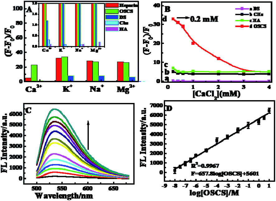 | ||
| Fig. 5 (A) Effect of different metal ions (Ca2+, Mg2+, K+ and Na+) in the presence of various anionic polysaccharides heparin (red), OSCS (green), DS (blue), Chs (light blue) and HA (pink). Inset: partial enlarged detail. (B) The concentration of Ca2+ ions on the (F − F0)/F0 value of probe 2 in the OSCS biosensing system in the presence of various anionic polysaccharides (DS (a), Chs (b), HA (c) and OSCS (d)). (C) Fluorescence emission spectra with increasing amounts of OSCS from 10−10% to 10% w/w (from bottom to top) in the OSCS biosensing system. (D) Relationship between the fluorescence intensity and the concentrations of OSCS. | ||
Under the optimal conditions (10 nM A10–MB–A10 and 0.2 mM Ca2+ ions), quantification of OSCS in heparin was performed using the proposed OSCS biosensing system (Scheme 1B). Standard heparin solutions (9 μg mL−1) were first mixed with a series of concentrations of OSCS ranging from 10−10 to 10% (w/w). Fig. 5C shows the fluorescence intensity changes upon different concentrations of OSCS. As the concentration of OSCS increased at a fixed concentration of heparin, the fluorescence intensity of the proposed probe 2 were progressively enhanced. Fig. 5D shows the linear relationship between the fluorescence responses and the concentrations of OSCS. The results illustrate that the proposed probe 2 is capable of detecting as low as 10−8% (w/w) OSCS in heparin. Compared with other assay methods for OSCS, we also found that the sensitivity of the proposed system was superior to that of other reported methods (Tables S3 and S4†). Furthermore, reproducibility and stability are also important in the biosensing field. Fig. S2† depicts the reproducibility and stability of the OSCS sensing system. The relative standard deviations (RSDs) at two concentration levels [10−7% (w/w) and 10% (w/w) OSCS in heparin] are less than 3% (2.8% and 2.3%), signifying that the proposed system is highly reproducible for detecting OSCS in heparin (Fig. S2A†). Fig. S2B† shows the stability of the proposed biosensing protocols, showing the fluorescence response of the biosensor to 10% (w/w) OSCS has no apparent change in ten continuous days of testing, verifying the good stability of the biosensing protocols.
4. Conclusions
In conclusion, we reported a “turn-on” ultrasensitive method for the detection of heparin or OSCS with HCR signal amplification and MnO2 nanosheet quenching-based background lowering. As results, the detection limit of 4 pM for heparin and 10−8% (w/w) for OSCS shows the ultrahigh sensitivity of the biosensing protocols. Furthermore, the similar DNA probe for the construction of both heparin and OSCS sensors shows the sensor possesses potential application value from an economic standpoint. The novel, simple, MnO2 nanosheet-SG fluorescence biosensing platform with lowered background holds great promise for other related analytes.Acknowledgements
This work was financially supported by the projects (20901003, 21073001 and 21005001) from National Natural Science Foundation of China, Natural Science Foundation of Anhui (KJ2009B013Z).References
- I. Capila and R. J. Linhardt, Angew. Chem., Int. Ed., 2002, 41, 390–412 CrossRef CAS.
- G. J. Despotis, G. Gravlee, K. Filos and J. Levy, Anesthesiology, 1999, 91, 1122–1151 CrossRef CAS PubMed.
- E. Theodore, M. D. Warkentin, L. Robert and M. B. A. Barkin, Pharmacotherapy, 1999, 19, 181–195 CrossRef.
- Q. Dai, W. M. Liu, X. Q. Zhuang, J. S. Wu, H. Y. Zhang and P. F. Wang, Anal. Chem., 2011, 83, 6559–6564 CrossRef CAS PubMed.
- S. L. Wang and Y. T. Chang, Chem. Commun., 2008, 10, 1173–1175 RSC.
- M. Wang, D. Zhang, G. Zhang and D. Zhu, Chem. Commun., 2008, 4469–4471 RSC.
- S. Beni, J. F. Limtiaco and C. K. Larive, Anal. Bioanal. Chem., 2011, 399, 527–539 CrossRef CAS PubMed.
- G. A. Crespo, M. G. Afshar and E. Bakker, Angew. Chem., Int. Ed., 2012, 51, 12575–12578 CrossRef CAS PubMed.
- A. Shvarev and E. Bakker, Anal. Chem., 2005, 77, 5221–5228 CrossRef CAS PubMed.
- A. Shvarev and E. Bakker, J. Am. Chem. Soc., 2003, 125, 11192–11193 CrossRef CAS PubMed.
- M. Guerrini, Z. Shriver, A. Naggi, B. Casu, R. J. Linhardt, G. Torri and R. Sasisekharan, Nat. Biotechnol., 2010, 28(3), 207–211 CrossRef CAS.
- M. Guerrini, D. Beccati, Z. Shriver, A. Naggi, K. Viswanathan, A. Bisio, I. Capila, J. C. Lansing, S. Guglieri, B. Fraser, A. Al-Hakim, N. S. Gunay, Z. Zhang, L. Robinson, L. Buhse, M. Nasr, J. Woodcock, R. Langer, G. Venkataraman, R. J. Linhardt, B. Casu, G. Torri and R. Sasisekharan, Nat. Biotechnol., 2008, 26, 669–675 CrossRef CAS PubMed.
- M. L. Trehy, J. C. Reepmeyer, R. E. Kolinski, B. J. Westenberger and L. F. Buhse, J. Pharm. Biomed. Anal., 2009, 49, 670–673 CrossRef CAS PubMed.
- N. Volpi, F. Maccari, F. J. Suwan and R. J. Linhardt, Electrophoresis, 2012, 33, 1531–1537 CrossRef CAS PubMed.
- Z. Zhang, B. Li, J. Suwan, F. Zhang, Z. Wang, H. Liu, B. Mulloy and R. J. Linhardt, J. Pharm. Sci., 2009, 98, 4017–4026 CrossRef CAS PubMed.
- G. Li, C. Cai, L. Li, L. Fu, Y. Chang, F. Zhang, T. Toida, C. Xue and R. J. Linhardt, Anal. Chem., 2013, 86, 326–330 CrossRef PubMed.
- L. Wang, S. Buchanan and M. E. Meyerhoff, Anal. Chem., 2008, 80, 9845–9847 CrossRef CAS PubMed.
- H. Yan and H. F. Wang, Anal. Chem., 2011, 83, 8589–8595 CrossRef CAS PubMed.
- C. Y. Kuo and W. L. Tseng, Chem. Commun., 2013, 49, 4607 RSC.
- S. Y. Hung and W. L. Tseng, Biosens. Bioelectron., 2014, 57, 186–191 CrossRef CAS PubMed.
- C. Y. Lee and W. L. Tseng, Anal. Chem., 2015, 87, 5031–5035 CrossRef CAS PubMed.
- L. M. Lu, X. B. Zhang, R. M. Kong, B. Yang and W. H. Tan, J. Am. Chem. Soc., 2011, 133, 11686–11691 CrossRef CAS PubMed.
- J. Huang, Y. Wu, Y. Chen, Z. Zhu, X. Yang, C. J. Yang, K. Wang and W. H. Tan, Angew. Chem., Int. Ed., 2011, 50, 401–404 CrossRef CAS PubMed.
- Y. Cheng, X. Zhang, Z. Li, X. Jiao, Y. Wang and Y. Zhang, Angew. Chem., Int. Ed. Engl., 2009, 48, 3268–3272 CrossRef CAS PubMed.
- C. Chen, D. A. Ridzon, A. J. Broomer, Z. Zhou, D. H. Lee, J. T. Nguyen, M. Barbisin, N. L. Xu, V. R. Mahuvakar, M. R. Andersen, K. Q. Lao, K. J. Livak and K. J. Guegler, Nucleic Acids Res., 2005, 33, e179 CrossRef PubMed.
- P. Liu, X. H. Yang, S. Sun, Q. Wang, K. M. Wang, J. Huang, J. B. Liu and L. L. He, Anal. Chem., 2013, 85, 7689–7695 CrossRef CAS PubMed.
- J. Ikbal, G. S. Lim and Z. Gao, TrAC, Trends Anal. Chem., 2015, 64, 86–99 CrossRef CAS.
- H. M. Deng, X. J. Yang and Z. Q. Gao, Analyst, 2015, 140, 3210–3215 RSC.
- J. Liu and Y. Lu, J. Am. Chem. Soc., 2007, 129, 9838–9839 CrossRef CAS PubMed.
- L. Zhou, Y. Omomo, N. Sakai, K. Fukuda, L. Nakai, Y. Ebina, K. Takada, M. Watanabe and T. Sasaki, J. Am. Chem. Soc., 2003, 15, 2873 Search PubMed.
- H. Zipper, H. Brunner, J. Bernhagen and F. Vitzthum, Nucleic Acids Res., 2004, 32, e103 CrossRef PubMed.
- F. Chevalier, R. Lucas, J. Angulo, M. Martin-Lomas and P. M. Nieto, Carbohydr. Res., 2004, 339, 975–983 CrossRef CAS PubMed.
- D. G. He, X. X. He, K. M. Wang, X. Yang, X. X. Yang, X. C. Li and Z. Zou, Chem. Commun., 2014, 50, 11049 RSC.
- B. A. Pinaud, Z. Chen, D. N. Abram and T. F. Jaramillo, J. Phys. Chem. C, 2011, 115, 11830–11838 CAS.
- D. L. Rabenstein, J. M. Robert and J. Peng, Carbohydr. Res., 1995, 278, 239–256 CrossRef CAS PubMed.
Footnote |
| † Electronic supplementary information (ESI) available. See DOI: 10.1039/c6ra15625h |
| This journal is © The Royal Society of Chemistry 2016 |