Individual and combined antioxidant effects of ginsenoside F2 and cyanidin-3-O-glucoside in human embryonic kidney 293 cells

Di Liu, Fengguang Pan, Jiyun Liu, Ying Wang, Ting Zhang, Erlei Wang and Jingbo Liu*
Laboratory of Nutrition and Functional Food, Jilin University, Changchun 130062, Jilin, People's Republic of China. E-mail: ljb168@sohu.com; Tel: +86-13944170656

Received 7th June 2016 , Accepted 9th August 2016

First published on 10th August 2016


Abstract

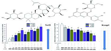
Ginsenosides and anthocyanins have already been reported to exhibit protective effects on intracellular defense against oxidative stress. Nuclear factor erythroid-derived 2 (NF-E2)-like 2 (Nrf2) plays a key role in signal pathways to counteract oxidative stress and controls expression of antioxidant enzymes. The present study is aimed at investigating the individual and combined antioxidant effects of ginsenoside F2 (F2) and cyanidin-3-O-glucoside (C3G) against H2O2-induced oxidative stress in Human Embryonic Kidney (HEK-293) cells. Cell viability, MDA level and activities of antioxidant enzymes were measured using corresponding assay kits. A DCFH-DA fluorescent probe assay was used to measure the level of intracellular reactive oxygen species (ROS). Quantitative real-time PCR and Western blotting were used to detect the expression of Nrf2 and Kelch-like ECH associated protein 1 (Keap1). The results showed that F2 and C3G significantly inhibited the generation of MDA and intracellular ROS, and increased the activities of antioxidant enzymes. Furthermore, F2 and C3G significantly enhanced the protein and mRNA expressions of Nrf2 and reduced the expressions of Keap1 in a concentration-dependent manner. On the basis of the stronger individual effects, synergistic effects were present in the combinations of F2 and C3G. In conclusion, these results demonstrated that F2 and C3G can protect HEK-293 cells against H2O2-induced oxidative stress and possibly act synergistically through reducing intracellular ROS, as well as activating the Nrf2/Keap1 signaling pathway and increasing antioxidant enzyme levels.


1. Introduction

Oxidative stress is caused by an imbalance between increased production of free radical and reactive oxygen species (ROS), and poorer antioxidant defenses, and is thought to be one of the main contributing factors to aging, inflammation and some chronic diseases. Many signal pathways are stimulated by ROS when cells respond to changes in extra- and intracellular environmental conditions.1 Nuclear factor erythroid-derived 2 (NF-E2)-like 2 (Nrf2) is a key transcription factor that controls an important adaptive pathway to counteract oxidative stress.2 Under normal physiological conditions, Nrf2 is kept in the cytoplasm, and is retained by the Kelch-like ECH associated protein 1 (Keap1).3 In response to oxidative stress induced by ROS, electrophilic or other insults, the ubiquitination and degradation of Nrf2 decreases and the free Nrf2 translocates to the nucleus, where it can be combined with antioxidant response element (ARE) and thus regulate the expressions of many cytoprotective genes, some antioxidant and detoxifying enzymes, such as catalase (CAT), superoxide dismutase (SOD), glutathione peroxidase (GSH-Px).4,5 The HEK-293 cell line is the immortalized cell with normal renal-specific properties, which has been demonstrated to display several features of renal distal tubular cells with the epithelial morphology of apical zonulae occludentes and less pronounced brush-border.6,7 The HEK-293 cells have been used as cellular model of oxidative stress.8,9

Ginseng (the roots of Panax ginseng C. A. Meyer) has been used as a traditional medicine for thousands of years in the Far East.10 Ginsenosides have been regarded as the major active components of ginseng, which were demonstrated to contribute to diverse pharmacological effects and to target a myriad of tissues.11,12 Ginsenoside F2 (F2) is one of the main metabolites of protopanaxadiol-type (PPD-type) ginsenosides, such as Rb1, Rb2, Rc and Rd, which are hydrolyzed to lose sugar moieties by intestinal microorganism in the body.13,14 The pharmacological effects of ginsenoside F2 have been focused on its anti-cancer and anti-alopecia activities.15–18 However, there is a dearth of information on the role of ginsenoside F2 in inhibiting oxidative stress induced by H2O2. Anthocyanins are natural colorants and antioxidants belonging to the classes of flavonoids. They seem to contribute to prevent human diseases related to oxidative stress.19 As one of the most ubiquitous anthocyanins, Cyanidin-3-O-glucoside (C3G) is probably the best known and investigated anthocyanin, which have potential beneficial effects in various human pathologies.20 Some previous studies have demonstrated that C3G is able to protect human endothelial cells against dysfunction induced by palmitate and TNF-α through activating the Nrf2 related pathway, further up-regulating antioxidant and detoxifying enzymes.21,22

In some cases there are probably synergic or antagonistic effects in common dietary antioxidants. It has been demonstrated that common anthocyanins (including cyanidin-3-O-glucosid) show the synergistic antioxidant effect against the peroxyl radicals with the most common dietary flavanols,23 and some pigments (betanin, lycopene, and β-carotene).24 Ginsenoside Rb1, precursor of ginsenoside F2, has been reported to exhibit synergistic effects with tanshinone IIA and salvianolic acid B in myocardial ischemia rats.25 However until now very few studies have focused on the ginsenosides–anthocyanins interactions in terms of anti-oxidative activity.

The aim of this study was to investigate the effects of ginsenoside F2 and C3G against H2O2-induced oxidative stress in HEK-293 cells, and to evaluate the impacts of combination on cell viability, intracellular ROS, antioxidant enzymes and other factors in antioxidant pathway.

2. Materials and methods

2.1. Materials and chemicals

F2 (Fig. 1A) and C3G (Fig. 1B) were purchased from the Chinese National Institute for the Control of Pharmaceutical and Biological Products, purity ≥ 98% (Beijing, PR China). The fluorescence probes 2′,7′-dichlorodihydrofluorescein diacetate (DCFH-DA) was obtained from Sigma (USA). HEK-293 cells were purchased from Chinese Infrastructure of Cell Line Resources. Dulbecco's Modified Eagle's Medium (DMEM), Fetal Bovine Serum (FBS), Penicillin-Streptomycin Solution (PSS) and MEM Nonessential Amino Acids (MEM NEAA) was obtained from Gibco (USA). The Cell Titer 96® AQueous One Solution Cell Proliferation Assay (MTS) and GoTaq Green Master Mix were purchased from Promega Biotechnology Co. Ltd. The MDA, SOD, GSH-Px, and CAT assay kits were purchased from Nanjing Jiancheng Bioengineering Co. (Nanjing, China, http://www.njjcbio.com). Rainbow protein standard marker and D2000 DNA marker were purchased from TIANGEN BIOTECH (BEIJING) CO. LTD. (http://www.tiangen.com). The enhanced chemiluminescence (ECL) detection kit and radio immunoprecipitation assay (RIPA) lysis buffer were purchased from Beyotime Institute of Biotechnology (http://www.beyotime.com). PrimeScriptTM RT Reagent Kit with gDNA Eraser and SYBR Premix were purchased from Takara Bioengineering Co. Ltd. Regular Agarose G-10 was obtained from Biowest. The antibodies against Nrf2, Keap1 and goat-anti-mouse second antibody were purchased from Wuhan Boster Biological Engineering Co., Ltd. (http://www.boster.com.cn). Polyvinylidene fluoride (PVDF, 0.2 μm) was purchased from Pall Corporation (America).
image file: c6ra14831j-f1.tif
Fig. 1 Structures and formula weights of ginsenoside F2 (A) and cyanidin-3-O-glucoside (B).

2.2. Cell culture and treatment

HEK-293 cells were cultivated in DMEM supplemented with 10% FBS, 1% PSS and 1% MEM NEAA at 37 °C in 5% CO2. HEK-293 cells were seeded in flat-bottom well plates and then incubated in a CO2 incubator overnight, until all cells adhered to the wall. The cells were incubated with 1.25, 5 and 20 μM of F2, C3G and their combination (volume proportion of 1[thin space (1/6-em)]:[thin space (1/6-em)]1) for 12 h and then were immediately exposed to 400 μM of H2O2 for 6 h.

2.3. Individual and combined protective effects of ginsenoside F2 and C3G on cell viability

Normal HEK-293 cells without ginsenoside F2, C3G and H2O2 treatment were used as a control. DMEM and H2O2 were added then to the treatment without the addition of ginsenoside F2, C3G as a damage group. Cell viability was determined by the MTS assay as previously mentioned with a few changes.26,27 MTS (20 μL) was added to the cultured cells incubated at 37 °C, 5% CO2 for 1 hour. Then the plates were read in a multi-mode microplate reader (Bio Tek Instruments, USA) at 490 nm wavelength. The results were expressed as the mean optical density (OD) of each group and dose. All the experiments were repeated at least 3 times.

2.4. Measurement of intracellular ROS

Production of intracellular ROS was assessed using DCFH-DA as described previously.28 The medium was removed and the cells were incubated with DCFH-DA (10 μM) for 20 min at 37 °C in the dark. The green fluorescence images of DCF in HEK-293 cells were collected by Laser Scanning Confocal Microscopy (LSCM) (Olympus, Japan). The fluorescence intensity was determined by a multi-mode microplate reader (BioTek Instruments, USA) at an excitation wavelength of 485 nm and an emission wavelength of 528 nm.

2.5. Measurement of antioxidant enzyme activities and MDA level

The cells were washed once with PBS and lysed in ice-cold RIPA lysis buffer containing 50 mM Tris–HCl (pH 7.4), 150 mM NaCl, 1% Triton X-100, 1% sodium deoxycholate, 0.1% SDS, sodium orthovanadate, sodium fluoride, EDTA, leupeptin and 1 mM PMSF for 10 min and then centrifuged at 12[thin space (1/6-em)]000g, 4 °C for 10 min. Activities of SOD, GSH-Px, CAT, MDA and BCA levels were measured according to the manufacturer's protocol of corresponding assay kit.

2.6. Western blot analysis

Briefly, 30 μg of the proteins in supernatant were separated by 12% sodium dodecyl sulphate-polyacrylamide (SDS-PAGE) gels electrophoresis and the blots were transferred onto PVDF membranes. The membranes were incubated in blocking buffer at room temperature for 1 h and then incubated with antibody against Nrf2, Keap1 and GAPDH in TBST (1[thin space (1/6-em)]:[thin space (1/6-em)]400 dilution) overnight at 4 °C. Then were incubated with horseradish peroxidase-conjugated secondary antibodies (1[thin space (1/6-em)]:[thin space (1/6-em)]4000 dilution) for 1 h and detected by DNR MiniBIS Pro Bio Imaging System (DNR, Israel) using ECL reagents. The images were collected and the bands were quantitated by densitometric analysis using the ImageJ software (http://rsb.info.nih.gov/ij/index.html). The data of Nrf2 and Keap1 were normalized on the basis of GAPDH level.

2.7. Quantitative real-time polymerase chain reaction (qRT-PCR) analysis

For the quantification of gene expression, qRT-PCR was conducted by a real-time system (CFX96; Bio-Rad) according to a previous publication with a few changes.29 Briefly, total RNA of cells was extracted with TRIzol reagent according to the manufacturer's instructions. The cDNA was reversely transcribed from total RNA after erasing gDNA by using a reverse transcriptase kit. The PCR reaction contained 1 μg of cDNA, primers, RNase-free water and SYBR premix. The sequences of the primer pairs used for the amplification of human Nrf2, Keap1 and GAPDH are as followed:

Nrf2 (forward primer: 5′-AACCCTTGTCACCATCTCAG-3′; reverse primer: 5′-GCAGCCACTTTATTCTTACC-3′),

Keap1 (forward primer: 5′-ACTCGTTGACGCCGAACTT-3′; reverse primer: 5′-GCAGGGCGACCACTGATT-3′),

GAPDH (forward primer: 5′-ATCCCATCACCATCTTCC-3′; reverse primer: 5′-CCATCACGCCACAGTTT-3′).

Amplification conditions were set as followed: 95 °C for 3 min, 40 cycles of 95 °C for 15 s, 51.6 °C (nrf2) or 58 °C (keap1) for 20 s and 72 °C for 30 s. The relative index (2−ΔΔCt) was calculated by comparing the average expression level to control samples which was defined as 1.00. Expression levels of nrf2 and keap1 genes were normalized by concurrent measurement of GAPDH levels.

2.8. Statistical analysis

All of the assays were carried out in triplicate. Data analyses were performed using the SPSS 21.0 software. The results were expressed as the mean ± standard deviation in the text and figures. The statistical significance of differences between two groups was determined by the one-way ANOVA program with the LSD test unless specified. Probability values of less than 0.05 were considered significant.

3. Results

3.1. Individual and combined protective effects of ginsenoside F2 and C3G against oxidative stress in HEK-293 cells

The cytotoxic and proliferative effects of ginsenoside F2, C3G and their combination on HEK-293 cells were measured by MTS assay. The results showed that the ginsenoside F2, C3G and their combination did not exhibit significant stimulation or inhibitation effect on the cell viability at the concentration from 1.25 to 20 μM (data not shown). The results implied that the F2 or C3G or their combination did not have cytotoxicity, and the inhibition effect of F2 or C3G or their combination on oxidative stress was not due to the cell proliferative effect. This concentration range was chosen to use for the pretreatment of F2, C3G and their combination in the subsequent experiments.

In our study, H2O2-induced HEK-293 cell model was constructed to carry on the biological research and to explore the synergistic protective effect of ginsenoside F2 and C3G against oxidative stress. 400 μM of H2O2 was chosen as the optimal damage concentration according to our previous study.30 In Fig. 2, when HEK-293 cells were exposed to 400 μM H2O2 without pretreatment of F2 or C3G or their combination, the cell viability decreased significantly compared to the control group (p < 0.05). In contrast, the cell viabilities of pretreatment groups were higher than that of the oxidative injury group. In these experiments, both ginsenoside F2 and C3G exhibited concentration-dependent alleviation of H2O2-induced cell damage, which showed significant protective effects at 1.25, 5 and 20 μM (p < 0.05) (Fig. 2). With the intervention of combination, which contains equal amounts of F2 and C3G, the cell viabilities increased significantly compared with a single F2 or C3G pretreatment at the corresponding concentration (1.25, 5 and 20 μM, p < 0.05) (Fig. 2). However, the combination of F2 and C3G exhibited significant enhanced inhibition on H2O2-induced damage.


image file: c6ra14831j-f2.tif
Fig. 2 Individual and combined protective effects of ginsenoside F2 and C3G against oxidative stress in HEK-293 cells. Cells were treated with ginsenoside F2, C3G and their combination at the indicated concentrations (1.25, 5 and 20 μM) for 12 h and H2O2 (400 μM) for 6 h. The combination of 1.25 μM contains 0.625 μM F2 and 0.625 μM C3G, the combination of 5 μM contains 2.5 μM F2 and 2.5 μM C3G, the combination of 20 μM contains 10 μM F2 and 10 μM C3G. Cell viability was assessed by MTS assay. Vertical bars indicate mean values ± SD (n = 3). Values with different letters indicate significant differences (P < 0.05).

3.2. Individual and combined effects of ginsenoside F2 and C3G on ameliorating H2O2-induced ROS generation in HEK-293 cells

To determine whether the ginsenoside F2 and C3G inhibit ROS production during oxidative stress induced by H2O2, HEK-293 cells were treated with ginsenoside F2, C3G and their combination at the indicated concentrations (1.25, 5 and 20 μM) for 12 h and were exposed to H2O2 (400 μM) and DCFH-DA. As shown in Fig. 3 A, compared with the control group (a), green fluorescence intensity of cells displayed considerable increase after incubated with H2O2 (b). However, the intensity of green fluorescence decreased markedly in the F2 group (c, d and e) and C3G group (f, g and h) with a concentration-dependent effect. Compared with the single F2 or C3G pretreatment group, it was to see that the fluorescence intensity of combination group decreased even more (i, j and k). However, following incubation with ginsensodie F2, C3G and their combination, the DCF fluorescence was decreased significantly in comparison with the injury group (only H2O2-treated) (Fig. 3A).
image file: c6ra14831j-f3.tif
Fig. 3 Individual and combined ameliorated effects of ginsenoside F2 and C3G against H2O2-induced ROS generation in HEK-293 cells. (A) Fluorescence images of DCF in HEK-293 cells were collected by Laser Scanning Confocal Microscopy (LSCM). (a) control group; (b) damage group (400 μM H2O2 treated); (c) 1.25 μM F2 + 400 μM H2O2; (d) 5 μM F2 + 400 μM H2O2; (e) 20 μM F2 + 400 μM H2O2; (f) 1.25 μM C3G + 400 μM H2O2; (g) 5 μM C3G + 400 μM H2O2; (h) 20 μM C3G + 400 μM H2O2; (i) 0.625 μM F2 + 0.625 μM C3G + 400 μM H2O2; (j) 2.5 μM F2 + 2.5 μM C3G + 400 μM H2O2; (k) 10 μM F2 + 10 μM C3G + 400 μM H2O2. (B) Production of intracellular ROS was measured using the fluorescence probe DCFH-DA. The level of ROS generation in the control group was designed as 1.0 and that was used to express the relative ROS production in other groups. Vertical bars indicate mean values ± SD (n = 3). Values with different letters indicate significant differences (P < 0.05).

In our study, when HEK-293 cells were treated with 400 μM H2O2, the relative ROS production significantly increased compared with the control group (p < 0.05). The increase of ROS was suppressed in a concentration-dependent fashion with pretreatment of F2 or C3G (Fig. 3B). Moreover, when ginsenoside F2 was added together with C3G, additional inhibitory effects on ROS generation were observed at concentrations of 1.25, 5 and 20 μM (p < 0.05). These data demonstrated that ginsenoside F2 and C3G possibly act synergistically protect HEK-293 cells from H2O2-induced oxidative stress by ameliorating intracellular ROS generation.

3.3. Individual and combined effects of ginsenoside F2 and C3G on MDA level and activities of antioxidant enzymes activities in HEK-293 cells

To evaluate the attenuation of consequences of oxidative stress, the content of lipid peroxidation product (MDA) and activities of antioxidant enzymes were measured in cultured HEK-293 cells. As shown in Fig. 4, H2O2 treatment markedly increased the level of MDA and decreased the activities of SOD, GSH-Px and CAT in the cultures. However, F2 or C3G pretreatment resulted in a concentration-dependent reduction of the MDA level (Fig. 4A). Moreover, when ginsenoside F2 was combined with C3G, significantly additional decreases of MDA were observed at doses of 1.25, 5 and 20 μM (p < 0.05) (Fig. 4A). Ginsenoside F2 pretreatment showed noticeably increased SOD and GSH-Px activities with a concentration-dependent manner in comparison with the injury group (P < 0.05), and C3G pretreatment also showed similar elevations at doses of 5 and 20 μM (p < 0.05) (Fig. 4B and C). However, with the intervention of combination, the activities of SOD and GSH-Px were significantly increased compared to a single F2 or C3G pretreatment at doses of 5 and 20 μM (p < 0.05) (Fig. 4B and C). Additionally, CAT activities were also significantly increased by the pretreatment with 5 and 20 μM of F2 or C3G (P < 0.05). And when ginsenoside F2 was combined with C3G, additional increases of CAT activity were observed at doses of 5 and 20 μM (p < 0.05) (Fig. 4D). Although the SOD, GSH-Px and CAT activities in 1.25 μM combination group showed no significant difference compared with the single F2 or C3G group, noticeably enhanced activities were observed compared with injury groups. These results also confirmed that ginsenoside F2 and C3G inhibited H2O2-induced oxidative stress by reducing MDA content, as well as elevating the activities of SOD, GSH-Px and CAT, and synergism between the individuals may be exist.
image file: c6ra14831j-f4.tif
Fig. 4 Individual and combined effects of ginsenoside F2 and C3G on MDA level and activities of SOD, GSH-Px and CAT after H2O2-induced oxidative injury in HEK-293 cells. Cells were treated with indicated concentrations (1.25, 5 and 20 μM) of ginsenoside F2, C3G and their combination for 12 h and H2O2 (400 μM) for 6 h. MDA level (A) and cellular SOD (B), GSH-Px (C), CAT (D) were measured with assay kits. Vertical bars indicate mean values ± SD (n = 3). Values with different letters indicate significant differences (P < 0.05).

3.4. Ginsenoside F2, C3G and their combination suppressed H2O2-induced oxidative stress in HEK-293 cells by activating Nrf2/Keap1 signaling pathway

To further investigate the potential mechanisms of individual ginsenoside F2, C3G and combination in ameliorating cellular oxidative stress, the protein and mRNA expression levels of Nrf2 and Keap1 were examined by qRT-PCR and western blot methods, respectively. As shown in Fig. 5, when cells were exposed to H2O2, the protein and mRNA expression levels of Nrf2 were up-regulated, followed by a down-regulation in Keap1 expression compared with control group. Ginsenoside F2 or C3G pretreatment resulted in significant increases in the protein and mRNA expressions of Nrf2 in a concentration-dependent manner (P < 0.05) (Fig. 5). However, the expressions of Keap1, the negative regulator of Nrf2, were noticeably decreased at concentration of 5 and 20 μM with the mediation of F2 or C3G (P < 0.05) (Fig. 5). In addition, we investigated the combined effects of F2 and C3G on Nrf2 and Keap1 expressions in H2O2-induced oxidative stress cells. The results showed that, when 0.625 μM ginsenoside F2 was combined with 0.625 μM C3G, the protein and mRNA expressions of Nrf2 were larger than those of both 1.25 μM F2 and 1.25 μM C3G (p < 0.05), as well as at the concentrations of 5 and 20 μM (p < 0.05) (Fig. 5). Meanwhile, with the intervention of combination, additional decreases of Keap1 expressions were observed at concentrations of 1.25, 5 and 20 μM (p < 0.05) (Fig. 5).
image file: c6ra14831j-f5.tif
Fig. 5 Individual and combined effects of ginsenoside F2 and C3G on protein and mRNA expressions of Nrf2 and Keap1. Cells were treated with indicated concentrations (1.25, 5 and 20 μM) of ginsenoside F2, C3G and their combination for 12 h and H2O2 (400 μM) for 6 h. The protein expression levels of Nrf2, Keap1 and GAPDH were determined by western blot analyses. The mRNA expression levels of Nrf2, Keap1 and GAPDH were determined by qRT-PCR. Vertical bars indicate mean values ± SD (n = 3). Values with different letters indicate significant differences (P < 0.05).

4. Discussion

Ginsenosides have been used together with drugs on treatment of inflammation or cancer, which is aimed to strengthen the shots, ameliorate side effects, minimize or slow down the development of drug resistance.31–34 There are some positive outcomes of synergism of anthocyanins or flavonoids on antioxidant activity, such as catechin and malvidin 3-glucoside, kaempferol and myricetin, epicatechin and quercetin-3-β-glucoside.23,35 However, this is the first report on antioxidant effect of ginsenoside F2 and C3G, and possibly synergistic antioxidant in HEK-293 cell type. In our study, individual and combined antioxidant effect of F2 and C3G was observed. And the synergistic antioxidant effects were analyzed by the direct comparison methodology which was reported by Kuei-long Liao.36 The results showed that the F2 and C3G inhibited oxidative stress induced by H2O2, reduced intracellular ROS levels and increased the activities of antioxidant enzymes including SOD, GSH-Px and CAT through regulation of Nrf2/Keap1 signaling pathway in HEK-293 cells. Moreover, the co-administrations of F2 with C3G possibly act synergistically antioxidant effects.

Ginsenosides share a similar sterol structure as basic, and they are characterized by the number and position of sugar moieties at positions C-3 and C-6 of the chemical structure, eventually companied by different activities.37 F2 (Fig. 1A) is one of the PPD-type classifications that differ from the structure of the PPD by the substituent of one glucose moiety at position C-3 and C-20. The chemical structure of ginsenoside Rb1 is differed from F2 by the addition of one glucose moiety at position C-2′ and C-6′′. Ginsenoside Rd is differed from F2 by the addition of one glucose moiety at position C-2′. The two ginsenosides have been reported to have protective effect against oxidative stress or oxidative injury induced by H2O2 in cultured cells.38,39 Several studies have shown that the ginsenoside metabolites possess superior pharmacological activities in vitro and in vivo compared with other PPD-type and PPT-type ginsenosides.12,40–42 The ginsenoside F2 attenuates H2O2-induced oxidative stress in our study. This result suggested that F2 exhibits antioxidant activity partly by breaking apart the glucosidic bond to lose the glucose moiety. The antioxidant activities of anthocyanins against hydroxyl radicals, superoxide radicals and proxyl radicals were affected by the different hydroxyl substitutions and methylations in ring structures. C3G (Fig. 1B) is one of the best known and investigated anthocyanins. Previous study has revealed that among the aglycons with same A and C rings, the ORAC activity of the compound with 3′-, 4′-OH substitution in the B ring (cyanidin) is higher than the compounds with only one 4′-OH group (pelargonidin, malvidin, and peonidin).43 Bors et al. demonstrated that the 3′- and 4′-OH in the B ring (catechol) structure is a critical determinant for the radical scavenging and/or anti-oxidative potential in flavonoids with a saturated 2,3-bond.44 The C3G attenuates H2O2-induced oxidative stress in our study. The results suggested that the antioxidant activity of C3G is partly by scavenging radicals, which attributes to the 3′- and 4′-OH substitutions in the B ring.

H2O2 is a potential source of hydroxyl radicals, and has been widely used as an exogenous trigger of oxidative stress in vitro models. Previous researches have reported that H2O2 was used as a stable source of free radicals to induce oxidative stress in HEK-293 cells.8,45 Based on the experimental results, we chose the concentrations (from 1.25 to 20 μM) of F2, C3G and their combination which did not affect the levels of cell viability. The following results showed that the F2, C3G and their combination significantly enhanced the cell viability in H2O2-stimulated HEK-293 cells. These results revealed the potential synergistic effect of F2 and C3G, and demonstrated that they can be combined to ameliorate oxidative damage.

The status of cellular redox can be indicated by the ability of cells to maintain the level of ROS. Oxidative stress is thought to be one of the main contributing factors to many diseases, which is associated with the accumulation of ROS. Previous studies have indicated that ginsenosides derived from Panax species and C3G target ROS, thereby they may prevent several diseases induced by oxidative stress.22,46 Data from our current study showed that the combined pretreatment of ginsenoside F2 and C3G significantly inhibited the generation of intracellular ROS stronger than either of the single pretreatments (Fig. 3). These results provide the evidences that ginsenoside F2 and C3G may synergistically protect HEK-293 cells from H2O2-induced oxidative stress, probably in part due to their improved abilities of scavenging intracellular ROS.

The ability to increase the activities of intracellular antioxidant enzymes has been recognized as one of the vital factors to investigate the cytoprotective properties of antioxidants.26 Lipid peroxidative injury has often been expressed by MDA content. SOD, GSH-Px and CAT are considered to be the key enzymes of antioxidant defense system. Some in vitro studies have demonstrated that the antioxidant effects of ginsenosides may not only be dependent on the direct scavenging of free radicals, but also be mediated by activation and completion of intracellular antioxidant systems.47 C3G has also been proved to be able to protect cells against oxidative stress by decreasing MDA level and enhancing the activities of SOD and GSH,21,22,48 which were in agreement with our results. In this study, when ginsenoside F2 was combined with C3G, the MDA content significantly decreased compared with the single pretreatments (Fig. 4A), confirming that F2 and C3G synergistically inhibited H2O2-induced lipid peroxidative injury. Furthermore, the combination of F2 and C3G increased the activities of SOD, GSH-Px and CAT in HEK-293 cells (Fig. 4), suggesting that the combination shows a synergistic effect. This activation of antioxidant enzymes appears to be inadequate to protect HEK-293 cells against H2O2-induced oxidative stress. Therefore, more potent activators of Nrf2/Keap1 pathway are essential to cytoprotection.

Several studies have shown that Nrf2/Keap1 signaling pathway plays a critical role in the cellular defenses against oxidative stress.49 Ginsenoside Rg3, precursor of ginsenoside F2, has been demonstrated to exhibit cardioprotection by significantly increasing the mRNA and protein expression levels of Nrf2 and HO-1, and inhibiting the over-expression of Keap1.50 Ginsneoside Rh3, another metabolite of PPD-type ginsenosides, has been reported exhibited anti-inflammatory ability in part by increasing the binding activity of Nrf2 to antioxidant response element.51 Some in vitro experiments also demonstrated that C3G is able to protect human endothelial cells by activating Nrf2 pathway that in turn activates antioxidant and detoxifying enzymes.21 Our study showed that, the protein and mRNA expressions of Nrf2 could be up-regulated and the expressions of Keap1 were down-regulated by administrating ginsenoside F2 or C3G respectively. Furthermore, the expressions of Nrf2 were increased and the expressions of Keap1 were decreased significantly while the HEK-293 cells were co-administrated with F2 and C3G. These results demonstrate that the potential synergistic cytoprotection of F2 and C3G in HEK-293 cells is mediated through activation of Nrf2/Keap1 signaling pathway as well as their intrinsic antioxidant activities.

5. Conclusion

Overall, our results demonstrate that F2 and C3G can protect HEK-293 cells from H2O2-induced oxidative stress through the elevation of cellular viability, the reduction of intracellular ROS, the induction of antioxidant enzymes' activities and the activation of Nrf2/Keap1 pathway. The potential synergistic antioxidant effects of combination treatments with ginsenoside F2 and C3G on cells indicate that this approach may be significant in terms that a variety of diseases are related to oxidative stress. In this regard, effective combination of natural antioxidants is of great importance. The combination assays in this study can be applicable to investigate the impact of antioxidant in inhibiting oxidative stress and thus preventing diseases implicitly. The combination of F2 and C3G seems to be a beneficial method in mitigation of oxidative stress. However, the key points of the synergistic mechanism and in vivo investigation require further studying, as well as other potential aspects of synergistic effects, such as anti-inflammation and anti-apoptosis.

Abbreviations

C3GCyanidin-3-O-glucoside
F2Ginsenoside F2
HEK-293Human embryonic kidney cell
H2O2Hydrogen peroxide
ROSReactive oxygen species
MDAMalonic dialdehyde
SODSuperoxide dismutase
GSH-PxGlutathione peroxidase
CATCatalase
Nrf2Nuclear factor erythroid-derived 2 (NF-E2)-like 2
Keap1Kelch-like ECH associated protein 1

Acknowledgements

This work was partially supported by the National Natural Science Foundation of China (no. 31271907 and no. 31471597).

References

  1. S. Reuter, S. C. Gupta, M. M. Chaturvedi and B. B. Aggarwal, Free Radicals Biol. Med., 2010, 49, 1603–1616 CrossRef CAS PubMed.
  2. J. Lee, D. G. Jo, D. Park, H. Y. Chung and M. P. Mattson, Pharmacol. Rev., 2014, 66, 815–868 CrossRef PubMed.
  3. N. Wakabayashi, K. Itoh, J. Wakabayashi, H. Motohashi, S. Noda, S. Takahashi, S. Imakado, T. Kotsuji, F. Otsuka, D. R. Roop, T. Harada, J. D. Engel and M. Yamamoto, Nat. Genet., 2003, 35, 238–245 CrossRef CAS PubMed.
  4. T. Prestera, P. Talalay, J. Alam, Y. I. Ahn, P. J. Lee and A. M. Choi, Mol. Med., 1995, 1, 827–837 CAS.
  5. T. W. Kensler, N. Wakabayash and S. Biswal, in Annual Review of Pharmacology and Toxicology, Annual Reviews, Palo Alto, 2007, vol. 47, pp. 89–116 Search PubMed.
  6. N. L. Simmons, Exp. Physiol., 1990, 75, 309–319 CrossRef CAS PubMed.
  7. A. I. Serban, L. Stanca, O. I. Geicu and A. Dinischiotu, Int. J. Mol. Sci., 2015, 16, 20100–20117 CrossRef CAS PubMed.
  8. J. B. Liu, Z. F. Chen, J. He, Y. Zhang, T. Zhang and Y. Q. Jiang, Food Funct., 2014, 5, 3179–3188 CAS.
  9. E. Bradshaw, G. Beggs and M. Cartafalsa, Mol. Biol. Cell, 2014, 25, 1 CrossRef PubMed.
  10. D. O. Kennedy and A. B. Scholey, Pharmacol., Biochem. Behav., 2003, 75, 687–700 CrossRef CAS.
  11. A. S. Attele, J. A. Wu and C. S. Yuan, Biochem. Pharmacol., 1999, 58, 1685–1693 CrossRef CAS PubMed.
  12. B. G. Kim, S. Y. Choi, M. R. Kim, H. J. Suh and H. J. Park, Process Biochem., 2010, 45, 1319–1324 CrossRef CAS.
  13. H. Hasegawa, J. H. Sung, S. Matsumiya and M. Uchiyama, Planta Med., 1996, 62, 453–457 CrossRef CAS PubMed.
  14. H.-d. Wan and D. Li, RSC Adv., 2015, 5, 78874–78879 RSC.
  15. J. Y. Shin, J. M. Lee, H. S. Shin, S. Y. Park, J. E. Yang, S. K. Cho and T. H. Yi, J. Ginseng Res., 2012, 36, 86–92 CrossRef CAS PubMed.
  16. T. T. Mai, J. Moon, Y. Song, P. Q. Viet, P. V. Phuc, J. M. Lee, T. H. Yi, M. Cho and S. K. Cho, Cancer Lett., 2012, 321, 144–153 CrossRef CAS PubMed.
  17. H. S. Shin, S. Y. Park, E. S. Hwang, D. G. Lee, G. T. Mavlonov and T. H. Yi, Biol. Pharm. Bull., 2014, 37, 755–763 CAS.
  18. H. S. Shin, S. Y. Park, E. S. Hwang, D. G. Lee, H. G. Song, G. T. Mavlonov and T. H. Yi, Eur. J. Pharmacol., 2014, 730, 82–89 CrossRef CAS PubMed.
  19. F. Cimino, R. Ambra, R. Canali, A. Saija and F. Virgili, J. Agric. Food Chem., 2006, 54, 4041–4047 CrossRef CAS PubMed.
  20. J. Kong, Phytochemistry, 2003, 64, 923–933 CrossRef CAS PubMed.
  21. A. Speciale, S. Anwar, R. Canali, J. Chirafisi, A. Saija, F. Virgili and F. Cimino, Mol. Nutr. Food Res., 2013, 57, 1979–1987 CAS.
  22. D. Fratantonio, A. Speciale, D. Ferrari, M. Cristani, A. Saija and F. Cimino, Toxicol. Lett., 2015, 239, 152–160 CrossRef CAS PubMed.
  23. M. Rossetto, P. Vanzani, F. Mattivi, M. Lunelli, M. Scarpa and A. Rigo, Arch. Biochem. Biophys., 2002, 408, 239–245 CrossRef CAS PubMed.
  24. M. K. Reddy, R. L. Alexander-Lindo and M. G. Nair, J. Agric. Food Chem., 2005, 53, 9268–9273 CrossRef CAS PubMed.
  25. Y. Lu, X. Liu, X. Liang, L. Xiang and W. Zhang, J. Ethnopharmacol., 2011, 134, 45–49 CrossRef CAS PubMed.
  26. M. S. Lee, B. Lee, K. E. Park, T. Utsuki, T. Shin, C. W. Oh and H. R. Kim, Food Chem., 2015, 174, 538–546 CrossRef CAS PubMed.
  27. A. H. Cory, T. C. Owen, J. A. Barltrop and J. G. Cory, Cancer Commun., 1991, 3, 207–212 CAS.
  28. J. M. Xu, Z. M. Hao, X. B. Gou, W. Tian, Y. L. Jin, S. X. Cui, J. Guo, Y. J. Sun, Y. Wang and Z. L. Xu, Microsc. Res. Tech., 2013, 76, 612–617 CrossRef CAS PubMed.
  29. S. Cao, D. Chao, H. Zhou, G. Balboni and Y. Xia, Br. J. Pharmacol., 2015, 172, 1869–1881 CrossRef CAS PubMed.
  30. D. Liu, T. Zhang, Z. Chen, Y. Wang, S. Ma, J. Liu and J. Liu, J. Ginseng Res., 2016 DOI:10.1016/j.jgr.2016.02.007.
  31. Y. Song, F. Zhao, L. Zhang, Y. Du, T. Wang and F. Fu, Fitoterapia, 2013, 91, 173–179 CrossRef CAS PubMed.
  32. Y. J. Lee, S. Lee, J. N. Ho, S. S. Byun, S. K. Hong, S. E. Lee and E. Lee, Oncol. Rep., 2014, 32, 1803–1808 CAS.
  33. J. W. Jiang, X. M. Chen, X. H. Chen and S. S. Zheng, World J. Gastroenterol., 2011, 17, 3605–3613 CrossRef CAS PubMed.
  34. D. Lu, A. Xu, H. Mai, J. Zhao, C. Zhang, R. Qi, H. Wang, D. Lu and L. Zhu, Oxid. Med. Cell. Longevity, 2015, 2015, 437127 Search PubMed.
  35. M. Hidalgo, C. Sánchez-Moreno and S. de Pascual-Teresa, Food Chem., 2010, 121, 691–696 CrossRef CAS.
  36. K. L. Liao and M. C. Yin, J. Agric. Food Chem., 2000, 48, 2266–2270 CrossRef CAS PubMed.
  37. D. G. Popovich and D. D. Kitts, Arch. Biochem. Biophys., 2002, 406, 1–8 CrossRef CAS PubMed.
  38. R. D. Ye, J. L. Han, X. W. Kong, L. Z. Zhao, R. Cao, Z. R. Rao and G. Zhao, Biol. Pharm. Bull., 2008, 31, 1923–1927 CAS.
  39. Z. M. Song, Y. Liu, B. S. Hao, S. J. Yu, H. Zhang, D. H. Liu, B. Zhou, L. Wu, M. Wang, Z. J. Xiong, C. D. Wu, J. M. Zhu and X. X. Qian, PLoS One, 2014, 9, 9 Search PubMed.
  40. L. W. Qi, C. Z. Wang and C. S. Yuan, Biochem. Pharmacol., 2010, 80, 947–954 CrossRef CAS PubMed.
  41. Z. Q. Liu, X. Y. Luo, G. Z. Liu, Y. P. Chen, Z. C. Wang and Y. X. Sun, J. Agric. Food Chem., 2003, 51, 2555–2558 CrossRef CAS PubMed.
  42. M. A. Tawab, U. Bahr, M. Karas, M. Wurglics and M. Schubert-Zsilavecz, Drug Metab. Dispos., 2003, 31, 1065–1071 CrossRef PubMed.
  43. G. C. Hong Wang and R. L. Prior, J. Agric. Food Chem., 1997, 45, 304–309 CrossRef.
  44. W. Bors, W. Heller, C. Michel and M. Saran, in Methods in Enzymology, Academic Press, 1990, vol. 186, pp. 343–355 Search PubMed.
  45. S. B. Hani and M. Bayachou, Asian Pac. J. Trop. Biomed., 2014, 4, 399–403 CrossRef CAS PubMed.
  46. L. Jia, Y. Q. Zhao and X. J. Liang, Curr. Med. Chem., 2009, 16, 2924–2942 CrossRef CAS PubMed.
  47. J. T. Xie, Z. H. Shao, T. L. Vanden Hoek, W. T. Chang, J. Li, S. Mehendale, C. Z. Wang, C. W. Hsu, L. B. Becker, J. J. Yin and C. S. Yuan, Eur. J. Pharmacol., 2006, 532, 201–207 CrossRef CAS PubMed.
  48. A. Speciale, R. Canali, J. Chirafisi, A. Saija, F. Virgili and F. Cimino, J. Agric. Food Chem., 2010, 58, 12048–12054 CrossRef CAS PubMed.
  49. Y. S. Keum, K. K. Park, J. M. Lee, K. S. Chun, J. H. Park, S. K. Lee, H. Kwon and Y. J. Surh, Cancer Lett., 2000, 150, 41–48 CrossRef CAS PubMed.
  50. X. Wang, L. Chen, T. Wang, X. Jiang, H. Zhang, P. Li, B. Lv and X. Gao, Phytomedicine, 2015, 22, 875–884 CrossRef CAS PubMed.
  51. Y. Y. Lee, J. S. Park, E. J. Lee, S. Y. Lee, D. H. Kim, J. L. Kang and H. S. Kim, J. Agric. Food Chem., 2015, 63, 3472–3480 CrossRef CAS PubMed.

Footnote

Electronic supplementary information (ESI) available. See DOI: 10.1039/c6ra14831j

This journal is © The Royal Society of Chemistry 2016