The role of the KRSIK motif of human angiogenin in heparin and DNA binding

Kwon Joo Yeo ad, Jun-Goo Jeeb, Jin-Wan Parka, Yu-Jin Leec, Kyoung-Seok Ryua, Byoung-Mog Kwonc, Young Ho Jeon*d and Hae-Kap Cheong*a
aProtein Structure Group, Korea Basic Science Institute, 162 Yeongudanji-ro, Ochang, Chungbuk 28119, Republic of Korea. E-mail: haekap@kbsi.re.kr
bCollege of Pharmacy, Kyungpook National University, 80 Daehak-ro, Buk-gu, Daegu 41566, Republic of Korea
cKorea Research Institute of Bioscience and Biotechnology, 125Gwahak-ro, Yuseong-gu, Daejeon, 34141, Republic of Korea
dCollege of Pharmacy, Korea University, 2511 Sejong-ro, Sejong, 30019, Republic of Korea. E-mail: yhjeon@korea.ac.kr

Received 5th June 2016 , Accepted 11th August 2016

First published on 24th August 2016


Abstract

The positively charged surface with a 50KRSIK54 motif is the main interaction site of hAng for both heparin and DNA binding, providing an insight into the potential role of the 50KRSIK54 motif for the internalization and promoter binding of hAng, which is essential for the regulation of angiogenesis.

Angiogenin (Ang), a ribonucleolytic enzyme, has been implicated in various biological processes, such as angiogenesis, cell proliferation, migration, and invasion in both normal and cancer cells.1 Cell binding and nuclear translocation of hAng, which are essential steps for the internalization and DNA binding of hAng, are critical processes for stimulating ribosomal RNA (rRNA) transcription and inducing other angiogenic factors.2 Therefore, inhibition of the cell binding of exogenous hAng and the nuclear translocation or the DNA binding of endogenous hAng in cancer cells might be a useful approach for the screening and development of hAng specific inhibitors. However, the mechanisms underlying the cell and DNA binding of hAng are still unclear.

Previous reports showed that the cell binding of hAng is inhibited by heparin, suggesting that heparan/chondroitin sulfate on the cell surface is involved in the cell binding of hAng.3 In addition, mutations at the heparin binding sites of rat Ang (rAng) decreased their internalization and nuclear translocation.4 Thus, a structural study of the heparin binding of hAng is a critical issue for understanding the cell binding and internalization of exogenous hAng and the inhibition mechanism.

In the cytoplasm, the hAng translocates into the nucleus, then, binds to DNA, stimulating rRNA synthesis.2c Thus, identification of the DNA binding site of hAng is also critical for understanding the regulation of angiogenic factors. Although the crystal structure and the solution structure of hAng have been solved,5 structural study with the substrates related to its cell and DNA bindings have not been studied.

In this report, using the NMR technique, an interaction study of hAng with a heparin fragment and DNA oligomer, which are its substrates, identified the heparin and DNA binding site, suggesting that the site has a critical role in the cell binding and internalization as well as the DNA binding of hAng. The site might be a target site for screening for hAng specific inhibitors.

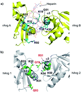
In a previous report, two rAng formed the complex (2rAng–heparin) with a single strand of tetrassacharide heparin in the crystal structure.4 To determine the stoichiometry of the hAng–heparin complex in the solution state, the molecular weights of the apo and complex forms were calculated using high-performance liquid chromatography (HPLC) combined multi-angle light scattering (MALS) spectra (ESI, Fig. S1). From the analysis of the spectra, the molecular weights of the hAng in the absence and presence of tetrasaccharide heparin were determined to be 12.3 kDa and 14.2 kDa, respectively, indicating that the hAng forms a complex with tetrasaccharide heparin at a ratio of 1 to 1. These results differ from the crystal structure of the 2rAng–heparin complex (rAng[thin space (1/6-em)]:[thin space (1/6-em)]heparin = 2[thin space (1/6-em)]:[thin space (1/6-em)]1) in the previous report.4 Thus, the binding property of hAng to heparin molecule is not identical to its rAng. To understand the differences in the binding properties of hAng and rAng, their sequences (ESI, Fig. S2) and 3D structures are compared in Fig. 1. Together with the 31RRR33 motif, the K19 and R92 residues of rAng seem to stabilize the interaction between the α2 helix of rAng A and the negatively charged heparin (Fig. 1a). However, the positively charged K19 and R92 residues of rAng are replaced with neutral Q19 and Q93 residues in the hAng, respectively (Fig. 1b and ESI, Fig. S2). These substitutions may become weak interactions between hAng 1 and the negatively charged heparin whereas the G51 residue on the α3 helix of rAng is replaced with a positively charged R51 residue in hAng. This R51 residue in hAng 2 may stabilize the interaction between the 50KRSIK54 motif of hAng 2 and the negatively charged heparin. It seems that these different residues in hAng reduce hAng 1–heparin interactions and enhance hAng 2–heparin interactions, resulting in the formation of 1 to 1 complex through interactions between the 50KRSIK54 motif of hAng 2 and the heparin.


image file: c6ra14599j-f1.tif
Fig. 1 (a) The crystal structure of the 2rAng–heparin complex.4 The residues interacting with the heparin are represented by sticks with a cyan color. (b) Structural comparison of hAng to the 2rAng–heparin complex (PDB: 4QFJ). hAng 1 and hAng 2 (PDB: 1ANG) are aligned to rAng A and rAng B, respectively. The residues, which might affect heparin binding in hAng based on the 2rAng–heparin complex, are represented by sticks with a green color. The Q19 and Q93 residues in hAng 1 corresponds to the K19 and R92 residues interacting with the heparin in rAng A. The R51 residue in hAng 2, which might be involved in the interaction with the heparin, corresponds to the G51 residue in rAng B. These residues are denoted by red text in the panel (b).

Although it is known that heparin inhibits the cell binding of hAng and the 31RRR33 motif affects the heparin binding of hAng,3b the specific heparin binding site of hAng is not clearly identified. Moreover, the heparin binding sites (31RRR33 and 50KGSIK54 motifs) are important for the internalization and nuclear translocation of rAng,4 Thus, identification of the specific heparin binding site of hAng is required to understand the cell binding and internalization of hAng and its inhibition mechanism. In order to identify the specific heparin binding site of hAng in the solution state, the NMR backbone amide resonances of hAng were assigned from 2D and 3D heteronuclear spectra using 13C, 15N double labeled samples (ESI, Fig. S3). The results of the backbone assignment were identical to the previous report.5b Heteronuclear single quantum correlation (HSQC) spectra for hAng showed significant changes in backbone signals with the addition of tetrasaccharide heparin, resulting in tetrasaccharide heparin binding to hAng (ESI, Fig. S4). Most of the amino acids located on the α1 and α3 helices of hAng were involved in the heparin binding (Fig. 2a and b). To examine the effect of heparin length, 1H–15N HSQC spectra were acquired for heparin fragments of different lengths and the differences in chemical shifts according to the fragments are presented along with the amino acid sequence of hAng (ESI, Fig. S5). Tetrasaccharide, hexasaccharide, and octasaccharide heparins showed similar patterns of chemical shift perturbations, whereas disaccharide heparin did not change the chemical shifts of the backbone signals, indicating that the binding properties of tetrasaccharide and octasaccharide heparins are similar and disaccharide heparin does not bind to hAng. These results suggest that the tetrasaccharide heparin has the minimal length necessary for the interaction with hAng. The results are similar to the previous report stating that the tetrasaccharide part of octasaccharide heparin is involved in the interaction with rAng.4 However, unlike the crystal structure of rAng, the 31RRR33 motif on the α2 helix of hAng, which is known as a major heparin binding site of rAng,4 was not significantly affected even in the presence of octasaccharide heparin, suggesting that the hAng forms the 1 to 1 complex with the tetrasaccharide heparin through interactions with the 50KRSIK54 motif, which is the most positively charged region on the α3 helix of hAng (Fig. 2c). The binding constant (Kd) of tetrasaccharide heparin to hAng was determined by measuring the chemical shift changes as a function of heparin concentration (ESI, Fig. S6a). Kd was 14.4 (±2.2) μM from the average values of three residues (D2, R51, K54), which are located on the binding surface of α1 and α3 helices highly perturbed by the tetrasaccharide heparin. In addition, ITC experiment showed that the tetrasaccharide heparin binds to hAng with Kd = 13.1 (±1.2) (ESI, Fig. S6b).


image file: c6ra14599j-f2.tif
Fig. 2 (a) Weighted average of 1H–15N chemical shift perturbations (Δδ = {δH2 + 0.2 × δN2}1/2) of the hAng residues upon binding to the tetrasaccharide heparin. Chemical shift perturbation data were calculated at 1 to 0.5 molar ratio of hAng to heparin. (b) Binding site mapping of the tetrasaccharide heparin on the hAng structure (PDB: 1ANG). Amino acids showing chemical shift perturbations with the addition of the tetrasaccharide heparin are displayed in yellow, orange, and red: yellow, 0.03 < Δδ; orange, 0.05 < Δδ < 0.1; red, Δδ > 0.1. (c) Surface charge for hAng. The red circle indicates the most positively charged region of the α3 helix hAng.

To confirm the major binding motif of hAng to the heparin molecule, two mutants were designed and their interaction studies with heparin were performed using NMR. The positively charged residues on the α2 and α3 helices of hAng were mutated because they are critical for the interaction with the negatively charged heparin from the above results. One is the hAng (R31A/R32A/R33A) mutant, which was mutated on the α2 helix and is an important sequence for the interaction in the 2rAng–heparin complex.4 The other one, the hAng (K50Q/R51A/K54Q) mutant, was mutated on the α3 helix suggested to be the major heparin binding site of hAng in this study. Similar to the wild type, chemical shifts of the backbone signals of the hAng (R31A/R32A/R33A) mutant were perturbed by the addition of tetrasaccharide heparin, whereas those of the hAng (K50Q/R51A/K54Q) mutant were not significantly affected by the addition of heparin (ESI, Fig. S7), suggesting that the hAng (K50Q/R51A/K54Q) does not strongly bind to the tetrasaccharide heparin. The Kd value for the hAng K50Q/R51A/K54Q mutant was significantly decreased compared to the wild type and the hAng R31A/R32A/R33A mutant (ESI, Table S1). These data suggested that the 50KRSIK54 motif on the α3 helix of hAng is a major site for heparin binding.

The positively charged 31RRR33 and 50KRSIK54 motifs may participate in the interaction with negatively charged DNA in the nucleolus. The (CT/GA)-repeat DNA sequence is known as the hAng DNA binding sequence.6 In this study, the (CT/GA)4 double strand (8-mer) DNA was useful for the identification of the DNA binding site of hAng. Electrophoretic mobility shift assay (EMSA) and 1H–15N HSQC spectra showed that the band of the (CT/GA)4 DNA fragment in the presence of hAng moved slowly and appeared smeared (ESI, Fig. S8a) and the backbone signals of hAng were significantly perturbed with the addition of the DNA fragment (ESI, Fig. S8b), respectively, reflecting the binding with the hAng. From the analysis of NMR spectra, the backbone signals of most amino acids on the α1 and α3 helices of hAng were shifted by the interaction with the (CT/GA)4 double strand DNA, suggesting that the negatively charged DNA binds to the positively charged 50KRSIK54 motif of hAng (ESI, Fig. S9). Unlike the heparin binding, the chemical shift perturbation of R33 residue is remarkably affected by DNA binding compared to those of R31 and R33 residues of the 31RRR33 motif (ESI, Fig. S9). It seems that the R33 residue is involved in the DNA binding. In the previous report, the R33 residue of hAng is essential for the accumulation of hAng in the nucleolus while the R31 and R32 residues modulate this process.7 Thus, the R33 residue in the 31RRR33 motif may affect DNA binding for the accumulation of hAng in the nucleolus. We also tested DNA binding for two mutants (ESI, Fig. S10) using NMR. HSQC spectra showed that the (CT/GA)4 DNA still interacts with the hAng (R31A/R32A/R33A) mutant, whereas it does not bind strongly to the hAng (K50Q/R51A/K54Q) mutant. These data suggest that although the R33 residue in the 31RRR33 motif participates in DNA binding, the 50KRSIK54 motif of hAng is more important than the 31RRR33 motif and is essential for the DNA binding. In other words, the 31RRR33 motif is not essential for DNA binding. Together with the heparin binding study results, we suggest that the 50KRSIK54 motif of hAng is the major cell and DNA binding site.

The effects of wild type and mutant hAng on nuclear translocation were examined (Fig. 3). Compared to non-treated cells, the wild type hAng in HeLa cells underwent nuclear translocation after incubation for 30 min, whereas both mutants were similar to non-treated cells, indicating that both mutants were not capable of translocating into the nuclei. Moreover, the levels of hAng (R31A/R32A/R33A) and hAng (K50Q/R51A/K54Q) mutants in the cytoplasm were similar to the non-treated cells, meaning that both mutants were not internalized into the cells. Thus, both 31RRR33 and the 50KRSIK54 motifs affect the internalization of hAng. Nevertheless, the 31RRR33 motif is not likely to critically affect the cell binding of hAng through the interaction with the heparan sulfate on the cell surface because the hAng (R31A/R32A/R33A) mutant still binds as strongly to the heparin molecule as the wild type from the results of Heparin-Sepharose chromatography in this study (ESI, Fig. S11). These data suggest that the binding affinity of the hAng (R31A/R32A/R33A) mutant is similar to that of the wild type. In contrast, the interaction between the hAng (K50Q/R51A/K54Q) mutant and the heparin is significantly lower. Together with the results of the cell experiment, the Heparin-Sepharose chromatography results suggest that the 50KRSIK54 motif of hAng is critical for not only heparin binding but also cell binding through binding to heparan sulfate on the cell surface. Although the previous report suggested that the 31RRR33 motif of hAng participates in heparin binding and cell adhesion,3b our data suggest that the 31RRR33 motif is less critical than the 50KRSIK54 motif and is not essential for cell binding through the interaction with heparan sulfate. Following the cell binding of hAng, which is an essential step for internalization, through the interaction between the 50KRSIK54 motif and heparan sulfate, other factors may be required for the internalization process of hAng. From this point of view, the 31RRR33 motif may affect the interactions with cell surface hAng-binding proteins, which promote the degradation of the basement membrane and extracellular matrix (ECM), facilitating the endocytosis of hAng.1e,f Indeed, binding of hAng to cell surface actin, which is known as hAng-binding protein, is required for the internalization of hAng.1f Thus, the 31RRR33 motif of hAng may be involved in the internalization process through interactions with cell surface hAng-binding proteins.


image file: c6ra14599j-f3.tif
Fig. 3 Nuclear translocation of hAng versus the mutants, R31A/R32A/R33A and K50Q/R51A/K54Q mutants in HeLa cells. Dual staining on the merged fluorescence image of hAng and DAPI shows nuclear translocation of the hAng wild type in HeLa cells, whereas the single blue strain shows no nuclear translocation of the mutants.

In conclusion, knowledge of the cell and DNA binding mechanisms of hAng is critical for understanding hAng's functions. In particular, it has been suggested that the cell binding of hAng could be an important process during the metastasis of tumor cells.3b However, the cell and DNA binding site of hAng remains unclear due to the lack of structural information about hAng complexed with its substrate involved in cell and DNA binding. In this study, first, we found that the 50KRSIK54 motif is the major cell binding site for hAng and is critical for the internalization of hAng through an interaction study of hAng with heparin molecules as well as cell experiments, elucidating the cell binding and inhibition mechanisms of hAng. Second, we also identified the DNA binding site of hAng by the interaction study with the (CT/GA)-repeat DNA known as the hAng binding sequence. We found that both 31RRR33 and 50KRSIK54 motifs are involved in the DNA binding, in particular, the 50KRSIK54 motif is the essential and major site for the DNA binding of hAng. Thus, this report suggests that the 50KRSIK54 motif of hAng might be critical for both the cell binding through the interaction with heparan sulfate on the cell surface and the regulation of the transcription of angiogenic factors through the interaction with DNA in the nucleolus.

The positively charged 50KRSIK54 motif of hAng may participate in interactions with other negatively charged proteins inside cells, such as the TAD2 domain of the p53 tumor suppressor.8 Therefore, the 50KRSIK54 motif of hAng may be a critical site for hAng's functions from the cell surface to the nucleolus. From this point of view, blocking the functions of the 50KRSIK54 motif of hAng might be critical to regulate hAng's functions. Our data provide an important clue for screening structure based hAng specific inhibitors for cancer therapy. Heparin derivatives,9 antibiotics,10 chemicals, nucleotides, or peptides that can block the binding of the 50KRSIK54 motif of hAng to other proteins or molecules could be useful drug candidates and designing their derivatives could be a good strategy for generating hAng specific inhibitors.

Acknowledgements

This work was supported by a Korea University Grant, the National Research Foundation of Korea (NRF) grant funded by the Korea government (Ministry of Science, ICT and Future Planning) (No. NRF-2014M3A9D9069710, NRF-2013M3A6A4045160 and NRF-2014R1A4A1007304 (Y. H. J.)), and the KBSI grant (T36412) to K. S. Ryu. NMR, ITC and MALS instruments were supported by Protein Structure Group in Korea Basic Science Institute (KBSI).

Notes and references

  1. (a) J. W. Fett, D. J. Strydom, R. R. Lobb, E. M. Alderman, J. L. Bethune, J. F. Riordan and B. L. Vallee, Biochemistry, 1985, 24, 5480 Search PubMed; (b) R. Shapiro, D. J. Strydom, K. A. Olson and B. L. Vallee, Biochemistry, 1987, 26, 5141 Search PubMed; (c) K. Kishimoto, S. Liu, T. Tsuji, K. A. Olson and G. F. Hu, Oncogene, 2005, 24, 445 Search PubMed; (d) K. A. Olson, T. C. French, B. L. Vallee and J. W. Fett, Cancer Res., 1994, 54, 4576 Search PubMed; (e) X. Gao and Z. Xu, Acta Biochim. Biophys. Sin., 2008, 40, 619 Search PubMed; (f) G. F. Hu, J. F. Riordan and B. L. Vallee, Proc. Natl. Acad. Sci. U. S. A., 1994, 91, 12096 CrossRef CAS PubMed.
  2. (a) S. Li and G. F. Hu, Int. J. Biochem. Mol. Biol., 2010, 1, 26 CAS; (b) J. Moroianu and J. F. Riordan, Proc. Natl. Acad. Sci. U. S. A., 1994, 91, 1677 CrossRef CAS PubMed; (c) Z. P. Xu, T. Tsuji, J. F. Riordan and G. F. Hu, Biochem. Biophys. Res. Commun., 2002, 294, 287 CrossRef CAS PubMed.
  3. (a) F. Soncin, R. Shapiro and J. W. Fett, J. Biol. Chem., 1994, 269, 8999 CAS; (b) F. Soncin, D. J. Strydom and R. Shapiro, J. Biol. Chem., 1997, 272, 9818 CrossRef CAS PubMed.
  4. K. J. Yeo, E. Hwang, K. M. Min, J. G. Jee, C. K. Lee, K. Y. Hwang, Y. H. Jeon, S. I. Chang and H. K. Cheong, Chem. Commun., 2014, 50, 12966 RSC.
  5. (a) K. R. Acharya, R. Shapiro, S. C. Allen, J. F. Riordan and B. L. Vallee, Proc. Natl. Acad. Sci. U. S. A., 1994, 91, 2915 CrossRef CAS PubMed; (b) O. Lequin, H. Thüring, M. Robin and J. Y. Lallemand, Eur. J. Biochem., 1997, 250, 712 CAS.
  6. Z. P. Xu, T. Tsuji, J. F. Riordan and G. F. Hu, Biochemistry, 2003, 42, 121 CrossRef CAS PubMed.
  7. J. Moroianu and J. F. Riordan, Biochem. Biophys. Res. Commun., 1994, 203, 1765 CrossRef CAS PubMed.
  8. S. Sadagopan, M. V. Veettil, S. Chakraborty, N. Sharma-Walia, N. Paudel, V. Bottero and B. Chandran, Oncogene, 2012, 31, 4835 CrossRef CAS PubMed.
  9. (a) Y. Huang, M. A. Shaw, E. S. Mullins, T. L. Kirley and N. Ayres, Biomacromolecules, 2014, 15, 4455 CrossRef CAS PubMed; (b) Y. Huang, L. Taylor, X. Chen and N. Ayres, J. Polym. Sci., Part A: Polym. Chem., 2013, 51, 5230 CrossRef CAS; (c) L. Taylor, X. Chen and N. Ayres, Polym. Int., 2014, 63, 127 CrossRef CAS.
  10. (a) N. Yoshioka, L. Wang, K. Kishimoto, T. Tsuji and G. F. Hu, Proc. Natl. Acad. Sci. U. S. A., 2006, 103, 14519 CrossRef CAS PubMed; (b) G. F. Hu, Proc. Natl. Acad. Sci. U. S. A., 1998, 95, 9791 CrossRef CAS PubMed; (c) S. Ibaragi, N. Yoshioka, S. Li, M. G Hu, S. Hirukawa, P. M. Sadow and G. F. Hu, Clin. Cancer Res., 2009, 15, 1981 CrossRef CAS PubMed; (d) S. Hirukawa, K. A. Olson, T. Tsuji and G. F. Hu, Clin. Cancer Res., 2005, 11, 8745 CrossRef CAS PubMed.

Footnotes

Electronic supplementary information (ESI) available. See DOI: 10.1039/c6ra14599j
These authors contributed equally to this work.

This journal is © The Royal Society of Chemistry 2016