DOI:
10.1039/C6RA14104H
(Paper)
RSC Adv., 2016,
6, 91192-91200
Effective tumor-targeted delivery of etoposide using chitosan nanoparticles conjugated with folic acid and sulfobetaine methacrylate†
Received
31st May 2016
, Accepted 8th September 2016
First published on 16th September 2016
Abstract
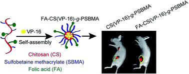
We demonstrated chitosan (CS)-based biocompatible nanoparticles coated with folic acid (FA) and poly(sulfobetaine methacrylate) (PSBMA) as an effective tumor-specific drug delivery system. The graft copolymer FA–CS-g-PSBMA could self-assemble into nanoparticles in an aqueous phase and maintain a spherical shape. Etoposide (VP-16), a widely-used chemotherapy drug with poor water solubility, could be incorporated into the inner core of hydrophobic CS to form FA–CS(VP-16)-g-PSBMA nanoparticles. The synthesis of the nanocarrier was verified by using zeta potential analysis, 1H nuclear magnetic resonance and Fourier transform infrared spectra. Next, both in vitro and in vivo experiments were performed to evaluate the release behavior, cellular uptake, cytotoxicity, biodistribution and therapeutic efficacy of the nanoparticles. Our results showed FA–CS(VP-16)-g-PSBMA nanoparticles released VP-16 more effectively in acidic phosphate-buffered saline than that under neutral conditions, and could be effectively internalized into HeLa cells. Compared to the nanoparticles without FA, FA–CS(VP-16)-g-PSBMA nanoparticles exhibited a more significant inhibitory effect on HeLa cell viability in vitro. When HeLa tumor-bearing mice were intravenously administrated with fluorescence-labelled nanoparticles, FA-conjugated nanoparticles accumulated more rapidly at the tumor site. Furthermore, FA–CS(VP-16)-g-PSBMA nanoparticles demonstrated more superior therapeutic efficacy than VP-16. These results suggest that FA–CS-g-PSBMA nanoparticles represent a promising nanocarrier for anti-tumor drug delivery.
Introduction
It has been recognized that the unparalleled development of nanomaterials and nanotechnology has and will continuously promote the diagnosis and treatment of diseases. Currently, nanoparticles are being investigated intensively for drug delivery and more specifically for cancer therapy.1–4 Because of the defective vascular endothelia and inadequate lymphatic drainage at tumor sites, certain sizes of nanoparticles and macromolecules could accumulate more in the tumor tissue than in normal tissues in a passive manner, which is known as the enhanced permeability and retention (EPR) effect.5,6 Therefore, nanoparticles could function as a suitable carrier for anti-tumor reagents to improve the in vivo pharmacokinetics, reduce the dosage for effective therapy, and suppress systemic toxicity by decreasing the off-target deposition of drugs in normal tissues. In addition, hydrophobic anti-tumor reagents could be encapsulated by nanoparticles to overcome their poor solubility in aqueous condition without the usage of toxic organic solvents or detergents which usually cause undesirable side effects.7
As a natural polysaccharide, chitosan (CS) is generally accepted as a non-toxic and biocompatible polymer and has been widely used for the development of safe and effective drug delivery.8–10 We previously demonstrated an approach to graft hydrophilic poly(sulfobetaine methacrylate) (PSBMA) onto the backbone of CS under γ-ray irradiation, and the resulting amphiphilic structure of the graft copolymer CS-g-PSBMA could self-assemble into micelles (Fig. 1).11 CS-g-PSBMA nanoparticles have superhydrophilic and ultralow biofouling properties. When a powerful antioxidant was loaded into the hydrophobic core of CS-g-PSBMA nanoparticles, its in vivo short half-life was greatly extended to nearly 10 hours, thus it exhibited remarkable protective effects against hemopoietic toxicity caused by whole body irradiation. The good biocompatibility and biodistribution of CS-g-PSBMA nanoparticles offer a satisfactory drug-encapsulated system.
 |
| Fig. 1 Schematic depiction of the synthesis of FA–CS-g-PSBMA nanoparticles for packaging VP-16. SBMA: sulfobetaine methacrylate; BDACT: S,S′-bis(R,R′-dimethyl-R′′-acetic acid) trithiocarbonate; EDC: N-(3-dimethylaminopropyl)-N′-ethylcarbodiimide hydrochloride; NHS: N-hydroxysuccinimide; FR: folate receptor. | |
In present study, we attempt to further explore the application of CS-g-PSBMA nanoparticles for effective delivery of anti-tumor reagents. To improve the tumor-selective targeting effects, a low molecular weight targeting agent, folic acid (FA), was used to graft onto CS-g-PSBMA to form FA–CS-g-PSBMA nanoparticles for active targeting (Fig. 1), since folate receptor (FR) is frequently overexpressed on the cell membrane of many epithelial tumor cells, including ovary, kidney, colon, prostate, lung and etc.12–14 The expression of FR, however, is highly restricted in most normal tissues. FA modification has been one of the promising strategies to improve the tumor-targeting effects of biomaterials.15–17 After binding with FR, FA-conjugated nanoparticles could be easily internalized into tumor cells via FR-mediated endocytosis.18,19 Subsequently, etoposide (VP-16), a semisynthetic derivative of podophyllotoxin that inhibits topoisomerase II,20 was loaded into FA–CS-g-PSBMA nanoparticles to study the tumor-targeting effect. Both in vitro and in vivo experiments were performed to evaluate the release behavior, cellular uptake, cytotoxicity, biodistribution and therapeutic efficacy of the nanoparticles. Our results showed the specific tumor-targeting effects and potent anti-tumor activity of VP-16-encapsulated FA–CS-g-PSBMA (FA–CS(VP-16)-g-PSBMA) nanoparticles, suggesting these FA–CS-g-PSBMA nanoparticles might be a promising nanocarrier for anti-tumor drug delivery.
Experimental
Chemicals and materials
Chitosan (deacetylation degree = 95.2%,
= 50
000 g mol−1) was kindly provided by Golden-Shell Biochemical Co. Ltd. (Zhejiang, China). VP-16, SBMA, N-(3-dimethylaminopropyl)-N′-ethylcarbodiimide hydrochloride (EDC), N-hydroxysuccinimide (NHS), fluorescein isothiocyanate (FITC), FA and 3-(4,5-dimethylthiazolyl-2)-2,5-diphenyltetrazolium bromide (MTT) were purchased from Sigma-Aldrich (Shanghai, China). Annexin V-FITC, 4,6-diamidino-2-phenylindole (DAPI), and propidium iodide (PI) were purchased from Thermo Fisher Scientific (Waltham, MA). Cy5.5 was purchased from Azco Biotech (Oceanside CA). Dulbecco's Modified Eagle Medium (DMEM) and fetal bovine serum (FBS) were obtained from Hyclone (Logan, UT). S,S′-Bis(R,R′-dimethyl-R′′-acetic acid) trithiocarbonate (BDACT) was synthesized for the polymerization.21
Synthesis of FA–CS-g-PSBMA nanoparticles
CS-g-PSBMA copolymer was prepared as previously described.8,11 A mixture of FA, EDC and NHS (1.0 M, molar ratio was 1
:
1
:
1) was prepared in 15 ml anhydrous DMSO. The mixture was then added slowly to 30 ml 0.37% (w/v) CS-g-PSBMA and stirred at room temperature in the dark for 16 h. Next, the reaction mixture was dialyzed against 1.0 mM NaOH aqueous with a 3500 Da molecular weight cutoff (MWCO) membrane for 72 h to remove un-reacted FA, and dialyzed against distilled water for another 72 h. Finally, the dialyzate was lyophilized to afford chitosan graft copolymer FA–CS-g-PSBMA. The structure of graft copolymer was characterized by 1H nuclear magnetic resonance (NMR) spectra using Varian INVOA-400 instrument (Palo Alto, CA) operated at 400 MHz. The particle size distribution and zeta potential value of nanoparticles were measured by using dynamic light scattering (DLS) (Zetasizer, Malvern, UK) with irradiation (He–Ne laser, 632.8 nm) at 1 mg ml−1 in phosphate-buffered saline (PBS, pH = 7.4). Field-emitting scanning electron microscopy (SEM) images were obtained by a Hitachi S-4700 microscope (Tokyo, Japan) working at an accelerating voltage of 15 kV. Fourier transform infrared (FT-IR) spectra were obtained on a Varian-1000 spectrometer, the samples were ground with KBr crystals, and the mixture was then pressed into a pellet for IR measurement.
Loading VP-16 onto FA–CS-g-PSBMA
VP-16 (30 mg) was dissolved in 1 ml of DMSO, and then was slowly added into FA–CS-g-PSBMA aqueous solution (10 mg ml−1, 10 ml) under strong stirring. The mixture solution was purged using nitrogen gas to eliminate DMSO and disperse VP-16 with FA–CS-g-PSBMA overnight. The suspension was then centrifuged at 12
000 rpm for 40 min to remove the unpackaged free VP-16. The supernatant was lyophilized to afford FA–CS(VP-16)-g-PSBMA nanoparticles. To measure the loading efficiency (LE) of VP-16, 5.0 mg FA–CS(VP-16)-g-PSBMA were redispersed in ethanol (3.0 ml) under ultrasonication for 2 h, and then was filtered under reduced pressure to remove the CS-g-PSBMA carrier by an organic membrane (pore size: 0.1 μm). VP-16 concentration in ethanol was measured by using UV/visible spectroscopy (UV-3600, Shimadzu, Japan) at 283 nm. The concentration of VP-16 in ethanol was then determined according to the standard curve. On the basis of the calculation of weight of VP-16 (WVP-16) and FA–CS(VP-16)-g-PSBMA (WFA–CS(VP-16)-g-PSBMA), VP-16 LE (in wt%) was estimated as LE = WVP-16/WFA–CS(VP-16)-g-PSBMA × 100%.
Measurement of VP-16 release from FA–CS(VP-16)-g-PSBMA
FA–CS(VP-16)-g-PSBMA were dispersed in 5 ml PBS (pH = 7.4) and transferred into a dialysis bag (MWCO = 3500 Da). The dialysis bag was then immersed in 145 ml of PBS (containing 0.6% sodium dodecyl sulfate) at pH 6.0 or 7.4. The release medium was continuously agitated with a stirrer at 37 °C. At predetermined time intervals, 2 ml of the external medium was collected and replaced with the same volume of fresh PBS. The amount of released VP-16 in the PBS was then determined by using the UV/visible spectroscopy at 283 nm. The concentration of VP-16 in PBS was then determined according to the standard curve.
Cell culture and MTT assay
Human cervical cancer HeLa cells were cultured in DMEM medium supplemented with 10% FBS, 100 μg ml−1 streptomycin and 100 units per ml penicillin at 37 °C under a humidified atmosphere containing 5% CO2. For MTT assay, HeLa cells were seeded at a density of 5 × 10³ cells per well in 96-well plates and treated with indicated formulations containing the same concentration of VP-16 for 2 days. Then cell were incubated with 50 μg/100 μl MTT at 37 °C for 4 h and 100 μl DMSO was then added to dissolve the formazan crystals. The absorbance was measured at 570 nm using a microplate reader (BioTek, Winooski, VT).
Cellular uptake of nanoparticles
To visualize the cellular uptake of nanoparticles, FA–CS(VP-16)-g-PSBMA or CS(VP-16)-g-PSBMA nanoparticles were labeled with FITC. Briefly, FITC was dissolved in ethanol (7.72 mg ml−1, 1 ml) and then slowly added into nanoparticle solution (10 mg ml−1, 0.5 ml, pH = 7.5) under strong stirring. The reaction was performed at 25 °C water bath in the dark for 4 h. Unreacted FITC were removed by dialysis (MWCO = 3500 Da). HeLa cells were incubated in a FBS-free DMEM medium containing FITC-labeled CS(VP-16)-g-PSBMA or FA–CS(VP-16)-g-PSBMA (10 μg ml−1 of VP-16) for 8 h. After washing twice with PBS, cells were fixed with 4% paraformaldehyde for 15 min, and then the cell nucleus was labeled with DAPI for 5 min. Cell images were obtained by using UltraView Vox confocal laser scanning microscope (PerkinElmer, Waltham, MA).
Flow cytometry analysis
After treatment of indicated formulations containing 10 μg ml−1 or 20 μg ml−1 VP-16 for 8 h, the FITC-positive HeLa cells were monitored by FACS Calibur flow cytometer (BD Biosciences, San Jose, CA). For apoptosis assay, Hela cells were treated with different formulations containing 10 μg ml−1 VP-16 for 2 days, and stained with Annexin V-FITC and PI. The percentage of apoptotic cells was monitored by FACS Calibur flow cytometer. At least 10
000 cells were measured for each sample.
Animals and tumor xenograft model
Eight-week-old male C57BL/6 mice and female athymic nude mice were purchased from Model Animal Research Center of Nanjing University (Nanjing, China) and maintained under a 12 h light/dark cycle at constant temperature and provided with water and food ad libitum. Mice were subcutaneously inoculated with the HeLa cells (2 × 10⁶ cells per mouse) to set up the tumor xenograft model. VP-16, CS(VP-16)-g-PSBMA, FA–CS(VP-16)-g-PSBMA (5 mg kg−1 of VP-16) or saline were administered via tail vein and the tumor size was monitored by a bench micrometer, and the tumor volume was calculated as volume = length × width2/2. Mouse tissues were fixed with 10% formalin and embedded in paraffin. Tissue sections (5 μm) were stained with hematoxylin and eosin (HE) for light microscopy. All animal experimental procedures were in accordance with the NIH guidelines for the care and use of laboratory animals. The Animal Ethical Committee of Soochow University approved all animal experimental protocols.
Pharmacokinetics of FA–CS(VP-16)-g-PSBMA and in vivo imaging of Cy5.5 labeled nanoparticles
In order to label nanoparticles with near-infrared fluorescence (NIRF) dye Cy5.5, FA–CS(VP-16)-g-PSBMA or CS(VP-16)-g-PSBMA solution (10 mg ml−1, 2 ml) was mixed with hydroxysuccinimide ester of Cy5.5 (1%, w/v), and the labeling reaction was performed at room temperature in the dark for 6 h. Byproducts and unreacted Cy5.5 molecules were removed by dialysis (MWCO = 3500 Da). The amount of Cy5.5 in the FA–CS(VP-16)-g-PSBMA and CS(VP-16)-g-PSBMA were similar as ∼0.6 wt%. Cy5.5-labeled nanoparticles were injected via the tail vein into C57BL/6 mice or HeLa tumor-bearing mice. Blood samples were collected from C57BL/6 mice at different time points. Then the blood samples were centrifuged at 5000 rpm for 10 min to obtain serum. The Cy5.5 fluorescence intensities in serum were measured by using a microplate reader (Biotek) with excitation at 675 nm and emission at 705 nm. The time-dependent accumulation of nanoparticles in tumor site were non-invasively imaged using the In-Vivo Fx Pro imaging system (Kodak, Rochester, NY).
Statistical analysis
Data were expressed as means ± the standard deviation (SD). Statistical analysis was performed by one-way ANOVA following multiple comparisons by using SPSS 18.0 software. Results with a p value less than 0.05 were considered statistically significant.
Results and discussion
Characterization of FA–CS(VP-16)-g-PSBMA
The CS-g-PSBMA copolymer was synthesized by graft polymerization of SBMA onto chitosan under γ-ray irradiation,11 and FA was grafted onto CS-g-PSBMA by the reaction between amino group and carboxyl group on the condition of catalysis of EDC and NHS (Fig. 1). The amphiphilic FA–CS-g-PSBMA copolymer can self-assemble into nanoparticles in an aqueous phase, and the diameter of FA–CS-g-PSBMA was 163.9 ± 32 nm with narrow size distribution (Fig. 2A). In this structure, zwitterionic PSBMA can prevent the aggregation of nanoparticles and VP-16 can be loaded into the inner core of hydrophobic CS with the LE of 27.3%. After loading of VP-16, the diameter of nanoparticles was swelled to 200.5 ± 17 nm while maintaining their spherical shapes (Fig. 2B). In addition, the zeta potential values of FA–CS-g-PSBMA and FA–CS(VP-16)-g-PSBMA were −6.70 mV and −5.36 mV in PBS (pH = 7.4), respectively. The structures of graft copolymers were characterized by 1H NMR spectra, and typical 1H NMR spectra of FA–CS-g-PSBMA, as well as CS-g-PSBMA and FA were shown in Fig. 2C and S1.† Besides the characteristic peaks of chitosan and SBMA, the characteristic resonances of FA were detected at δ = 7.15–8.64 ppm, suggesting that FA was successfully grafted onto chitosan. The structure was further confirmed by FT-IR spectra, in comparison with chitosan and CS-g-PSBMA, the characteristic peaks occurred for FA–CS-g-PSBMA copolymer at 1608 cm−1, 1510 cm−1 and 1043 cm−1 corresponding to benzene ring stretching vibration, N–H stretching vibration and COO− stretching vibration, respectively (Fig. 2D).
 |
| Fig. 2 Characterization of nanoparticles. SEM images (scale bar = 200 nm), particle size distributions in distilled water and zeta potentials of FA–CS-g-PSBMA nanoparticles (A) and FA–CS(VP-16)-g-PSBMA nanoparticles (B). (C) 1H NMR spectrum (400 M, CF3COOD/D2O (v/v = 1/9)) of FA–CS-g-PSBMA. (D) FT-IR spectra of (a) chitosan and (b) CS-g-PSBMA and (c) FA–CS-g-PSBMA. | |
Cellular uptake and cytotoxicity of FA–CS(VP-16)-g-PSBMA
In order to evaluate of the cellular uptake of the nanoparticles, FA–CS(VP-16)-g-PSBMA and CS(VP-16)-g-PSBMA were labelled with FITC. After incubation with FITC-labelled nanoparticles for 8 h, HeLa cells were examined by confocal laser scanning microscope and flow cytometer. As shown in Fig. 3A, cells treated with FA–CS(VP-16)-g-PSBMA nanoparticles demonstrated a higher intracellular fluorescence signal compare to CS(VP-16)-g-PSBMA-treated group. On the other hand, FA–CS(VP-16)-g-PSBMA nanoparticles did not show the improved targeting capacity when treating a FR-negative L929 cell line (Fig. S2†). These data indicated the conjugated FA could facilitate the internalization of nanoparticles in HeLa cells with high FR expression.22
 |
| Fig. 3 In vitro effects of FA–CS(VP-16)-g-PSBMA nanoparticles. (A) Cellular uptake of FITC-labelled CS(VP-16)-g-PSBMA and FA–CS(VP-16)-g-PSBMA nanoparticles in HeLa tumor cells. The nuclei of the cells were stained with DAPI. (B) Time-dependent VP-16 release from FA–CS(VP-16)-g-PSBMA nanoparticles at pH = 6.0 and pH = 7.4, respectively. (C) HeLa cells were treated with different concentration of VP-16 and nanoparticles containing the same amount of VP-16 for 48 h. MTT assay was performed to measure the decrease of cell viability. (D) After treated with the nanoparticles containing 10 μg ml−1 VP-16 for 24 h, flow cytometer analysis was performed to examine the percentage of cells under apoptosis. *, p < 0.05 vs. VP-16-treated group, n = 5. | |
The dynamic process of VP-16 release from FA–CS(VP-16)-g-PSBMA was investigated at pH 6.0 (similar pH of the environment around the tumor) and pH 7.4 (similar pH of physiological blood).23,24 As shown in Fig. 3B, the percentage of VP-16 cumulative release was monitored as a function of time at 37 °C. The cumulative release curve of VP-16 from the nanoparticles reached a plateau within 120 h. Importantly, the release profile of VP-16 was dependent on the pH value. The releasing ratio of VP-16 at pH 7.4 was 24.2% and 72.7% within 24 h and 120 h, respectively. Whereas it increased to approximately 59.6% and 91.0% at pH 6.0 within 24 h and 120 h, suggesting FA–CS-g-PSBMA nanoparticles could preserve the encapsulated drug for a considerable period of time in the blood circulation, and release drug effectively when they approach the solid tumor with acidic pH microenvironment. The protonation of amino groups on the CS chain at low pH could modify the network electrical state and induce the insoluble–soluble transition of CS, thus facilitating the drug release process.25 The favorable pH-triggered releasing behavior might increase the drug accumulation in the targeting site and limit the toxic effect on normal tissues.
Next, cell viability analysis was performed to assess the cytotoxic effects of the VP-16-encapsulated nanoparticles. VP-16 treatment decreased the cell viability in a dose-dependent manner (Fig. 3C). The cell viability of CS(VP-16)-g-PSBMA-treated group was equivalent to that of VP-16. In accordance with the amount of the internalized nanoparticles, FA–CS(VP-16)-g-PSBMA exerted the highest cytotoxic effects when HeLa cells were treated the nanoparticles containing the corresponding amount of VP-16. And we did not observe any cytotoxic effects when HeLa cells were treated with the same amount of nanocarriers without VP-16, implicating the biocompatibility of FA–CS-g-PSBMA nanoparticles as drug carriers.
VP-16 impedes DNA synthesis through forming a ternary complex with DNA and topoisomerase II, thus induces double stranded DNA breaks and cell cycle arrest, and ultimately promotes cell apoptosis.26,27 Treatment of 10 μg ml−1 VP-16 for 24 h significantly increased the percentage of apoptotic cell numbers (Fig. 3D). The apoptosis-inducing capacity of CS(VP-16)-g-PSBMA nanoparticles showed no statistical significance with VP-16. However, treatment of FA-conjugated graft copolymer containing 10 μg ml−1 VP-16 for 24 h induced more apoptotic cells. These data suggested FA–chitosan conjugation was capable to improve the targeting properties of the nanoparticles for FA receptor-positive tumor cells in vitro, therefore delivery more amount of drug to tumor cells and exert more potent anti-tumor activity.
In vivo pharmacokinetics and biodistribution of FA–CS(VP-16)-g-PSBMA nanoparticles
To determine the in vivo effects of FA–CS(VP-16)-g-PSBMA, we first evaluated the pharmacokinetics of the nanocarriers by measuring the blood retention time. Cy5.5-labeled FA–CS(VP-16)-g-PSBMA (LE = 27.3%) or CS(VP-16)-g-PSBMA (LE = 28.6%) nanoparticles were intravenously administrated to C57BL/6 mice (containing 5 mg kg−1 of VP-16 in 100 μl of saline). As shown in Fig. 4A, both nanoparticles showed similar pattern of retention behavior in blood with a half-life of nearly 10 h calculated by a one-compartment open model.
 |
| Fig. 4 In vivo biodistribution of nanoparticles in HeLa tumor-bearing mice. (A) Blood retention kinetics of Cy5.5-labelled nanoparticles. (B) NIRF imaging and quantification of HeLa tumor-bearing mice treated with Cy5.5-labelled nanoparticles for different times. Tumor tissues were marked with yellow dotted lines. (C) NIRF imaging and quantification of heart, liver, kidney, spleen and tumor tissues after treated with Cy5.5-labelled nanoparticles for 48 h. Upper panel is merged NIRF and white-light imaging. Lower panel is NIRF imaging. | |
HeLa tumor-bearing nude mice were treated with the same amount of nanoparticles as C57BL/6 mice. In time-dependent NIRF images, the tumor tissues were delineated from surrounding normal tissues by yellow dotted lines (Fig. 4B). Interestingly, tumor site of mice treated with FA–CS(VP-16)-g-PSBMA nanoparticles exhibited fluorescence signaling as early as 2 h, whereas the accumulation of CS(VP-16)-g-PSBMA nanoparticles in tumor site was observed at 24 h, suggesting FA grafting reinforce the tumor-targeting effect of the nanoparticles. The tumor-bearing mice were sacrificed 48 h post-injection of nanoparticles, and different organs and tumor tissues were collected for NIRF imaging. As shown in Fig. 4C, the tumor tissues from FA–CS(VP-16)-g-PSBMA-treated mice showed a more intensive fluorescence compared to CS(VP-16)-g-PSBMA-treated mice. A mild accumulation of fluorescence was observed in both liver and spleen, indicating the clearance of nanoparticles by the mononuclear phagocytic system located in the liver and spleen.
The PSBMA-grafted surfaces that resist the non-specific adsorption of protein and the adhesion of cells prolonged the blood retention period of the nanoparticles.28,29 By contrast, it has been reported that the half-life of VP-16 was less than 1 h in rodent models through intravenous injection.30,31 A longer circulating half-life of the nanoparticles could enhance the pharmacological activity and allow enough time for effective drug delivery to the tumor site. The EPR effects permitted the accumulation of nanoparticles at the tumor site where the vascular permeability is high due to the abnormal angiogenesis,32 and the FA grafting greatly accelerated the accumulating process. It is worthwhile to note that more cytotoxic effects might be induced by simply extending the biological half-life of intact VP-16. Nevertheless, once VP-16 were encapsulated by FA–CS-g-PSBMA nanoparticles, both active and passive tumor-targeting capabilities mediated by FA-conjugation and EPR effects could prevent the side effects of VP-16 in normal tissues. After accumulation at tumor site, sustained release of VP-16 from FA–CS(VP-16)-g-PSBMA nanoparticles was triggered under the relatively acidic circumstances, or in endosomes and lysosomes by hydrolytic degradation of the nanocarriers after they were taken up by the endocytic pathway.33,34
Anti-tumor effect of FA–CS(VP-16)-g-PSBMA nanoparticles
We compared the therapeutic efficacy of FA–CS(VP-16)-g-PSBMA to that of VP-16. HeLa tumor-bearing nude mice were intravenously treated with FA–CS(VP-16)-g-PSBMA (containing 5 mg kg−1 of VP-16 in 100 μl of saline) or VP-16 for nine treatments in three weeks. Forty-three days post-injection, the tumor size of mice treated with saline was substantially enlarged with the average volume of 1665.32 ± 198.32 mm3 and average weight of 1.09 ± 0.13 g (Fig. 5A–C). VP-16 treatment succeed in postponing the growth of tumor tissue, and the tumor average volume and weight were 850.41 ± 71.12 mm3 and 0.57 ± 0.10 g, respectively, which were about half of that of saline-treated mice. As expected, the tumor size and weight of FA–CS(VP-16)-g-PSBMA-treated mice were statistically significantly less than that of VP-16-treated mice. The tumor average volume and weight were 492.25 ± 69.81 mm3 (57.88% of VP-16-treated group) and 0.29 ± 0.08 g (50.88% of VP-16-treated group), respectively. Mice treated with CS(VP-16)-g-PSBMA did not show statistical difference in tumor size or weight as compared with control mice. Histological examination did not reveal any lesions in heart, liver and spleen (Fig. 5D). However, renal interstitial haemorrhage was observed in VP-16-treated mice, but not in the nanoparticle-treated mice. By effective tumor-targeting VP-16 delivery in vivo, FA–CS(VP-16)-g-PSBMA nanoparticles showed more potent anti-tumor activity and lower systemic toxicity.
 |
| Fig. 5 Inhibition of tumor growth of FA–CS(VP-16)-g-PSBMA nanoparticles. (A) Black arrowheads indicated the time points when HeLa tumor-bearing mice were treated with VP-16, CS(VP-16)-g-PSBMA or FA–CS(VP-16)-g-PSBMA nanoparticles. The volumes of tumors were measured at the indicated time internals. (B) Tumor images after nanoparticle therapy. (C) The weight of tumors were measure at day 43. *, p < 0.05 vs. VP-16-treated group, n = 6. (D) Histological examination of heart, liver, spleen and kidney. Glomerulus and interstitial haemorrhage in the kidney were indicated by white and black arrowheads, respectively. | |
Conclusions
In summary, FA–CS-g-PSBMA nanoparticles were synthesized for tumor-targeting drug delivery with the following distinguishing features: (1) excellent biocompatibility and biodegradation. (2) Higher release of drug in acidic tumor environment. (3) Passive tumor-targeting via EPR effects. (4) Active tumor-targeting via high FA-binding affinity of FR on the tumor cell surface. (5) Relative longer biological half-life in vivo. In vitro assay confirmed the internalizing effectiveness of the nanoparticles, as well as the cytotoxic effect and apoptosis-inducing function. When intravenously injected into HeLa xenograft mice, FA–CS(VP-16)-g-PSBMA nanoparticles showed more effective tumor-targeting and anti-tumor ability. Based on these results, we concluded that FA–CS-g-PSBMA nanoparticles may hold the promising prospects in the area of targeted tumor therapy.
Acknowledgements
This work is supported by Natural Science Foundation of China (No. 31270897, 81271682 and 91326202), ITER program (2014GB112006), Jiangsu Provincial Key Laboratory of Radiation Medicine and Protection and the Priority Academic Program Development of Jiangsu Higher Education Institutions (PAPD).
References
- M. Gupta, G. P. Agrawal and S. P. Vyas, Curr. Mol. Med., 2013, 13, 179–204 CrossRef CAS PubMed
. - E. Pérez-Herrero and A. Fernández-Medarde, Eur. J. Pharm. Biopharm., 2015, 93, 52–79 CrossRef PubMed
. - B. B. Cerqueira, A. Lasham, A. N. Shelling and R. Al-Kassas, Eur. J. Pharm. Biopharm., 2015, 97, 140–151 CrossRef CAS PubMed
. - Z. Cheng, A. Al Zaki, J. Z. Hui, V. R. Muzykantov and A. Tsourkas, Science, 2012, 338, 903–910 CrossRef CAS PubMed
. - H. Maeda, Adv. Drug Delivery Rev., 2015, 91, 3–6 CrossRef CAS PubMed
. - H. Nakamura, F. Jun and H. Maeda, Expert Opin. Drug Delivery, 2015, 12, 53–64 CrossRef CAS PubMed
. - J. H. Park, G. Saravanakumar, K. Kim and I. C. Kwon, Adv. Drug Delivery Rev., 2010, 62, 28–41 CrossRef CAS PubMed
. - W. Huang, Y. J. Wang, S. Zhang, L. Huang, D. B. Hua and X. L. Zhu, Macromolecules, 2013, 46, 814–818 CrossRef CAS
. - D. B. Hua, J. L. Jiang, L. J. Kuang, J. Jiang, W. Zheng and H. J. Liang, Macromolecules, 2011, 44, 1298–1302 CrossRef CAS
. - J. Y. Yhee, S. Son, S. H. Kim, K. Park, K. Choi and I. C. Kwon, J. Controlled Release, 2014, 193, 202–213 CrossRef CAS PubMed
. - Y. Zhou, S. Hua, J. Yu, P. Dong, F. Liu and D. Hua, J. Mater. Chem. B, 2015, 3, 2931–2934 RSC
. - N. Parker, M. J. Turk, E. Westrick, J. D. Lewis, P. S. Low and C. P. Leamon, Anal. Biochem., 2005, 338, 284–293 CrossRef CAS PubMed
. - L. Teng, J. Xie, L. Teng and R. J. Lee, Expert Opin. Drug Delivery, 2012, 9, 901–908 CrossRef CAS PubMed
. - B. Bahrami, M. Mohammadnia-Afrouzi, P. Bakhshaei, Y. Yazdani, G. Ghalamfarsa, M. Yousefi, S. Sadreddini, F. Jadidi-Niaragh and M. Hojjat-Farsangi, Tumor Biol., 2015, 36, 5727–5742 CrossRef CAS PubMed
. - K. L. Nair, S. Jagadeeshan, S. A. Nair and G. S. Kumar, PLoS One, 2013, 8, e70697 Search PubMed
. - J. Varshosaz, F. Hassanzadeh, H. Sadeghi-Aliabadi and F. Firozian, BioMed Res. Int., 2014, 2014, 708593 Search PubMed
. - Y. C. Kuo and C. H. Lee, J. Pharm. Sci., 2015, 104, 1804–1814 CrossRef CAS PubMed
. - L. H. Matherly and D. I. Goldman, Vitam. Horm., 2003, 66, 403–456 CAS
. - A. Chaudhury and S. Das, Curr. Pharm. Biotechnol., 2015, 16, 333–343 CAS
. - A. Montecucco and G. Biamonti, Cancer Lett., 2007, 252, 9–18 CrossRef CAS PubMed
. - J. T. Lai, D. Filla and R. Shea, Macromolecules, 2002, 35, 6754–6756 CrossRef CAS
. - J. You, X. Li, F. de Cui, Y. Z. Du, H. Yuan and F. Q. Hu, Nanotechnology, 2008, 19, 045102 CrossRef PubMed
. - E. S. Lee, Z. Gao and Y. H. Bae, J. Controlled Release, 2008, 132, 164–170 CrossRef CAS PubMed
. - R. Vivek, V. Nipun Babu, R. Thangam, K. S. Subramanian and S. Kannan, Colloids Surf., B, 2013, 111, 117–123 CrossRef CAS PubMed
. - C. Pillai, W. Paul and C. P. Sharma, Prog. Polym. Sci., 2009, 34, 641–678 CrossRef CAS
. - Y. Pommier, E. Leo, H. Zhang and C. Marchand, Chem. Biol., 2010, 17, 421–433 CrossRef CAS PubMed
. - A. Montecucco and G. Biamonti, Cancer Lett., 2007, 252, 9–18 CrossRef CAS PubMed
. - Z. Zhang, T. Chao, S. Chen and S. Jiang, Langmuir, 2006, 22, 10072–10077 CrossRef CAS PubMed
. - H. W. Chien, C. C. Tsai, W. B. Tsai, M. J. Wang, W. H. Kuo, T. C. Wei and S. T. Huang, Colloids Surf., B, 2013, 107, 152–159 CrossRef CAS PubMed
. - M. Snehalatha, V. Kolachina, R. N. Saha, A. K. Babbar, N. Sharma and R. K. Sharma, J. Pharm. BioAllied Sci., 2013, 5, 290–297 CrossRef PubMed
. - L. Chen, L. Tan, X. Zhang, J. Li, Z. Qian, M. Xiang and Y. Wei, Int. J. Pharm., 2015, 495, 265–275 CrossRef CAS PubMed
. - J. Fang, H. Nakamura and H. Maeda, Adv. Drug Delivery Rev., 2011, 63, 136–151 CrossRef CAS PubMed
. - A. K. Varkouhi, M. Scholte, G. Storm and H. J. Haisma, J. Controlled Release, 2011, 151, 220–228 CrossRef CAS PubMed
. - K. J. Chen, Y. L. Chiu, Y. M. Chen, Y. C. Ho and H. W. Sung, Biomaterials, 2011, 32, 2586–2592 CrossRef CAS PubMed
.
Footnotes |
† Electronic supplementary information (ESI) available. See DOI: 10.1039/c6ra14104h |
‡ These authors contributed equally to this publication. |
|
This journal is © The Royal Society of Chemistry 2016 |
Click here to see how this site uses Cookies. View our privacy policy here.