In vitro toxicity, apoptosis and antimicrobial effects of phyto-mediated copper oxide nanoparticles

V. Gopinath*a, S. Priyadarshinia, A. R. Al-Malekia, M. Alagirib, Rosiyah Yahyac, S. Saravanand and Jamuna Vadivelua
aDepartment of Medical Microbiology, Faculty of Medicine, University of Malaya, Kuala Lumpur – 50603, Malaysia. E-mail: yele.gopi@gmail.com; Tel: +60-12-9678151
bCenter for Material Science and Nanodevices, Department of Physics and Nanotechnology, SRM University, Kattankulathur – 603 203, Tamil Nadu, India
cDepartment of Chemistry, University of Malaya, Kuala Lumpur, 50603, Malaysia
dDepartment of Physiology and Pathophysiology, St-Boniface Hospital Albrechtsen Research Centre, University of Manitoba, Winnipeg, R2H A26, Canada

Received 28th May 2016 , Accepted 23rd October 2016

First published on 8th November 2016


Abstract

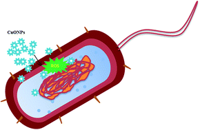
Herein, copper oxide nanoparticles (CuONPs) are proposed for widespread use in emerging biomedical applications. The aim of this study is to synthesize and characterize CuONPs using the aqueous dried fruit extract of Tribulus terrestris and assess their potential in vitro cytotoxicity and antibacterial activity. The CuONPs bio-physical and morphological properties are characterized via different analytical techniques. The synthesized particles are highly stable and spherical with particle sizes in the range of 5–22 nm. Confocal microscopy reveals that ROS generation is the potent mechanism behind the role of CuONPs mediated antimicrobial toxicity. In addition, MTT and apoptotic analyses illustrate that the CuONPs are relatively cytofriendly toward human mesenchymal stem cells and also exhibit anti-cancer activity on human adenocarcinoma AGS cell lines. These results clearly indicate that the prepared copper oxide nanoparticles are safe and their application can be expanded as effectual antimicrobial and anticancer agents for pharmaceutical applications.


1. Introduction

There has been a global increase in the cooperative interests toward the nanomaterials field with the development of functional materials, which are extensively used in multidisciplinary applications. Most of the traditional methods for the synthesis of nanoparticles utilize various chemicals to fuel material growth, which result in toxic products. In addition, some industrial synthetic processes for the synthesis of nanoparticles require highly carcinogenic chemicals for polymerization.1 The usage of such methods is strictly limited due to the introduction of hazardous chemicals.2 The existing metal oxide nanoparticles have limited benefits for biomedical and environmental use, therefore the economic viable biogenic experimentation of metal and metal oxide nanoparticles has evolved using biological entities, including microorganisms and plants,3–5 which offer sustainable and eco-friendly products.

Among the numerous types of nanomaterials, there have been conflicting results that metal oxide nanoparticles demonstrate a highly toxic effect to cells and microorganisms. Due to their potential antimicrobial properties, CuONPs are used in textiles, paints, plastics and food containers.6 Aqueous A. indica leaf extract mediated CuONPs have potent antimicrobial and anticancer activity.7 However, there is limited evidence available on the possible antimicrobial effect of CuONPs. CuO is a cheaper material than silver, easily mixed with polymers and relatively stable in terms of both chemical and physical properties.8 CuO nanoparticulates may be valuable antimicrobial agents since they can be prepared with extremely high surface areas and unusual crystal morphologies.9

To utilize CuO nanoparticles for biomedical applications, attention must be paid to their biocompatibility. The liver is one of the target organs for CuO nanoparticles after they gain entry into the body through one of the many possible routes.10 Using the toxicological approach, a comparative proteomic study revealed that CuO is highly toxic to macrophage cells compared with TiO2 nanoparticles.11 The cytotoxic effects of CuONPs are dose12–14 and size dependent. Moreover, nanoparticles are more toxic than micrometer particles of the same metal oxide,15,16 which is likely due to the damage that CuONPs cause in the mitochondria.

Apoptotic cell death is a key process in cancer development and progression. The ability of cancer cells to avoid apoptosis and continue to propagate is one of the basic characteristics of cancer.17 The best fundamental way to carry out cancer therapy is to control apoptosis during cancerous growth.18 It is known that the apoptosis of cells is generally accompanied by characteristic biochemical changes, such as the translocation of phosphatidyl serine, DNA breakdown, and protease signalling cascades.19

Herein, we present the T. terrestris fruit mediated synthesis of CuONPs for the first time via a green chemistry approach. T. terrestris is a traditional medicinal plant.20 It contains alkaloids, flavonoids, tannins, ascorbic acid and phenolic compounds, which are mainly involved in the synthesis of metal and metal oxide nanoparticles.21 In addition, nanoparticles with multiple biological effects could serve as a potent candidate for numerous biomedical applications. Hence, we synthesized and characterized high quality CuONPs via a biogenic route using the extract of T. terrestris. The antimicrobial potential, anticancer activity and cytotoxicity of the as-synthesized CuONPs were investigated to support their potential for biomedical applications.

2. Materials and methods

2.1. Experimental methods

All chemicals and media components (analytical grade) were purchased from Sigma Aldrich Chemicals, India. Bacterial strains were obtained from the Department of Medical Microbiology, University of Malaya. Bacterial cultures, namely Escherichia coli, Pseudomonas aeruginosa, Staphylococcus aureus and Bacillus cereus, were maintained in the respective medium. Gastric adenocarcinoma AGS cells (ATCC 43504) were procured from American Type Culture Collection (ATCC, USA). Human bone marrow mesenchymal stem cells were obtained from the National Centre for Cell Science (NCCS), Pune, India.

2.2. Preparation of T. terrestris fruit extract

T. terrestris fruits were purchased from the local area market, Chennai, Tamil Nadu, India. 10 g of dried fruit was washed thrice with deionized (DI) water and dried under direct sunlight for 5 days. Then the materials were minced well with a kitchen blender. 3 g of obtained powder was boiled with 100 mL of de-ionized water for 10 min. Finally the boiled extract was filtered using a 0.45 μm filter.

2.3. Biosynthesis of CuO nanoparticles

In order to synthesize CuONPs, the T. terrestris filtrate was mixed with the volume concentration of 5 mM aqueous CuSO4·5H2O. The reaction mixture was vigorously stirred and heated at 90 °C for approximately 120 min. After the reaction mixture cooled to room temperature, a brown-black colored product was obtained and collected by centrifugation at 16[thin space (1/6-em)]000 rpm for 15 min and washed with DI water. The resultant product was heated at 80 °C for 2 h to obtain the final CuONPs which were stored in a vacuum desiccator for further use. The bioactive protein responsible for the CuONPs is described in the ESI.

2.4. Characterization of CuO nanoparticles

UV-Vis spectrum scanning was performed for the fruit extract with CuSO4 reaction mixture using a Perkin-Elmer double beam spectrophotometer (Lambda 45, Perkin-Elmer, USA). The sample was scanned in the range of 200–700 cm−1 at a resolution of 1 nm. The functional biomolecules in the fruit extract and the synthesized nanoparticles were analyzed via FTIR (Lambda 45, Perkin-Elmer, USA). The FTIR sample was prepared by mixing the materials with KBr to make a pellet and spectra were recorded at a resolution of 4 cm−1 in the range of 450 to 4000 cm−1. The resultant nanoparticles' crystalline nature and purity were analyzed via XRD using an X'Pert Pro A analytical X-ray diffractometer operated at 40 keV with Cu Kα radiation in the θ–2θ configuration. The zeta potential of the nanoparticles was measured using a Zetasizer Nano ZS (Malvern Instruments, UK) with an He–Ne laser of 633 nm at 25 °C. Topographical images of the nanoparticles were recorded via HRTEM. The sample was prepared by loading a drop of the nanoparticles on a carbon-coated copper grid, followed by air drying for 30 min.

2.5. Antimicrobial assays

To determine the nanoparticles toxicity to bacteria, the Clinical and Laboratory Standards Institute (CLSI) standard broth microdilution method was followed. The MIC assay was performed in Muller Hinton Broth (MHB) or Muller Hinton agar plates. Typically 18–24 h grown bacterial suspensions were adjusted at a cell density of 108 CFU mL−1, as measured by OD at 600 nm. Broth was dispensed into the wells followed by the addition of the CuONPs solution and inoculum. Serial dilution was performed by the addition of various quantities of nanoparticles and the T. terrestris extract to a 96-well microtitre plate with MHB to reach the concentration of 2.5–50 μg mL−1. Microtitre plate wells containing MHB served as a negative control and the wells containing bacterial suspension served as a positive control. MIC50 was determined as the lowest concentration of the CuONPs that reduces growth by more than 50%. The bacterial suspensions in the plate showing no visible growth were sub-cultured on LB agar and incubated at 37 °C for 24 h. After the incubation period, the plates showing no visible growth were considered as the minimum bactericidal concentration (MBC99). The highest dilution showing at least 99% reduction in growth was considered as the MBC.

The bacterial morphological changes caused by the CuONPs were examined by field emission scanning electron microscopy (FESEM). Freshly grown bacterial suspensions were mixed with CuONPs and incubated for 30 min at 37 °C. The bacteria were centrifuged at 6000 rpm for 10 min at 4 °C and the pellets were washed twice with PBS at pH 7 before the samples were fixed with 8% glutaraldehyde (1[thin space (1/6-em)]:[thin space (1/6-em)]1 v/v) and Sorensen's phosphate buffer (SPB) for 60 min. A 1[thin space (1/6-em)]:[thin space (1/6-em)]1 (v/v) of SPB and water mixture was applied before the samples were fixed with 4% osmium tetroxide (OsO4) mixed with 1[thin space (1/6-em)]:[thin space (1/6-em)]1 (v/v) H2O. After overnight incubation, the samples were washed with deionized water for 15 min and then dehydrated in increasing concentrations of ethanol. Dehydration with an ethanol–acetone mixture followed by pure acetone was applied before critical point drying (CPD).22 The samples were then viewed under an FESEM (JEOL JSM-7001F, Germany) to study the bacterial morphological changes before and after CuONPs treatment.

In order to differentiate the live and adhered bacterial cells under confocal imaging, the Live/Dead Backlight L-7012 kit (Invitrogen SA, Spain) was used. The cell density of the overnight grown culture of E. coli, P. aeruginosa, S. aureus and B. cereus was measured using optical methods at 540 nm and a concentration of 106 CFU mL−1 was used. The bacterial cells were then exposed to CuONPs at their MIC concentration (17, 16, 19.5 and 21 μg mL−1) for 30 min and 60 min. Then, the cells were collected by centrifugation and the pellets were washed with PBS. The cells were then stained with SYTO 9 and propidium iodide (PI) for 15 min in the dark to avoid light exposure. The stained bacterial cells were observed under a CLSM. A Zeiss CLSM 710-META confocal laser scanning microscope was used to record bacterial cell images. Fluorophores were excited using an argon laser at 488 nm and an HeNe laser at 543 nm and observed with a 63× objective lens using two filters (505–550 nm and 560 nm). The image size was 1024 × 1024 pixels, which corresponded to an area of 143 mm × 143 mm.

The oxidative stress induced by the CuONPs to the bacterial cells is reflected by the quantity of reactive oxygen species (ROS). The effects of CuONPs on intracellular ROS generation in test bacteria were monitored by 2′,7′-dichlorofluorescein diacetate DCF-DA assay. Briefly, the bacterial cells were harvested by centrifugation, washed twice with PBS, and incubated with 10 μM DCF-DA in PBS at 37 °C for 30 min. The cells were then incubated with the nanoparticles at 37 °C for 24 h. The intracellular ROS level was examined under a fluorescence microscope with the excitation and emission wavelengths at 495 nm and 525 nm, respectively, and the ROS level was examined by confocal laser scanning microscopy (CLSM) images.

2.6. Cell culture studies-cytotoxicity assay

Gastric adenocarcinoma AGS cells and human BM-MSCs were maintained in culture in Dulbecco's modified essential medium (DMEM) supplemented with 10% FBS under standard culture conditions of 5% CO2 at 37 °C. The MTT based cell viability assay was carried out following our earlier report with minor modifications (Gopinath et al., 2015). In brief, 100 μL of cell suspension was seeded in a 96-well tissue culture plate (5 × 10⁴ cells per well) and incubated at 37 °C in a humidified 5% CO2 incubator. After 24 h, the growth medium was removed and freshly prepared CuONPs in 5% DMEM were serially diluted by a two-fold dilution (2.5–100 μg mL−1) and each sample with a concentration of 100 μL was added in triplicate to the respective wells and incubated for 24 h. The plate was observed for any changes in morphology and cell density by inverted phase contrast microscopy (Nikon, Japan). The content of the wells was then removed and 10 μL of the reconstituted MTT solution was added to all the test and control wells. The plate was gently shaken and incubated at 37 °C in a humidified 5% CO2 incubator for 4 h, following which the supernatant was removed and 100 μL of an MTT solubilization solution (10% Triton X-100) in acidic isopropanol (0.1 N HCl) was added to the formazan crystals. Absorbance was measured using a microplate reader at a wavelength of 570 nm. Cells treated with 0.1% Triton X-100 served as the positive control and untreated cells were considered as the control. The experiment was performed in triplicate. Results are expressed as the mean of five replicates in each experimental group ± SD. Statistical analysis was carried out using the Student's t test. A p value less than 0.05 is considered significant.

The non-toxic concentrations, as observed by the MTT assay towards hBM-MSCs, were further subjected to qualitative assessment of cytotoxicity by FDA staining via esterase activity. 10 μg mL−1 of CuONPs was incubated in appropriate volumes of DMEM for 48 h and the as-obtained conditioned medium was filter sterilized and treated to hBM-MSCs for 72 h as per international guidelines of ISO 10993-5:2009 with minor modifications. In the post treatment period, cells were washed with 1× PBS, flooded with fluorescein diacetate (FDA) solution (30 μg mL−1) and observed under a fluorescent microscope with a 20× objective to observe the cell morphology.

2.7. Apoptotic assay by Annexin-V/propidium iodide (PI) staining

AGS cells after exposure to CuONPs were subjected to the Tali apoptosis kit, which (USA) consists of Annexin V Alexa Fluor 488 and propidium iodide (Invitrogen), to assess the populations of apoptotic and necrotic cells. The Tali image-based cytometer is based on the cell images and their interactions with the reagents Annexin V Alexa Fluor 488 (phosphatidyl serine dye) and propidium iodide (DNA binding dye), which make it possible to distinguish the percentage of viable and apoptotic cells. AGS cells were exposed to the as-synthesized CuONPs at the concentrations of 2.5–100 μg mL−1 for 24 h. Untreated cells served as a positive control for viable cells, whereas staurosporine treated cells served as a positive control for dead cells. Thereafter, the cells were centrifuged at 3000 g for 5 min and the pellet was suspended with binding buffer followed by staining with Annexin-V and left to stand for 20 min in the dark. Finally, the cells were centrifuged at 3000 × g for 5 min and the pellet was suspended with binding buffer followed by PI for 5 min in the dark at 37 °C. The cells stained with Annexin V/PI were loaded into the slide chamber through capillary action and the slide was analyzed by the Tali cytometer, and this test was performed in triplicate. The slide was observed under a FLoid Cell Imaging station (Molecular Probes Life Technologies, USA) as per the manufacturer's instructions.

3. Result and discussion

CuONPs were effectively synthesized using T. terrestris fruit extract exposed to a 10 mM cupric sulphate (CuSO4·5H2O) containing solution at 90 °C. After the salt precursor exposure of 120 min the reaction solution colour changed to blackish brown color, which indicates the formation of CuONPs. This was monitored by UV-Vis spectrum analysis. After 120 min, the colour change of the reaction solution showed a surface plasmon resonance (SPR) peak at 295 nm,23 which is characteristic of CuONPs (Fig. 1). The control T. terrestris fruit extract alone showed the SPR peak at 280 nm, which is characteristic of plant bioactive molecules.24 The protein molecule responsible for the CuONPs synthesis is preliminary identified and described in the ESI.
image file: c6ra13871c-f1.tif
Fig. 1 UV-Vis spectra of T. terrestris fruit extract alone and the fruit extract exposed to 5 mM aqueous CuSO4 at the time intervals of 60 and 120 min at 90 °C.

The FTIR spectrum of the T. terrestris fruit extract alone shows three distinct peaks at 3282, 1627, and 570 cm−1 (Fig. 2b). The peak at 3282 cm−1 is attributed to the strong stretching vibrations of the OH– functional group.25 The peak at 1627 cm−1 is attributed to the (C[double bond, length as m-dash]O) NH2 carbonyl stretching of the amide functional group,26 and 570 cm−1 is assigned to the C–O functional groups present in the fruit extract.27 The FTIR spectrum of the as-synthesized CuONPs shows peaks at 3271, 1624 and 568 cm−1 (Fig. 2b). Comparison of the IR spectra of the fruit extract and synthesized CuONPs displays major shifts in the hydroxyl and carbonyl amide functional groups which may be responsible for the CuONPs synthesis. The preliminary IR results suggest that the carbonyl and hydroxyl functional groups present in the fruit extract may be responsible for the bioreduction of CuONPs.


image file: c6ra13871c-f2.tif
Fig. 2 FTIR spectrum analyses of functional groups on CuONPs (a) and T. terrestris fruit extract (b).

The X-ray diffraction pattern of the as-synthesized CuONPs is represented in Fig. 3. The observed sharp diffraction peaks at 2θ = 32.95°, 35.93°, 39.16°, 49.16°, 58.68°, 61.93°, 66.53°, 68.40°, 72.83° and 75.50° are assigned to (1 1 0), (−1 1 1), (1 1 1), (−2 0 2), (2 0 2), (1 1 3), (3 1 1), (2 2 1), (3 1 1) and (2 2 2) and are highly consistent with JCPDS standard no. 01-080-0076 of CuONPs with a monoclinic phase.28 The zeta potential and particle size distribution endorses that the biological metabolites present in the T. terrestris fruit extract were adsorbed on the surface of the copper oxide nanoparticles. Dynamic light scattering (DLS) data show that the CuONPs size was around 98 nm (ESI Fig. S1(a)). The absorbed zeta potential result exhibits that the CuONPs are highly negative with a zeta potential value of about −23.1 mV (ESI Fig. S1(b)).


image file: c6ra13871c-f3.tif
Fig. 3 X-ray diffraction spectrum of fruit extract synthesized CuONPs, JCPDS card number 01-080-0076.

The topographical structure observed from the HRTEM results reveal that the as-synthesized CuONPs particles using T. terrestris are in well dispersed and found to be in the nanometer size. The TEM morphological image illustrates a spherical structure in the size range of 5–22 nm, as shown in Fig. 4a and b.


image file: c6ra13871c-f4.tif
Fig. 4 HRTEM topographical images showing monodispersed, spherical and irregular shaped CuONPs with the size range of 5 to 22 nm (a and b).

To obtain the MIC of the biogenic CuONPs the bacterial pathogens E. coli, P. aeruginosa, S. aureus and B. cereus were exposed to different concentrations of nanoparticles. From the observed results, the CuONPs exhibit a considerable inhibitory effect towards the test strains of E. coli and P. aeruginosa (16 and 17.5 μg mL−1, respectively) with MIC less than that of B. cereus and S. aureus (19.5 and 21 μg mL−1, respectively). The λmax600 value gradually decreased with an increase in the amount of nanoparticles exposed to the bacteria.29 However, no growth inhibition is observed for the bacteria exposed to T. terrestris extract.25 The mean of three experimental values are listed in Table 1. Under normal circumstances, biogenic CuONPs are readily lethal to bacterial pathogens, such as E. coli, and less toxic for Proteus vulgaris.7 A recent report suggests that CuO and Cu2O nanoparticles cause nanometer sized pores on the E. coli cell membrane.30 The differently nanosized particles penetrate the membrane and induce cell death either by the production of ROS or by the interruption of cell function thereby inhibiting the synthesis of proteins and DNA.31

Table 1 MIC of T. terrestris fruit extract and CuONPs against tested bacterial pathogensa
S. no. Bacteria T. terrestris CuONPs
MIC > 50 (μg mL−1) MBC > 50 (μg mL−1) MIC (μg mL−1) MBC (μg mL−1)
a NI: No inhibition was observed when the cells were exposed to T. terrestris fruit extract.
1 E. coli NI NI 16 18.5
2 P. aeruginosa NI NI 17.5 19
3 S. aureus NI NI 19.5 22.5
4 B. cereus NI NI 21 24


The morphological changes in the CuONPs-treated bacteria were extensively studied by SEM. The control bacterial strains of S. aureus, B. cereus, E. coli and P. aeruginosa showed smooth and healthy cells (Fig. 5a–d). After 60 min of exposure to the CuONPs, almost all the cells showed membrane disruption or loss in their original morphological structure (Fig. 5e–h).32 This indicates that the copper oxide nanoparticles exhibit antibacterial activity by disrupting the cell integrity.


image file: c6ra13871c-f5.tif
Fig. 5 FE-SEM image of bacteria before and after CuONPs exposure. S. aureus, B. cereus, E. coli and P. aeruginosa (a–d, respectively) and CuONPs treatment showing the disruption of the cell membrane (e–h, respectively).

In order to determine the bacterial survival after nanoparticle exposure at various concentrations, fluorescent dye based assays for E. coli, P. aeruginosa, S. aureus, and B. cereus were performed. The fluorescent dye SYTO 9 only binds with viable bacterial cells, whereas the membrane-impermeable dye PI is commonly used to stain damaged or compromised cells and emits red fluorescence, which usually indicates dead cells.33 The bacterial cells not exposed to the nanoparticles showed green fluorescent colour, which indicates that the control cells were viable (Fig. 6ai–di). The test strains of E. coli, P. aeruginosa, S. aureus and B. cereus were exposed at their bactericidal concentrations to 18.5, 19, 22.5 and 24 μg mL−1 of CuONPs, and after 30 min, a few yellow-red spots were observed (Fig. 6aii–dii) with almost 35% of dead cells.34 After 60 min of CuONPs exposure, almost all the bacterial cells were fluorescent red (Fig. 6aiii–diii), which indicates complete cell death.35


image file: c6ra13871c-f6.tif
Fig. 6 CLSM images of bacterial cells E. coli, P. aeruginosa, S. aureus and B. cereus without nanoparticles treatment (ai–di, respectively), in which the observed green spots (SYTO9) are healthy viable cells. The bacterial cells exposed with CuONPs after 30 min, in which there is a yellow-red color observed (aii–dii, respectively), and 60 min incubation cells, in which the observed red fluorescent (PI) are identified as damaged or dead cells.

Regarding the antibacterial mechanistic approach, cellular functional changes in the bacteria after exposure to CuO and oxidative stress are observed. DCF-DA was used as an intracellular ROS indicator for the CuONPs treated cells.36 After exposure to CuONPs for 1 h, bacterial cells were stained with DCFH-DA for 60 min. It was observed that the nanoparticles treated bacteria became DCF+, (Fig. 7aii–dii) whereby the green fluorescent cells indicate that ROS were generated and participated in the nanoparticle mediated cell death.37 The control cells without nanoparticle exposure did not display the DCF+ green fluorescent stain, but showed a black colour (Fig. 7ai–di). Oxidative stress is a highly recognized mechanism for the antimicrobial activity of various NPs.38 Metal oxide nanoparticles inhibit the growth of Vibrio fischeri, microcrustaceans Daphnia magna and Thamnocephalus platyurus, where the particles do not necessarily have to enter the cells, but suffice with only bacterial cell wall surface contact. This may cause changes of the microenvironment in the vicinity of the organism-particle contact area and also may either increase the solubilization of metals or generate extracellular ROS, which may damage cell membranes.39 In normal conditions, the exposure of living cells to environmental stresses, such as metal toxicity, triggers the cellular defense machinery to cope with elevated levels of cellular ROS.40,41 In particular, for E. coli, this involves the utilization of peroxide and/or superoxide stimulons, the activation of which leads to the expression of enzymatic antioxidants as ROS scavengers.42 With respect to the abovementioned findings, we conclude that the antimicrobial activity of the biogenic CuONPs occurred through (i) disruption of the bacterial membrane and (ii) generation of intracellular ROS.


image file: c6ra13871c-f7.tif
Fig. 7 Detection of intracellular ROS generation in the bacterial cells E. coli, P. aeruginosa, S. aureus and B. cereus without nanoparticles (ai–di, respectively) and with CuONPs the cells were observed to have DCF+ green fluorescence (aii–dii, respectively).

Nanoparticles exhibit cytotoxicity by affecting intracellular processes. In order to address the cytotoxicity of the as-synthesized CuO nanoparticles, they were exposed to cancerous and normal cells at different dose levels. Intracellular function is highly dependent on the metabolic activity of exposed cells. Hence, the cytotoxic effects of AGS cells exposed to CuO nanoparticles at the concentrations of 2.5, 5, 10, 25, 50 and 100 μg mL−1 for 24 h were determined using the MTT assay. The results show that the CuONPs expressively decreased the cell viability in a dose dependent manner.43 The cell viability significantly decreased to 97.3%, 91.8%, 81%, 62.3%, 41.5% and 17.1% at the nanoparticles concentrations of 2.5–100 μg mL−1, respectively (Fig. 8a). Nanoparticles intended for anti-cancer therapy should not express any toxicity towards normal mammalian cells. In this context, the CuO nanoparticles were exposed to human mesenchymal stem cells for a period of 24 h and the results clearly indicate that the nanoparticles reduced their viability at a concentration of 25 μg mL−1 and above (Fig. 8b). Dissolute ions from the nanoparticles are one of the key components in determining the toxicity, apart from the nanoparticles themselves. To further evaluate the nontoxic concentration on mesenchymal stem cells, qualitative staining for hBM-BMCs exposed to CuONPs was performed using the FDA stain indirectly using the conditioned medium (Fig. 8ci and cii). Both the control and CuONPs treated cells were found to be metabolically active with clear cytoplasmic extensions, which indicates the cytofriendly nature of the CuONPs. Overall, it is clear that the CuONPs exert anti-cancerous activity from 10 μg mL−1, and in contrast this concentration has no detrimental effects on normal mesenchymal cells. Therefore, the CuONPs are safe and effective at this concentration as anti-cancer candidates. Furthermore, the nanoparticles toxicity on AGS cells was assessed for their apoptotic effects.


image file: c6ra13871c-f8.tif
Fig. 8 In vitro cytotoxicity effects of CuONPs on AGS cell lines and human mesenchymal stem cells. CuONPs at ≤10 μg mL−1 and above exhibit discrete cytotoxicity to AGS cell lines (a), significant reduction in human bone marrow derived mesenchymal stem cells viability was observed with CuONPs ≤25 μg mL−1 (b), and FDA stained micrographs of hBM-MSCs treated with conditioned medium with ≤10 μg mL−1 CuONPs for 72 h (c). * indicates significant differences compared to the control (p ≤ 0.05; n = 5).

Copper materials have been utilized for the treatment of cancer and some diseases for thousands of years44 and also copper contains similar properties to arsenic trioxide, which is another anti-tumor compound.45 Herein, AGS cell lines exposed to CuONPs at different concentrations of 2.5–100 μg mL−1 for 24 h were stained with Annexin V and PI. At the dosage of 10 μg mL−1 nanoparticles, the treated cells were observed for viable, apoptotic and necrotic cells (Fig. 9ai). For differentiation, Annexin V positive cells are considered apoptotic and PI positive cells are identified as necrotic cells (Fig. 9aii and aiii, respectively). Our data show that the CuONPs could induce apoptosis and necrosis to AGS cells in a dose dependent manner (Fig. 9b and c, respectively). It is noteworthy to mention that the anti-proliferative activity of the CuONPs was found to be higher compared to other metal oxide nanoparticles. Interestingly our findings agree well with the earlier report that CuO has very different surface properties and thus surface reactivity.46 Another possibility is that the CuO nanoparticles release Cu ions which are toxic to the cells. One ecotoxicological study concluded that the toxicity of CuO nanoparticles is largely explained by soluble Cu ions,39 whereas another study showed that dissolution of Cu nanoparticles is insufficient to explain this toxicity.47 The biogenic CuONPs toxicity level to AGS cells was effective for all the test bacterial pathogens. Low toxicity is a principal characteristic of potential medicines. However, only few studies have reported that certain metal and metal oxide nanoparticles can just be cleared from animal organs, as a result, it is considered that the toxicity and clearance of inorganic nanoparticles constitute a bottle neck for their biomedical use.48


image file: c6ra13871c-f9.tif
Fig. 9 Toxicity effects of 24 h of CuONPs (2.5–100 μg mL−1) exposure on human adenocarcinoma AGS cell lines (ai) (Annexin V positive) apoptosis (aii) and (PI positive) necrosis (aiii). Bars denote the mean percentage of three independent experimental results (b and c).

4. Conclusions

Our findings shed light on the origin of bacterial and cell cytotoxicity of CuONPs synthesized using T. terrestris fruits. The assessment of the antibacterial responses triggered by CuONPs treatment suggests that CuONPs could be highly effective for inhibiting all the tested bacterial strains. Furthermore, the mechanistic understanding of the physical contact and nanoparticles interactions with bacterial intracellular ROS implies the potential use these nanoparticles for the control of bacterial infections. The CuONPs also induce a high level of apoptosis and toxicity in a dose dependent manner to cancerous cells, but are relatively safe towards normal mammalian cells. These preliminary cell cytotoxicity studies of CuONPs provide a way to understand the holistic cell–nanoparticles interaction mechanism. Finally, our results suggest that the CuONPs could be used as potential antimicrobial products.

Acknowledgements

This research was supported by High Impact Research Chancellery Grant UM.C/625/1/HIR/MoE/CHAN/02 (Account No. H-50001-A000013) from the University of Malaya-Ministry of Education, Malaysia.

References

  1. V. K. Patel and S. Bhattacharya, ACS Appl. Mater. Interfaces, 2013, 5, 13364–13374 CAS.
  2. F. Duman, L. Ocsoy and F. O. Kup, Mater. Sci. Eng., C, 2016, 60, 333–338 CrossRef CAS PubMed.
  3. M. R. Salvadori, R. A. Ando, C. A. Oller do Nascimento and B. Corrêa, PLoS One, 2014, 9(1), 87968 Search PubMed.
  4. K. Ali, S. Dwivedi, A. Azam, Q. Saquib, M. S. Al-Said, A. A. Alkhedhairy and J. Musarrat, J. Colloid Interface Sci., 2016, 472, 145–156 CrossRef CAS PubMed.
  5. V. Gopinath, S. Priyadarshini, N. M. Priyadharshini, K. Pandian and P. Velusamy, Mater. Lett., 2013, 91, 224–227 CrossRef CAS.
  6. R. Dastjerdi and M. Montazer, Colloids Surf., B, 2010, 79, 5–18 CrossRef CAS PubMed.
  7. R. Sivaraj, P. K. S. M. Rahman, P. Rajiv, H. A. Salam and R. Venckatesh, Spectrochim. Acta, Part A, 2014, 133, 178–181 CrossRef CAS PubMed.
  8. G. Ren, D. Hu, E. W. C. Cheng, M. A. Vargas-Reus, P. Reip and R. P. Allaker, Int. J. Antimicrob. Agents, 2009, 33, 587–590 CrossRef CAS PubMed.
  9. P. K. Stoimenov, R. L. Klinger, G. L. Marchin and K. J. Klabunde, Langmuir, 2002, 18, 6679–6686 CrossRef CAS.
  10. K. Delgado, R. Quijada, R. Palma and H. Palza, Lett. Appl. Microbiol., 2011, 53, 50–54 CrossRef CAS PubMed.
  11. S. Triboulet, C. Aude-Garcia, L. Armand, V. Collin-Faure and M. Chevallet, et al., PLoS One, 2015, 10(4), 0124496 Search PubMed.
  12. A. M. Studer, L. K. Limbach, L. VanDuc, F. Krumeich and E. K. Athanassiou, et al., Toxicol. Lett., 2010, 197, 169–174 CrossRef CAS PubMed.
  13. B. Fahmy and S. A. Cormier, Toxicol. In Vitro, 2009, 23, 1365–1371 CrossRef CAS PubMed.
  14. P. Berntsen, C. Y. Park, B. Rothen-Rutishauser, A. Tsuda and T. M. Sager, et al., J. R. Soc., Interface, 2010, 7, 331–340 CrossRef PubMed.
  15. K. Midander, P. Cronholm, H. L. Karlsson, K. Elihn and L. Moller, et al., Small, 2009, 389–399 CrossRef CAS PubMed.
  16. M. Aufderheide, B. Halter, N. Mohle and D. Hochrainer, BioMed Res. Int., 2013, 734137 Search PubMed.
  17. A. Ashkenazi, Nat. Rev. Cancer, 2002, 2, 420–430 CrossRef CAS PubMed.
  18. W. H. Chen, G. F. Luo, X. D. Xu, H. Z. Jia, Q. Lei, K. Han and X. Z. Zhang, Nanoscale, 2014, 6, 9531–9535 RSC.
  19. Y. Shi, Nat. Struct. Biol., 2001, 8(5), 394–401 CrossRef CAS PubMed.
  20. K. Gauthaman, P. G. Adaikan and R. N. V. Prasad, Life Sci., 2002, 71, 1385–1396 CrossRef CAS PubMed.
  21. Z. Y. Zhao, M. H. Wang and T. T. Liu, Mater. Lett., 2015, 158, 274–277 CrossRef CAS.
  22. V. Gopinath, S. Priyadarshini, M. F. Loke, J. Arunkumar, E. Marsili, D. MubarakAli, P. Velusamy and J. Vadivelu, Arabian J. Chem., 2015 DOI:10.1016/j.arabjc.2015.11.011.
  23. R. Sankar, R. Maheswari, S. Karthik, K. S. Shivashangari and V. Ravikumar, Mater. Sci. Eng., C, 2014, 44, 234–239 CrossRef CAS PubMed.
  24. V. Gopinath, S. Priyadarshini, G. Venkatkumar, M. Saravanan and D. MubarakAli, Pharm. Nanotechnol., 2015, 3, 26–34 CrossRef CAS.
  25. V. Gopinath, D. MubarakAli, S. Priyadarshini, N. M. Priyadarshini, N. Thajuddin and P. Velusamy, Colloids Surf., B, 2012, 96, 69–74 CrossRef CAS PubMed.
  26. V. Gopinath, S. Priyadarshini, D. MubarakAli, M. F. Loke, N. Thajuddin, N. S. Alharbi, T. Yadavalli, M. Alagiri and J. Vadivelu, Arabian J. Chem., 2016 DOI:10.1016/j.arabjc.2016.02.005.
  27. M. Saravanan, A. K. Vemu and S. K. Barik, Colloids Surf., B, 2011, 88, 325–331 CrossRef CAS PubMed.
  28. P. C. Nagajyothi, P. Muthuraman, T. V. M. Sreekanth, D. H. Kim and J. Shim, Arabian J. Chem., 2016 DOI:10.1016/j.arabjc.2016.01.011.
  29. C. Yang, S. Jung and H. Yi, Biochem. Eng. J., 2014, 89, 10–20 CrossRef CAS.
  30. S. Meghana, K. Prachi, S. Chakraborty and P. Nagarajan, RSC Adv., 2015, 5(16), 12293–12299 RSC.
  31. C. Z. Chen and S. L. Cooper, Biomaterials, 2002, 23, 3359–3368 CrossRef CAS PubMed.
  32. R. Pati, R. K. Mehta, S. Mohanty, A. Padhi, M. Sengupta, B. Vaseeharan, C. Goswami and A. Sonawane, Nanomedicine, 2014, 10, 1195–1208 CAS.
  33. S. Kang, M. Pinault, L. D. Pfefferle and M. Elimelech, Langmuir, 2007, 23, 8670–8673 CrossRef CAS PubMed.
  34. L. Qian, H. Xiao, G. Zhao and B. He, ACS Appl. Mater. Interfaces, 2011, 3, 1895–1901 CAS.
  35. M. C. Wu, A. R. Deokar, J. H. Liao, P. Y. Shih and Y. C. Ling, ACS Nano, 2013, 7, 1281–1290 CrossRef CAS PubMed.
  36. H. L. Su, C. C. Chou, D. J. Hung, S. H. Lin, I. Pao, J. H. Lin, F. L. Huang, R. X. Dong and J. J. Lin, Biomaterials, 2009, 30, 5979–5987 CrossRef CAS PubMed.
  37. C. Gunawan, W. Y. Teoh, C. P. Marquis and R. Amal, ACS Nano, 2011, 5, 7214–7225 CrossRef CAS PubMed.
  38. A. R. Chowdhuri, S. Tripathy, S. Chandra, S. Roy and S. K. Sahu, RSC Adv., 2015, 5, 49420–49428 RSC.
  39. M. Heinlaan, A. Ivask, I. Blinova, H. C. Dubourguier and A. Kahru, Chemosphere, 2008, 71, 1308–1316 CrossRef CAS PubMed.
  40. C. H. Foyer and G. Noctor, Plant, Cell Environ., 2005, 28, 1056–1071 CrossRef CAS.
  41. S. S. Leonard, G. K. Harris and X. L. Shi, Free Radical Biol. Med., 2004, 37, 1921–1942 CrossRef CAS PubMed.
  42. S. B. Farr and T. Kogoma, Microbiol. Rev., 1991, 55, 561–585 CAS.
  43. M. A. Siddiqui, H. A. Alhadlaq, J. Ahmad, A. A. Al-Khedhairy and J. Musarrat, et al., PLoS One, 2013, 8(8), 69534 Search PubMed.
  44. K. M. Hajra and J. R. Liu, Apoptosis, 2004, 6, 691–704 CrossRef PubMed.
  45. Z. Y. Wang and Z. Chen, Lancet Oncol., 2000, 1, 101–106 CrossRef CAS PubMed.
  46. H. L. Karlsson, P. Cronholm, J. Gustafsson and L. Möller, Chem. Res. Toxicol., 2008, 21, 1726–1732 CrossRef CAS PubMed.
  47. R. J. Griffitt, R. Weil, K. A. Hyndman, N. D. Denslow, K. Powers, D. Taylor and D. S. Barber, Environ. Sci. Technol., 2007, 41, 8178–8186 CrossRef CAS PubMed.
  48. S. J. Soenen, P. Rivera-Gil, J. M. Montenegro, W. J. Parak, S. C. DeSmedt and K. Braeckmans, Nano Today, 2011, 6, 446–465 CrossRef CAS.

Footnote

Electronic supplementary information (ESI) available. See DOI: 10.1039/c6ra13871c

This journal is © The Royal Society of Chemistry 2016