DOI:
10.1039/C6RA13567F
(Paper)
RSC Adv., 2016,
6, 75741-75748
Simultaneous determination of guanine and adenine in the presence of uric acid by a poly(para toluene sulfonic acid) mediated electrochemical sensor in alkaline medium†
Received
25th May 2016
, Accepted 28th July 2016
First published on 3rd August 2016
Abstract
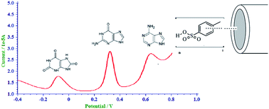
Rapid and sensitive determination of purine bases is vital in clinical analysis. An electrochemical sensor for the determination of the purine bases, guanine, adenine and uric acid, fabricated using a glassy carbon electrode modified with poly(para toluene sulfonic acid), is reported here. In the square wave mode, the modified glassy carbon electrode was able to produce well defined and well separated oxidation peaks for guanine, adenine and uric acid with 0.1 M sodium hydroxide as the supporting electrolyte. The oxidation peak currents for guanine and adenine, showed a dynamic range from 10–100 μM and 20–800 μM with a limit of detection of 0.35 μM and 0.78 μM respectively when determined simultaneously in the presence of uric acid. In simultaneous determination, uric acid also showed a linear increase of oxidation peak current in the concentration range of 10–100 μM, with a limit of detection of 5.88 μM. The variation of peak parameters with scan rate was studied to determine the nature of electro-oxidation and the number of electrons involved in the electrode process. The simultaneous determination of guanine, adenine and uric acid in acid denatured Herring sperm using the fabricated sensor is described. The excellent results obtained indicate that the sensor can be applied for the simultaneous as well as individual determination of guanine, adenine and uric acid in real samples.
Introduction
The purine bases guanine (G) and adenine (A) are two of the four major building blocks of nucleic acids.1 They play major roles in life processes like blood circulation, neurotransmitter release, maintenance of cardiac rhythm etc.1 The concentration of these compounds in blood are indicative of conditions like cancer and myocardial cellular energy status.2 Any abnormal change in the levels of G and A in nucleic acids is linked to deficiencies and/or mutations in the immune mechanism of the body.3 A monitoring of the oxidation signals of G and A is reported to have been used to study the changes in double stranded and single stranded DNA as well as their interaction with other molecules.4 The presence of G and A in physiological fluids may be the result of nucleic acid metabolism by degradation of tissues or dietary intake or malfunction of enzymes in other metabolic mechanisms.5 Hence detection of G and A in physiological fluids is indicative of certain diseases. Uric Acid (UA) is the principal oxidation product of purine metabolism.6 It is excreted through urine in a typical range of 250 mg dL−1 to 720 mg dL−1 and an anomalous change in its level is symptomatic for gout, hyperuricemia, Lesch–Nyan disease or cardiovascular abnormalities.6 G, A and UA co-exist in physiological fluids5 as well as in mutated DNA and hence their simultaneous determination is of much significance.
Electrochemical methods based on the oxidation of G, A and UA at unmodified electrodes7–10 suffer from disadvantages like high over potential, sluggish direct electron transfer, poor reproducibility and significant fouling of electrode surface due to adsorption of oxidation products. Modification of the electrode surface with suitable electron transfer mediators can overcome these disadvantages and improve sensitivity. In this regard, conducting polymeric film of aromatic and hetero aromatic compounds electropolymerised on the bare electrode are valuable. By adjusting the parameters of electropolymerisation, control over the polymer film thickness and its charge transfer properties could be achieved. Such polymer modified electrodes offer advantages like ease of preparation, strongly adhering uniform film on electrode surface, better sensitivity and selectivity.11 Polymelamine modified electrode had been fabricated by Liu et al.12 which was able to simultaneously determine G and A together with epinephrine. Li et al.5 reported a polymelamine and nano Ag hybridized film-modified glassy carbon electrode (GCE) for the simultaneous determination of dopamine, UA, G and A. A poly safranine modified GCE with three dimensionally distributed Au nanoparticles was devised by Niu et al. for the determination of the same molecules.6 A polyimidazole and graphene oxide based sensor was fabricated by Liu et al. for the simultaneous determination of A, G, UA, dopamine and ascorbic acid.13 There has been a report of a poly(para toluene sulfonic acid) (p-PTSA) modified GCE for the simultaneous determination of UA and ascorbic acid in neutral medium.14 However the simultaneous determination of G, A and UA in alkaline medium has not been reported afore; to the best of our knowledge.
Based on our recent works on sensors for biological molecules15 and polymer modified electrodes,11,16 here we report the simultaneous electrochemical determination of G and A in the presence of UA on a p-PTSA modified GCE (p-PTSA/GCE) in basic medium.
Experimental
Reagents and instrumentation
All the reagents used in the present study were of analytical grade and were used without any further purification. All the solutions were made in Millipore water. NaOH was supplied by Merck Life Science Pvt. Ltd, India. PTSA and G were procured from S.D. Fine Chemicals, India. A, and UA were supplied by Himedia laboratories Pvt. Ltd, India and Alfa Aesar, England respectively. Herring Sperm (HS) DNA was purchased from Sisco Research Laboratories Pvt. Ltd. India. The solutions were purged with nitrogen before each experiment and the experiments were carried out at room temperature.
CHI600C electrochemical analyser (CH Instruments Inc. USA) integrated to a desk top computer was used to perform the electrochemical experiments and impedance measurements. A three electrode set up with Ag/AgCl reference electrode, platinum wire auxiliary electrode and the GCE (3 mm diameter) or p-PTSA/GCE as the working electrode was employed for the measurements. Metrohm pH meter was used to measure the pH of the solutions.
Preparation of p-PTSA/GCE
The GCE was mechanically polished with alumina slurries of powder size 0.05 μm to a mirror finish and ultra-sonicated successively in methanol, 50% nitric acid, acetone and water for 5 minutes each so as to remove any adsorbed alumina. The electropolymerisation of PTSA on GCE was performed by a method reported elsewhere.11 The cleaned GCE was then immersed in a 1 mM solution of PTSA in 0.1 M NaCl and potential scanned between −2.0 and +2.5 V for 28 segments at 0.1 V s−1. The fabrication of the modified electrode is represented in Scheme 1. The modified electrode was washed with Millipore water and dried in air. Prior to voltammetric measurement, the electrode was activated by scanning the potential from −1.0 V to +1.0 V (vs. Ag/AgCl) in 0.1 M NaOH solution.
 |
| | Scheme 1 Fabrication of p-PTSA/GCE. | |
Preparation of DNA sample
Acid denatured DNA solution was prepared by the procedure reported by Brett and Serrano.17 In brief, 7 mg of the HS DNA was dissolved in 0.5 mL of perchloric acid with the aid of ultrasonication, followed by neutralisation with 0.5 mL of 9 M sodium hydroxide solution. The resulting solution was made up to 50 mL with the supporting electrolyte.
Results and discussion
Characterisation of the modified electrode
The effective surface area of the modified electrode was calculated from cyclic voltammetry studies with 2 mM K4[Fe(CN)6] as probe at different scan rates. According to Randles Sevcik equation,11 for a reversible electrochemical reaction involving n electrons for a species of concentration C, with diffusion coefficient D,| | |
Ip = 2.69 × 10⁵An3/2D1/2Cυ1/2
| (1) |
From the slope of peak current (Ip) versus square root of scan rate (υ) the effective surface area A of the modified electrode was found to be 0.624 cm2 as opposed to 0.451 cm2 at the bare electrodes indicating that, electro polymerisation has resulted in 38% increase in surface area.
Electrochemical oxidation of G, A and UA
A comparison of square wave voltammograms (SWV) of the bare and modified electrodes recorded in a solution of 0.1 M NaOH containing 300 μM each of G, A and UA (Fig. 1) shows that the electro oxidation of G, A and UA is catalysed by PTSA film on GCE. In the bare GCE, oxidation peaks for G, A and UA appear at 348 mV, 732 mV and 28 mV respectively against Ag/AgCl electrode with peak currents 6.72 μA, 2.09 μA and 0.743 μA respectively whereas on p-PTSA modified electrode the same appears at 304 mV, 608 mV and −108 mV with peak currents 21.28 μA, 12.38 μA and 9.52 μA respectively. Well defined peaks with peak separations of 196 mV for UA–G, 304 mV for G–A and 500 mV for UA–A were obtained with square wave voltammetry. The increased effective surface area of the GCE on modification with the polymer combined with the interaction between the sulfonic acid group of the polymer and the purine bases may be the reason for this enhancement in the electrochemical response.
 |
| | Fig. 1 SWV of (a) bare GCE (b) p-PTSA GCE in a solution of 0.1 M NaOH containing 300 μM each of G, A and UA. | |
Optimisation of experimental conditions
Choice of supporting electrolyte is an experimental parameter that can influence the electro-oxidation process. The electrochemical oxidation of G, A and UA was studied in 0.1 M concentration of various supporting electrolytes such as sulfuric acid, hydrochloric acid, acetate buffer, citrate buffer, sodium chloride, potassium chloride, phosphate buffer, NaOH and potassium hydroxide. The lowest oxidation potential was obtained for 0.1 M NaOH and hence it was selected as the supporting electrolyte.
The thickness of polymer film, best suited for the determination of G, A and UA was optimised by varying the number of cycles of polymerisation. The best result in 0.1 M NaOH was obtained when the number of cycles of polymerisation was 14 and hence further studies were conducted after the polymerising PTSA over GCE in 14 cycles.
Mechanistic study by varying scan rate
The variation of the oxidation peak parameters with scan rate (υ) in linear sweep (LS) voltammetric mode was used to study the mechanism of electro-oxidation. The oxidation peak of G, A and UA showed a positive shift with increasing scan rate pointing to the irreversibility of oxidation process. The oxidation peak currents for G, A and UA were found to vary linearly with square root of scan rate in the range 10–1000 mV s−1, 10–100 mV s−1 and 20–200 mV s−1 respectively according to the following equations indicative of a diffusion controlled process.18
For guanine:
| | |
Ip(μA) = −4.98 + 3.39υ1/2(V1/2s−1/2) [R2 = 0.9946]
| (2) |
For adenine:
| | |
Ip(μA) = −0.72 + 3.33υ1/2(V1/2s−1/2) [R2 = 0.9895]
| (3) |
For uric acid:
| | |
Ip(μA) = +0.80 + 0.15υ1/2(V1/2s−1/2) [R2 = 0.9845]
| (4) |
Further the plot of log
Ip vs. log
υ was found to have slopes of 0.43, 0.52 and 0. 36 for G, A and UA respectively which are closer to the theoretical value of 0.5 for a diffusion controlled process.19 These indicate that all the three electro oxidation processes are diffusion controlled. That is, the electrochemical kinetics is influenced by diffusion of electroactive species from bulk of the solution to the surface of the electrode.
According to Laviron's theory,20
| |
 | (5) |
and the plot of
Ep vs. natural logarithm of scan rate (ln
υ) should be linear for an irreversible process with heterogeneous rate constant
ks and the slope of the curve gives the value of
RT/
αnF where
α is the charge transfer coefficient (0.5 for a perfectly irreversible process),
n is the number of electrons involved in the electrochemical reaction and the other alphabets represent the accepted scientific terms. Using the slope obtained from the plot of
Ep vs. ln
υ, the number of electrons involved in each of the oxidation process was calculated to be 4 for G and 2 each for A and UA.
20 The mechanisms of the electro-oxidation processes in accordance with this data can be depicted as in
Scheme 2.
21
 |
| | Scheme 2 Possible reactions taking place on the electrode. | |
Determination of G, A and UA
Square wave voltammetry was used for the individual determination of G, A and UA in 0.1 M NaOH solution. The lower LOD for the assay was calculated using the following equation.22where S is the standard deviation of the lowest response and b is the slope of the calibration curve. Fig. 2a–c show the variation of oxidation peak current with concentration of G, A and UA in 0.1 M NaOH.
 |
| | Fig. 2 (a) Overlay of SWV obtained for the oxidation of (a) 10 μM to 100 μM of G (b) 1 μM to 130 μM of A and (c) 1 μM to 130 μM UA in 0.1 M NaOH. Inset shows calibration graph in the corresponding dynamic range. | |
The anodic peak current of G was found to vary linearly in the range 3 μM to 10 μM and from 10 μM to 300 μM with the limit of detection (LOD) as low as 0.0079 μM. For A, the variation was linear in the range 5 μM to 45 μM and 50 μM to 1500 μM and the LOD obtained was 0.94 μM. The variation of peak current for electro oxidation of UA was found to be linear in the range 20 μM to 100 μM and in the range 90 μM to 1500 μM. The LOD was calculated to be 3.31 μM.
Individual determinations of G, A and UA on the p-PTSA/GCE was also conducted in the presence of other two (Fig. 3). For G, in presence of 100 μM concentrations of A and UA, the peak current varied linearly in the range from 20 μM to 200 μM and from 250 μM to 1000 μM with a limit of detection of 3.08 μM. The presence of 50 μM concentration of G and UA did not interfere in the determination of A in the range from 10 μM to 120 μM and gave a LOD of 3.16 μM. For UA determination in the presence of 100 μM concentrations of G and A, the variation of current was linear in the range 20–110 μM and 100–1000 μM with a LOD of 7.49 μM.
 |
| | Fig. 3 Variation of peak current with concentration for the electro-oxidation of (a) G in the presence of 100 μM of A and UA (b) A in presence of 50 μM of G and UA (c) UA in the presence of 100 μM of G and A. Inset shows calibration graph in the corresponding dynamic range. | |
In a solution containing equimolar concentrations of G, A and UA, the oxidation peak current varied linearly from 10–1000 μM with a dynamic range 10 μM to 100 μM for both G and UA (Fig. 4) and LOD of 0.35 μM and 5.88 μM respectively. For A, the variation although linear from 20–800 μM the dynamic range was from 20–100 μM with a detection limit of 0.78 μM (Table 1).
 |
| | Fig. 4 (a)Overlay of SWV of electro-oxidation of a solution containing 5 μM to 600 μM each of G, A and UA (b) calibration graph. | |
Table 1 Statistical parameters for simultaneous determination
| Analyte |
Linear range (μM) |
Regression equation |
(R2) |
LOD (μM) |
| G |
10–100 |
Ip (μA) = +1.00 + 0.15C (μM) |
0.9978 |
0.35 |
| A |
20–100 |
Ip (μA) = +0.91 + 0.04C (μM) |
0.9932 |
0.78 |
| UA |
10–100 |
Ip (μA) = +1.68 + 0.04C (μM) |
0.9874 |
5.88 |
Although there has been reports of polymer modified electrodes being used for the simultaneous determination of G and A in the presence of UA and also with other targets, the determination occurs at higher overpotentials. It is under such a status quo that, a p-PTSA/GCE, which is easily fabricated via electropolymerisation of the very prevalent and available PTSA, becomes significant. It not only decreases the overpotential for the electro-oxidation process, but also gives LODs comparable to that of analogues sensors.
Stability, sensitivity, reproducibility and repeatability
The products of the oxidation reaction can adsorb on the electrode surface, producing fouling of the electrode surface and thereby affecting the stability of the electrode.23 In order to evaluate the antifouling property of the modified electrode, amperometric response of the electrode was studied in 100 μM solution of the analytes. The amperometric response was found to be stable for more than 5 minutes (Fig. 5).
 |
| | Fig. 5 Amperometric response of 50 μM G, A and UA in 0.1 M NaOH at 304 mV, 608 mV and −108 mV respectively. | |
The sensitivity of the measurement for the simultaneous determination of G, A and UA was calculated using the formula,22
. The electrode was found to be more sensitive for the determination of G than A and UA for the simultaneous as well as individual determinations (Fig. 6).
 |
| | Fig. 6 A comparison of sensitivities of G, A and UA on the p-PTSA/GCE for simultaneous and individual determinations. | |
In order to explore the reproducibility and repeatability of the developed sensor, relative standard deviation (RSD) of the intra and inter-assay was calculated. To conduct the intra assay five different p-PTSA/GCE was prepared and the peak currents for a solution containing 10 μM each of G, A and UA were measured. The RSD for the measurement for G, A and UA was found to be 0.25%, 0.30% and 0.84% respectively. For the inter assay, the peak current measured on a p-PTSA/GCE was noted five times in a solution containing 10 μM each of G, A and UA. The RSD (n = 5) for the inter assay for G, A and UA was found to be 0.32%, 2.08% and 3.36% respectively. The RSD values for intra and inter-assays indicate that the p-PTSA/GCE show good reproducibility and repeatability.
Influence of possible co-existing species
Influence of other DNA bases as well as some other biologically relevant molecules on the voltammetric signals of G, A and UA was explored in a 10 μM solution. The results obtained indicate that the signal change produced by molecules like cytosine, uracil and glutathione was within the tolerance limit when present in the same concentration. A fivefold excess of thymine and homovanillic acid produced interference in the determination of A in the mixture even though the signal change for G and UA was within the tolerance range. Dopamine interfered in the determination of G when present in tenfold excess concentration whereas it did not produce any significant interference in the signals for A and UA.
Application studies
p-TSA/GCE was used for the determination of G and A in acid denatured HS DNA. Two peaks appear at 308 mV and 608 mV corresponding to the oxidation of G and A respectively. As the volume of the DNA solution increases the peak currents vary linearly. The method of standard addition was used to determine the molar concentrations of G and A. The value of (G + C)/(A + T) was found to be 0.754 which is close to the theoretical value of 0.770.24
The acid denatured HS DNA solution did not show any signal corresponding to the electro-oxidation of UA, indicating that UA was not present in the sample. In order to demonstrate the applicability of the sensor for simultaneous determination of G and A in the presence of UA, the prepared acid denatured HS DNA was spiked with different concentrations of UA and the determination was carried out using standard addition method. Simultaneous determination of G and A in the presence of UA in the spiked HS DNA gave the value of (G + C)/(A + T) as 0.760 (RSD = 1.88% for n = 6) with good recoveries for the determination of UA (Table 2) with a RSD value of 3.63%. This indicates that the sensor can be applied for the determination of G and A in the presence of UA in real samples effectively.
Table 2 Determination of UA in spiked DNA
| Sample |
UA |
Recovery% |
RSDa% |
| Added (μM) |
Found (μM) |
| For 6 measurements. |
| Herring sperm DNA (acid denatured) |
10–120 |
10.5–123 |
91–105 |
3.63 |
To check the feasibility of the developed sensor for real sample analysis, the simultaneous determination of G, A and UA was conducted in spiked synthetic blood serum25 and synthetic urine samples. The results obtained are presented in Table 3. Good recoveries are obtained for the determination of G and A in the presence of UA which demonstrate the feasibility of the sensor for real sample analysis.
Table 3 Determination of G, A and UA in spiked samples
| Analyte |
Urine |
Serum |
| Added (μM) |
Found (μM) |
Recovery |
RSDa% |
Added (μM) |
Found (μM) |
Recovery |
RSDa% |
| For 6 measurements. |
| G |
20–100 |
20.3–99.2 |
98.5–102.0 |
1.23 |
15–100 |
15.3–102.0 |
100.3–106.0 |
1.89 |
| A |
25–100 |
24.9–99.7 |
95.9–99.8 |
1.40 |
25–100 |
24.0–98.7 |
96.0–100.5 |
1.73 |
| UA |
25–100 |
25.4–100.2 |
95.1–102.0 |
2.63 |
25–100 |
26.3–97.9 |
97.9–105.2 |
2.93 |
Conclusions
The simultaneous electrochemical oxidation of G and A in the presence of UA, is studied in the square wave mode on a p-PTSA/GCE with NaOH as the supporting electrolyte. In highly alkaline medium, well defined and well separated irreversible peaks were obtained for the electro oxidation of G, A and UA, which facilitate their simultaneous determination even in alkaline medium. The p-PTSA/GCE sensor gave good working linear range and low detection limits for the determination of these analytes in alkaline medium. The method was successfully applied to the determination of purine bases in HS DNA. The determination of UA in spiked HS DNA with the developed sensor gave reasonably good recoveries with acceptable RSD values. The utility of the sensor for the determination of G, A and UA in synthetic urine and synthetic blood serum has also been demonstrated.
Conflict of interests
The authors declare that there is no conflict of interests regarding the publication of this paper.
Human and animal testing
This research work does not include any live animal or human subjects; thus approval statements are not required.
Acknowledgements
The authors, Krishnapillai Girish Kumar and Siri Jagan Jesny, would like to thank the University Grants Commission, Government of India for financial assistance in the form of One Time Research Grant (Grant No. F.19-133/2014) and teacher fellowship under Faculty Development Programme respectively. Shalini Menon acknowledges the Kerala State Council for Science Technology and Environment for the Junior Research Fellowship. The authors thank Anuja Elevathoor Vikraman and Sreelakshmi for their contributions during the preliminary stages of work described here.
Notes and references
- A. A. Ensafi, M. Jafari-Asl, B. Rezaei and A. R. Allafchian, Sens. Actuators, B, 2013, 177, 634 CrossRef CAS.
- F. Q. Yang, J. Guan and S. P. Li, Talanta, 2007, 73, 269 CrossRef CAS PubMed.
- T. Liu, X. Zhu, L. Cui, P. Ju, X. Qu and S. Ai, J. Electroanal. Chem., 2011, 651, 216 CrossRef CAS.
- Y. Wei, Q. Huang, M. Li, X. Huang, B. Fang and L. Wang, Electrochim. Acta, 2011, 56, 8571 CrossRef CAS.
- H. Li, X. Wang and Z. Yu, J. Solid State Electrochem., 2014, 18, 105 CrossRef CAS.
- L. M. Niu, K. Q. Lian, H. M. Shi, Y. B. Wu, W. J. Kang and S. Y. Bi, Sens. Actuators, B, 2013, 178, 10 CrossRef CAS.
- E. Palecek, Nature, 1960, 188, 656 CrossRef CAS PubMed.
- J. Wang, X. Cai, G. Rivas, H. Shiraishi, P. A. M. Farias and N. Dontha, Anal. Chem., 1996, 68, 2629 CrossRef CAS PubMed.
- H. S. Wang, H. X. Ju and H. Y. Chen, Electroanalysis, 2001, 13, 1105 CrossRef CAS.
- E. Ferapontova and E. Dominguez, Electroanalysis, 2003, 15, 629 CrossRef CAS.
- S. Issac and K. Girish Kumar, Anal. Methods, 2010, 2, 1484 RSC.
- X. Liu, L. Luo, Y. Ding, Q. Wu, Y. Wei and D. Ye, J. Electroanal. Chem., 2012, 675, 47 CrossRef CAS.
- X. Liu, L. Zhang, S. Wei, S. Chen, X. Ou and Q. Lu, Biosens. Bioelectron., 2014, 57, 232 CrossRef CAS PubMed.
- L. Wang, P. Huang, J. Bai, H. Wang, X. Wu and Y. Zhao, Int. J. Electrochem. Sci., 2006, 1, 334 CAS.
- A. E. Vikraman, A. R. Jose, M. Jacob and K. Girish Kumar, Anal. Methods, 2015, 7, 6791 RSC.
- D. Thomas, Z. Rasheed, J. Sirijagan and K. Girish Kumar, J. Food Sci. Technol., 2015, 52, 6719 CrossRef CAS PubMed.
- A. M. O. Brett and S. H. P. Serrano, J. Braz. Chem. Soc., 1995, 1, 97 CrossRef.
- A. Thomas, A. E. Vikraman, D. Thomas and K. Girish Kumar, Food Analytical Methods, 2015, 8, 2028 CrossRef.
- R. Gupta, S. K. Guin and S. K. Aggarwal, RSC Adv., 2012, 2, 1810 RSC.
- E. Laviron, J. Electroanal. Chem., 1979, 101, 19 CrossRef CAS.
- V. K. Sharma, F. Jelen and L. Trnkova, Sensors, 2015, 15, 1564 CrossRef CAS PubMed.
- A. Shrivastava and V. B. Gupta, Chron. Young Sci., 2011, 2, 21 CrossRef.
- M. U. A. Prathap, R. Srivastava and B. Satpathi, Electrochim. Acta, 2013, 114, 285 CrossRef.
- H. Tang, G. Hu, S. Jiang and X. Liu, J. Appl. Electrochem., 2009, 39, 2323 CrossRef CAS.
- S. Menon, A. R. Jose, S. Jesny and K. Girish Kumar, Anal. Methods, 2016, 8, 5801 RSC.
Footnote |
| † Electronic supplementary information (ESI) available. See DOI: 10.1039/c6ra13567f |
|
| This journal is © The Royal Society of Chemistry 2016 |
Click here to see how this site uses Cookies. View our privacy policy here.