
Exfoliation of graphite and graphite oxide in water by chlorin e6†
Dania Hernández-Sáncheza,
Mattia Scardamagliab,
Sonia Saucedo-Anayaac,
Carla Bittencourtb and
Mildred Quintana*a
aInstituto de Física, Universidad Autónoma de San Luis Potosí, Manuel Nava 6, Zona Universitaria 78290, SLP, San Luis Potosí, Mexico. E-mail: mildred@ifisica.uaslp.mx; Tel: +52 444 8262362 ext. 130
bChimie des Interactions Plasma-Surface, University of Mons, Av. Nicolas Copernic, 1, 7000 Mons, Belgium
cUnidad Académica de Estudios Nucleares, Universidad Autónoma de Zacatecas, Ciprés Núm. 10, Fracc. La Peñuela, C.P. 98068, Zac., Zacatecas, Mexico
First published on 6th July 2016
Abstract
Few layer graphene (FLG) and graphene oxide (GO) are considered important materials for the development of future technological applications. Diverse strategies are followed for their synthesis and production resulting in graphene materials with different processability, electronic, optical, mechanical, and chemical properties. In particular, many efforts are directed at the integration of FLG or GO with water dispersable functional molecules. Recent advances in ultrasonication techniques have led to the control over the synthesis of carbon nanostructures using a versatile synthetic tool. Herein, we demonstrate the facile preparation of two different types of chlorin e6 (Ce6) nanohybrids in biocompatible media: few-layer graphene (FLG–Ce6) and graphene oxide (GO–Ce6) in deionized water (DW) and phosphate buffered saline (PBS). The exfoliation is energetically driven by acoustic cavitation while molecular interactions are responsible for the stabilization of FLG and GO in water by Ce6. The nanohybrid materials might find applications in energy and biomedicine fields since the main photophysical features of Ce6, such as its efficient use of energy in the near infrared region, its light harvesting properties, and its capacity for energy and electron transferring processes, are well preserved.
Introduction
The chemical production of graphene and its controlled chemical manipulation are expected to prompt graphene-based technology. It is well-known that graphene offers an exceptional combination of electronic, optical, mechanical, and chemical properties that make it an excellent candidate for the development of innovative sensors,1 highly efficient fuel cells,2 and exciting biomedical applications.3 An economical route to obtain graphene is derived from the chemical exfoliation of graphite.4 In principle, following this strategy graphene should be produced in large quantities in organic solvents with different polarities and in biocompatible media. However, the strong π–π stacking interactions between graphene layers in graphite are responsible for the delay in achieving this goal. Until now, the exfoliation of graphite can be accomplished by diverse methodologies such as the use of strong oxidizing agents yielding to graphene oxide (GO).5 Alternatively, graphite can be exfoliated by the energetically assisted intercalation of solvents or small molecules using ultrasonication,6 ball milling processing7 or microwave irradiation8 resulting in few-layer graphene (FLG). Both strategies present advantages and disadvantages depending on the application demand. For example, GO is a readily processable material easily dispersible in water and polar organic solvents. The presence of functional groups on its surface allows its direct integration into composite materials or polymeric matrices.9 Unfortunately, most of the important properties associated to graphene such as high electrical conductivity and mechanical strength are lacking in GO. Instead, FLG maintains the key properties of graphene, but solvents with high boiling points and large surface tensions are used during the exfoliation of graphite making difficult their evaporation and complete elimination.10 To undertake this problem, molecular additives such as surfactants,11 ionic liquids,12 planar molecules13 and polymers14 are used for the stabilization of FLG in aqueous and organic media, yielding functionalized-FLG. Herein, we demonstrate the straightforward exfoliation of graphite and graphite oxide in water by using chlorin e6 (Ce6) achieving FLG–Ce6 and GO–Ce6 nanohybrids, respectively, as shown in Scheme 1. | ||
| Scheme 1 Schematic representation of graphite and graphite oxide exfoliation using chlorin e6 (Ce6) as stabilizing agent. | ||
Ce6 is a tetrapyrrole and a chlorophyll analogue used in dye-sensitized solar cells improving the photoenergy conversion,15 additionally, Ce6 is effectively used as a photosensitizer molecule improving cancer cell photodynamic destruction.16 For these applications, it is a key factor to preserve the main photophysical features of Ce6 i.e. its efficient use of energy at the near infrared region, its light harvesting properties, and its capacity for energy and electron transferring processes.17 In addition, it is desirable that Ce6 composites and nanohybrid materials can be easily dispersible in water or biocompatible media. In this work, the exfoliation of graphite and graphite oxide to obtain FLG and GO, respectively, was performed in distilled water (DW) and in a different set of experiments in phosphate buffered saline (PBS), a buffer solution commonly used in biological research. This exfoliation is possible as result of the physical and chemical interactions between Ce6 and carbon sheets. We show a comparative study based on the spectroscopic features of FLG–Ce6 and GO–Ce6. It is expected that the understanding of the interactions between functional molecules such as Ce6 and nanostructured materials as FLG and GO pave the way for further development of advanced nanohybrids with applications in energy and biomedicine.
Experimental
Chemicals
Chlorin e6 (Ce6) was purchased from Frontier scientific Logan, Ut. Graphite was purchased from Bay Carbon, Inc. (SP-1 graphite powder, batch N° 04100, lot. N° 011705, www.baycarbon.com). All solvents and chemicals were obtained from commercial suppliers and used without further purification.Samples preparation
A stock solution of Ce6 (1 mg mL−1) was prepared in methanol. Exfoliation of graphite and graphite oxide were carried out in two different solvents: distilled water (DW) and phosphate buffered saline (PBS). Control experiments were performed under the same experimental conditions without the addition of Ce6. As expected relaying on the chemical structures of FLG and GO, exfoliation of graphite was not observed in PBS or DW. Instead GO dispersed very well in both solvents.Few layer graphene–Ce6 nanohybrid (FLG–Ce6)
Graphite powder (1 mg) was mixed in 7.5 mL of DW and in a different set of experiments in 7.5 mL of PBS. Then, 2.5 mL of Ce6 stock solution was added to each assay. This concentration was chosen in order to maintain the UV-vis absorption low enough to avoid deviations from the Beer–Lambert law and light scattering processes during the spectroscopic analysis. However, experiments at higher concentrations results in stable dispersions, as shown in S1.† Graphite crystals were ultrasonicated using a Branson 2510 ultrasonic bath in solutions of Ce6 in PBS or DW during 195 min taking the absorption and emission spectra every 15 min by the exhaustively mixing of 3 μL of the sonicated dispersion into 3 mL of the correspondent solvent. This procedure was performed in order to avoid saturation of the spectroscopic signals whilst finding the sonication time required for the maximum exfoliation of graphite. To avoid the excessive raise of temperature, ice was poured into the sonication bath during the whole process. The absorption and fluorescence (λexc = 404 nm) spectra of the mixture were recorded every 15 minutes. Sample at t = 0 was taken before the beginning of the bath sonication. At the end of the ultrasonication process a dark liquid dispersion consisting of a homogeneous phase with the presence of some macroscopic aggregates was obtained. These aggregates were removed from the mixture by mild centrifugation (500 RPM, 90 min) giving a dark green homogeneous dispersion. The supernatant was washed over a PTFE Advantech membrane filter with a 0.22 μm pore size until the absorption signals of Ce6 were not detected in the filtered solution. The resulting solid was suspended in 10 mL of DW or PBS, respectively. All the products were thoroughly characterized by UV-vis, fluorescence, transmission electron microscopy (TEM), Raman and X-ray photoemission spectroscopies (XPS). All the experiments and their corresponded analytical characterizations were performed at least 3 times.Graphene oxide–Ce6 (GO–Ce6) nanohybrid
Graphite oxide was prepared following the improved Hummers methodology reported by Marcano et al.5 Then, 1 mg of graphite oxide was well dispersed in 7.5 mL of DW or PBS by brief bath sonication and mixed with 2.5 mL of the Ce6 stock solution. Again, these concentrations were chosen to avoid deviations from the Beer–Lambert law and light scattering processes during spectroscopic analysis. The GO–Ce6 mixtures were light protected and left under magnetic stirring overnight (18 h). The macroscopic aggregates were removed by mild centrifugation (500 RPM-90 minutes) giving a dark green homogeneous dispersion. The supernatant was washed over a PTFE membrane with a 0.22 μm pore size until the absorption signals of Ce6 were not detected in the filtered solutions. The clean solid was suspended in 10 mL of DW or PBS respectively. All the products were thoroughly characterized by UV-vis, fluorescence, TEM, Raman, and XPS. All the experiments and their corresponded analytical characterizations were performed at least 3 times.Characterization techniques
The optical absorbance of the samples was measured by UV-vis spectroscopy with a Cary 60 spectrophotometer using 10 mm path length quartz cuvettes. The optical absorbance was measured at 404 nm. Fluorescence spectroscopy was carried out with a Cary Eclipse fluorescence spectrophotometer, software version 1.2 (146, Agilent technologies). Raman spectra were obtained by means of a Thermo Scientific DXR Raman Microscope equipped with a diode-pumped solid-state laser (DPSS) at wavelength of 532 nm as excitation source. A 10× objective with a 50 μM slit aperture and 5 s of exposure time. The laser power impinging on the sample was between 5 and 10 mW, 2 cm−1 spatial resolution and spot size ∼ 1 m2. The samples were recorded from drops of the dispersions deposited over clean silicon wafers and left dry under vacuum. TEM measurements were performed on a JEOL JEM-2100, using an accelerating voltage of 200 kV. The samples were prepared by drop casting from dispersions onto a TEM grid (200 mesh, cooper, carbon only). The chemical composition of the samples was investigated using XPS VERSAPROBE PHI 5000 from Physical Electronics, equipped with a Monochromatic Al Kα X-ray in UHV conditions. The energy resolution was 0.7 eV. For the compensation of built-up charge on the sample surface during the measurements, dual beam charge neutralization composed of an electron gun (∼1 eV) and the argon ion gun (≤10 eV) was used. The XPS spectra were deconvoluted into different chemical surroundings using commercially available software (CASA-XPS).Results and discussion
Ce6 is barely soluble in water but totally soluble in methanol. Using a significant concentration of Ce6 in methanol, a water miscible solvent, and injecting this solution in the proper amount of PBS or DW the lack of solubility of Ce6 in water was avoided. The chemical structure of Ce6 is shown in Scheme 1 and in S2.† Following this protocol, instead of using high surface tension and high boiling point solvents, such as dimethyl formamide (DMF) or N-methyl pyrrolidone (NMP), the ultrasonication of graphite crystals was performed in mixtures of water![[thin space (1/6-em)]](https://www.rsc.org/images/entities/char_2009.gif) :methanol 75:25 (v/v). The absorption spectra of Ce6 in the solvents used during the present study are shown in S3† for solubility comparison.
:methanol 75:25 (v/v). The absorption spectra of Ce6 in the solvents used during the present study are shown in S3† for solubility comparison.
FLG–Ce6 nanohybrid was obtained following the methodology described in ref. 6. After 45 min of sonication, the higher absorption and emission spectra were found for PBS dispersion, as shown in S4(a and c†). Instead in DW, the higher absorption and emission signals were found before the sonication treatment. Within the first 15 minutes of sonication both absorption and emission spectra were considerably diminished. After 45 min the maximum of emission of FLG–Ce6 in DW shifted from 661 nm to 652 nm, as reported in S4(b and d†). Thus, graphite was sonicated for 45 min in both PBS and DW.
In order to avoid further oxidation of GO and since it disperses particularly well in water, instead of using ultrasonication, GO–Ce6 nanohybrids were produced by the magnetic stirring of GO in the solutions of Ce6 in DW or PBS.
FLG–Ce6 and GO–Ce6 were carefully washed and dispersed in PBS or DW, correspondingly. All dispersions were stable during the time, as observed in Fig. 1. Images were taken after 30 days of preparation. Dispersions of FLG–Ce6 and GO–Ce6 follow the Beer–Lambert behavior, as shown in S5(a).†
 | ||
| Fig. 1 Representative photographs of the dispersibility and stability of the FLG–Ce6 and GO–Ce6 in PBS and DW. | ||
For the sake of comparison, the UV-vis absorption spectra of FLG–Ce6 and GO–Ce6 were normalized to Ce6 in the corresponding solvent, as shown in Fig. 2. The unmodified spectra are presented in S6.† The careful washing of FLG–Ce6 and GO–Ce6 nanohybrids avoids the presence of Ce6 molecules in the solvent. The increases in the absorption intensity through the entire absorption interval for FLG–Ce6 and GO–Ce6 nanohybrids in both PBS and DW, resulted from the absorption of FLG or GO, respectively.11 The dashed lines in Fig. 2 indicate the pondered absorption due to FLG or GO in the nanohybrid.
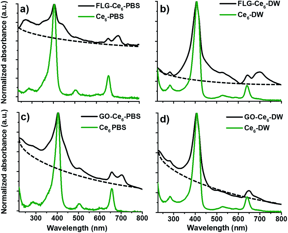 | ||
| Fig. 2 UV-vis absorption of FLG–Ce6 and GO–Ce6 nanohybrids in PBS (a and c) and DW (b and d). Free Ce6 (green line) is included as a control. | ||
The absorbance at 660 nm divided by cell length is plotted versus concentration following the protocol performed by Hernández et al.10 showing Lambert–Beer behavior for all the solvents. The concentrations of FLG in FLG–Ce6 were calculated from the calibration curves at ∼660 nm in PBS and DW, resulting in 0.01 and 0.02 mg mL−1, respectively.11 Likewise, the concentration of GO in GO–Ce6 calculated from the calibration curves at 227 nm in PBS and DW, resulted 0.02 and 0.05 mg mL−1, respectively, as shown in S5(a).†
Considering the higher dispersibility of GO in water, these results corresponded to the expected values.10,11 The concentration of Ce6 in each nanohybrid was also plotted and calculated at ∼404 nm in PBS and DW. See S5(b).†
The absorption band at 268 nm for FLG–Ce6 is related to π–π transitions of aromatic C![[double bond, length as m-dash]](https://www.rsc.org/images/entities/char_e001.gif) C bonds. This absorption signal is more evident in PBS where a wide band in this region is observed, while in DW the featured absorption band of Ce6 is clearly preserved, see Table 1. For GO–Ce6 the spectrum exhibits two characteristic peaks of GO, a maximum at 235 nm corresponding π–π transitions of aromatic CC bonds, and a shoulder at 279 nm attributed to n → π transitions of CO bonds18 as shown in Fig. 2c and d. In both, FLG–Ce6 and GO–Ce6, an intense peak in the blue wavelength region, due to an electron dipole movement that allows π–π* transitions commonly found in porphyrin compounds25 namely the Soret signal (∼404 nm), appears as broadened bands, Table 1, while Q bands are considerably reduced when compared with the spectrum of Ce6 in PBS or DW. The Q bands arise from a linear combination of the one-electron transitions. Importantly, a new absorption band (NAB) appears at ∼701 nm for FLG–Ce6 in both solvents and only in PBS for GO–Ce6, as shown in Fig. 2a–c and summarized in Table 1.
C bonds. This absorption signal is more evident in PBS where a wide band in this region is observed, while in DW the featured absorption band of Ce6 is clearly preserved, see Table 1. For GO–Ce6 the spectrum exhibits two characteristic peaks of GO, a maximum at 235 nm corresponding π–π transitions of aromatic CC bonds, and a shoulder at 279 nm attributed to n → π transitions of CO bonds18 as shown in Fig. 2c and d. In both, FLG–Ce6 and GO–Ce6, an intense peak in the blue wavelength region, due to an electron dipole movement that allows π–π* transitions commonly found in porphyrin compounds25 namely the Soret signal (∼404 nm), appears as broadened bands, Table 1, while Q bands are considerably reduced when compared with the spectrum of Ce6 in PBS or DW. The Q bands arise from a linear combination of the one-electron transitions. Importantly, a new absorption band (NAB) appears at ∼701 nm for FLG–Ce6 in both solvents and only in PBS for GO–Ce6, as shown in Fig. 2a–c and summarized in Table 1.
| Nanohybrid | π transitions | Soret band (nm) | Q band (1) (nm) | Q band (2) (nm) | NAB (nm) | *Soret band broadening (nm) |
|---|---|---|---|---|---|---|
| Ce6 PBS | — | 401 | 503 | 656 | — | 32 |
| Ce6 DW | — | 408 | 526 | 642 | — | 32 |
| FLG–Ce6 PBS | 268 | 402 | 505 | 654 | 701 | 100 |
| FLG–Ce6 DW | 281 | 405 | 521 | 640 | 700 | 45 |
| GO–Ce6 PBS | 235, 279 | 403 | 498 | 656 | 703 | 49 |
| GO–Ce6 DW | 235, 279 | 405 | 503 | 649 | — | 39 |
The intermolecular forces that hold together Ce6 molecules to FLG or GO are based on the Ce6–Ce6 transition dipole, hydrogen bonding formation, hydrophobic, π–π stacking, and electron-donor interactions. The absorption spectra of FLG–Ce6 and GO–Ce6 nanohybrids compared to Ce6 in the corresponding solvent display significant changes, Fig. 2. Additionally to the solvent properties such as dielectric constant and refraction index, the difference in morphology and chemical structure of FLG and GO might explain the differences in the spectroscopic features.
Two equivalent sub-lattices of four carbon atoms bonded together with σ bonds compose the graphene honeycomb lattice. Each carbon atom in the lattice has a π orbital that contribute to a delocalized network of electrons facilitating π–π stacking interactions within graphene sheets and Ce6 molecules. In FLG the main structure of graphene is maintained to a significant extent.19 Instead, for GO the polar oxygen functional groups are spread all over its surface rendering it hydrophilic. All the nanohybrids spectra show an increase in the full width at the half high of the maximum absorption (FWHM) in the Soret band at ∼404 nm and the formation of a new absorption band at ∼701 nm, excluding GO–Ce6 in DW. These observations provide evidence for the existence of π–π stacking interactions between graphene and Ce6 molecule, see Table 1. Changes in the peak shape in the blue region of the spectra of chlorophyll analogues have been related with changes in the solvation of the macrocycle, while the emergence of new signals in the red region of the spectra indicates aggregation processes.20 This phenomenon could explain the exfoliation of graphite into FLG through π–π interactions between FLG and Ce6 molecule, while solvation of Ce6 adsorbed molecules might be responsible for the stabilization of the nanohybrid in aqueous media. The presence of charged species in GO–Ce6 in PBS induced aggregation of the Ce6 on GO as observed in Fig. 2c. The absence of the new band at ∼701 nm for GO–Ce6 spectrum in DW suggests a different type of interaction, probably guided by H-bonding formation between the Ce6 carboxylic pendants and the oxidized functional moieties in GO. Ultrasonication process energetically drives all these interactions.
DW is a better solvent for the exfoliation of graphite and graphite oxide since higher concentration of the products is achieved. Theoretical calculations have demonstrated that the intercalation of electropositive or electronegative intercalants such as K, Cl, present in PBS, result in a 1.5–5-fold exfoliation energy than pristine graphite due to the additional binding forces from charge transfer between intercalants and graphene sheets.21
The fluorescence emission spectra (λext = 404 nm) of the nanohybrids were compared with the emission spectra of Ce6 in the corresponding solvent. These control dispersions were prepared at the concentration calculated from absorption intensity of the Soret band in the nanohybrid (Fig. 2). When compared fluorescence quenching is observed for all the nanohybrids through either energy transfer or electron transfer processes between the Ce6 and FLG or GO and the local environment, as observed in Fig. 3.22 These processes might follow diverse mechanism in each nanohybrid. For comparison, all the spectra are reported in the same graph in S7.†
 | ||
| Fig. 3 Fluorescence emission spectra of FLG–Ce6 and GO–Ce6 nanohybrids in PBS and DW. Free Ce6 (green line) is included as a control. | ||
Different processes may come into play to account for the fluorescence emission shifts of Ce6 in the nanohybrids. A direct effect of the carboxylic groups closely attached to the macrocycle on the spectroscopic properties might be supposed. The excited states of tetrapyrrolic compounds involved increased electronic distribution at the macrocycle periphery, changes in the carboxylic groups would most likely lead to a spectral red shift.23 This process is observed for nanohybrids in PBS. Alternatively, inner nitrogen groups can be protonated. The blue shift of Ce6 emission of nanohybrids in DW might be due to the protonation of inner nitrogen. Further investigation must be performed.
In order to characterize the morphology of the FLG–Ce6 and GO–Ce6 nanohybrids TEM was carried out. Representative micrographs are shown in Fig. 4. Large FLG and GO sheets were observed for all nanohybrids. The average lateral size computed from of 30 TEM micrograph for FLG and GO are ∼750 ± 100 nm and 1000 ± 200 nm, respectively.
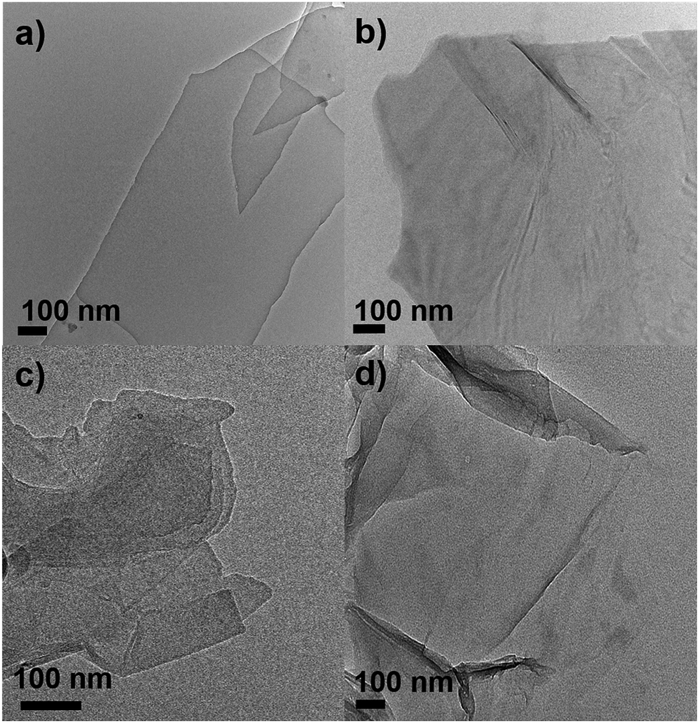 | ||
| Fig. 4 Representative TEM micrographs of (a) FLG–Ce6 in PBS, (b) FLG–Ce6 in DW, (c) GO–Ce6 in PBS, (d) GO–Ce6 in DW. | ||
Nanohybrids were characterized by using Raman spectroscopy (Fig. 5). Despite the strong fluorescence of Ce6, Raman spectra (λext = 532 nm) were recorded. For the nanohybrids, the collection of data was facilitated due to the graphene-induced fluorescence quenching. These results indicate an energy transfer from Ce6 to FLG and GO in agreement with experiments performed with different fluorescence molecules, demonstrating the efficiency of Ce6 in the exfoliation of graphite and graphite oxide.24–26
 | ||
| Fig. 5 Raman spectra (λexc = 532 nm) of (a) FLG–Ce6 and (b) GO–Ce6 nanohybrids in PBS and DW. Free Ce6 (green line) is included as a control. | ||
The Raman spectra were normalized to the intensity of the G band at ∼1580 cm−1. The Raman spectra of FLG–Ce6 exfoliated in PBS shows a symmetric 2D band at ∼2715 cm−1 indicative of FLG (<5 layers). The low intensity of the 2D band could be related to the electron-donor behaviour of Ce6 when hybridized with carbon nanostructures. For graphene, this effect becomes more markedly as the layer number decreases.27 In DW, this band is less intense and symmetric, indicating larger aggregation. The D band at ∼1350 cm−1, related to in-plane defects of graphene edges, absent in the Raman features of pristine graphite, is also observed for FLG–Ce6 in PBS. The ratio of the intensities of the D and G bands (ID/IG) is often used as indication of the quality of the material. For FLG–Ce6, the ID/IG = 0.13 accounts for low-density defect FLG. The strong signal of Ce6 on FLG–Ce6 in DW hinders the D band peak.
The low ID/IG ratio found from Raman spectroscopy is in complete agreement with the TEM images since defects are mainly expected on the graphene edges, larger sheets present low density of defects when compare with smaller sheets.
For GO–Ce6 nanohybrids the presence of a prominent D band at ∼1351 cm−1 corresponds to a disorder in the graphene basal structure owing to the oxidized functional groups, Fig. 5b. The Raman signals of Ce6 are observed in all nanohybrids. The spectral features of Ce6 are reported in S8.†28 Interestingly, the Raman signals of Ce6 present higher intensity in the nanohybrids prepared in DW. This result could indicate higher molecular adsorption due to hydrophobic and hydrogen bonding interactions in complete agreement with concentrations calculated from UV-vis absorption.
Finally, all products were characterized by X-ray photoelectron spectroscopy. This technique allows the direct determination of the surface elemental composition. The C1s core levels recorded for Ce6 powder and nanohybrids in different environments are shown in Fig. 6.
 | ||
| Fig. 6 XPS of C1s of (a) and (b) FLG–Ce6 and (c) and (d) GO–Ce6 nanohybrids in PBS and DW. | ||
The C1s core level of the Ce6 powder is mainly composed by a peak at 284.7 eV due to the carbon atoms in the aromatic rings and in C–H bonds; four minor peaks at higher binding energy are related to CN, OH, C–N, COOH and O–CO bonds, respectively. The nanohybrids still present contribution from the molecules in addition to the graphitic peak at 284.4 eV. The enhancement of the peaks at higher binding energy, in particular the one at ∼286.6 eV indicates the oxidation occurred due to the interaction within the molecule. The N1s core levels recorded for Ce6 powder and nanohybrids are shown in S8.†
Conclusions
Here, we described a facile methodology to produce FLG–Ce6 and GO–Ce6 nanohybrids by the direct exfoliation of graphite and graphite oxide in aqueous media using Ce6 as stabilizing molecule. Ce6 and FLG interactions are based on the synergy of its strong noncovalent π–π interactions, while for GO the oxidized carbon atoms contribute for the hybrid stabilization by the formation of hydrogen bonds. All these interactions are energetically driven by acoustic cavitation validating ultrasonication processes as a robust technique for the production of carbon nanostructures and carbon nanohybrids.29 Ce6 works as a stabilizer against aggregation of graphitic materials producing stable dispersions in aqueous media enhancing its main spectroscopic properties. In particular, FLG–Ce6 might mimics the photosynthetic antenna in green photosynthetic bacteria.30Acknowledgements
This research was supported by CONACYT CB-166014, I-225984, the project 193257 of Bilateral Cooperation Mexico (CONACYT) – Belgium (F.R.S.-FNRS) and PROMEP 9490. Belgian Fund for Scientific Research (FRS-FNRS) under FRFC contract “Chemographene” (2.4577.11) is acknowledged. DHS is grateful to CONACYT for the PhD fellowship 23755. CB is Research Associate of the FRS-FNRS. MS is FRS-FNRS Postdoctoral Researcher. Thanks are given to Drs Aurora Robledo, and MC. Lourdes González-González for technical support.Notes and references
- Y. Shao, J. Wang, H. Wu, J. Liu, I. A. Aksay and Y. Lin, Electroanalysis, 2010, 22, 1027 CrossRef CAS.
- J. Hou, Y. Shao, M. W. Ellis, R. B. Moore and B. Yi, Phys. Chem. Chem. Phys., 2011, 13, 15384 RSC.
- N. Mohanty and V. Berry, Nano Lett., 2008, 8, 4469 CrossRef CAS PubMed.
- M. Quintana, J. I. Tapia and M. P. Beilstein, J. Nanotechnol., 2014, 5, 2328 CAS.
- D. C. Marcano, D. V. Kosynkin, J. M. Berlin, A. Sinitskii, Z. Sun, A. Slesarev, L. B. Alemany, W. Lu and J. M. Tour, ACS Nano, 2010, 4, 4806 CrossRef CAS PubMed.
- M. Quintana, M. Grzelczak, K. Spyrou, B. Kooi, S. Bals, G. Van Tendeloo, P. Rudolf and M. Prato, Chem. Commun., 2012, 48, 12159 RSC.
- V. León, M. Quintana, M. A. Herrero, J. L. Fierro, A. de la Hoz, M. Prato and E. Vázquez, Chem. Commun., 2011, 47, 10936 RSC.
- M. Matsumoto, Y. Saito, C. Park, T. Fukushima and T. Aida, Nat. Chem., 2015, 7, 730 CrossRef CAS PubMed.
- S. Stankovich, D. A. Dikin, G. H. B. Dommett, K. M. Kohlhaas, E. J. Zimney, E. A. Stach, R. D. Piner, S. T. Nguyen and R. S. Ruoff, Nature, 2006, 442, 282 CrossRef CAS PubMed.
- Y. Hernández, V. Nicolosi, M. Lotya, F. M. Blighe, Z. Sun, S. De, I. T. McGovern, B. Holland, M. Byrne, Y. K. Gun'Ko, J. J. Boland, P. Niraj, G. Duesberg, S. Krishnamurthy, R. Goodhue, J. Hutchison, V. Scardaci, A. C. Ferrari and J. N. Coleman, Nat. Nanotechnol., 2008, 3, 563 CrossRef PubMed.
- M. Lotya, Y. Hernandez, P. J. King, R. J. Smith, V. Nicolosi, L. S. Karlsson, F. M. Blighe, S. De, Z. Wang, I. T. McGovern, G. S. Duesberg and J. N. Coleman, J. Am. Chem. Soc., 2009, 131, 3611 CrossRef CAS PubMed.
- X. Wang, P. F. Fulvio, G. A. Baker, G. M. Veith, R. R. Unocic, S. M. Mahurin, M. Chi and S. Dai, Chem. Commun., 2010, 46, 4487 RSC.
- Q. Su, S. Pang, V. Alijani, C. Li, X. Feng and K. Müllen, Adv. Mater., 2009, 21, 3191 CrossRef CAS.
- A. B. Bourlinos, V. Georgakilas, R. Zboril, T. A. Steriotis, A. K. Stubos and C. Trapalis, Solid State Commun., 2009, 149, 2172 CrossRef CAS.
- X. F. Wang, H. Tamiaki, O. K. Ikeuchi and S. Sasaki, J. Power Sources, 2013, 242, 860 CrossRef CAS.
- B. Tian, C. Wang, S. Zhang, L. Feng and Z. Liu, ACS Nano, 2011, 5, 7000 CrossRef CAS PubMed.
- D. Gust, T. A. Moore and A. L. Moore, Acc. Chem. Res., 2001, 34, 40 CrossRef CAS PubMed.
- Y. Liu, C.-Y. Liu and Y. Liu, Appl. Surf. Sci., 2011, 257, 5513 CrossRef CAS.
- M. Quintana, E. Vázquez and M. Prato, Acc. Chem. Res., 2013, 46, 138 CrossRef CAS PubMed.
- A. Agostiano, P. Cosma, M. Trotta, L. Monsù-Scolaro and N. Micali, J. Phys. Chem. B, 2002, 106, 12820–12829 CrossRef CAS.
- G. Yoon, D.-H. Seo, K. Ku, J. Kim, S. Jeon and K. Kang, Chem. Mater., 2015, 27, 2067–2073 CrossRef CAS.
- J. Geng, B. Kong, S. Bo and H. Jung, Chem. Commun., 2010, 46, 5091 RSC.
- C. Weiss, Electronic absorption spectra of chlorophylls, in The Porphyrins, ed. D. Dolphin, Academic Press, New York, 1978, pp. 211–223 Search PubMed.
- S. Sampath, A. N. Basuray, K. J. Hartlieb, T. Aytun, S. I. Stupp and J. F. Stoddart, Adv. Mater., 2013, 25, 2740 CrossRef CAS PubMed.
- J. M. Englert, J. Röhrl, C. D. Schmidt, R. Graupner, M. Hundhausen, F. Hauke and A. Hirsch, Adv. Mater., 2009, 21, 4265 CrossRef CAS.
- N. V. Kozhemyakina, J. M. Englert, G. Yang, E. Spieker, C. D. Schmidt, F. Hauke and A. Hirsch, Adv. Mater., 2010, 22, 5483 CrossRef CAS PubMed.
- A. C. Ferrari, J. C. Meyer, V. Scardaci, C. Casiraghi, M. Lazzeri, F. Mauri, S. Piscanec, D. Jiang, K. S. Novoselov, S. Roth and A. K. Geim, Phys. Rev. Lett., 2006, 97, 187401 CrossRef CAS PubMed.
- S. N. Terekhov, L. L. Gladkov, O. L. Gladkova, M. V. Parkhats, I. A. Khodasevich, A. Y. Panarin and P. Y. Turpin, Opt. Spectrosc., 2009, 106, 813 CrossRef CAS.
- J. I. Tapia, E. Larios, C. Bittencourt, M. J. Yacamán and M. Quintana, Carbon, 2016, 99, 541–546 CrossRef CAS.
- D. Gust, T. A. Moore and A. L. Moore, Acc. Chem. Res., 2009, 42, 1890 CrossRef CAS PubMed.
Footnote |
| † Electronic supplementary information (ESI) available. See DOI: 10.1039/c6ra13501c |
| This journal is © The Royal Society of Chemistry 2016 |