DOI:
10.1039/C6RA13144A
(Paper)
RSC Adv., 2016,
6, 93801-93814
Density functional theory and liquid chromatography-multistage mass spectrometry to characterize raltitrexed photo-degradation mechanisms†
Received
20th May 2016
, Accepted 25th September 2016
First published on 26th September 2016
Abstract
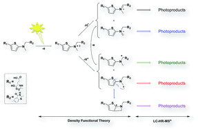
Used in cancer chemotherapy, raltitrexed (RALTI) is an antimetabolite drug acting as thymidylate synthase inhibitor. A photostability study of the antineoplastic drug in aqueous solution and under aerobic conditions is presented. The investigation involved its decomposition under homogenous photolysis making use of the simulated sunlight as exposition source and the identification of the photoproducts formed by high performance liquid chromatography-high-resolution multistage mass spectrometry (LC-HR-MSn). Structural information, combined to density functional theory calculations, has enabled elucidating the main photodegradation pathways of aqueous RALTI. One-electron oxidation was shown mainly responsible for the initiation of RALTI photodegradation in pure water involving specifically the amino thiophenate moiety. In the absence of photocatalysts, electron-transfer seems to mainly proceed through RALTI autoionization, responsible for the formation of RALTI α-amino radicals and RALTI iminium ions, intermediates able to perfectly substantiate the origin formation of most of the photoproducts generated under solar irradiation.
1. Introduction
Raltitrexed or RALTI (N-{5-[3,4-dihydro-2-methyl-4-oxoquinazolin-6-ylmethyl(methyl)amino]-2-thenoyl}-L-glutamic acid) is a thymidylate synthase inhibitor used in intravenous chemotherapy for many kinds of cancers.1–3 The licensed drug, formulated as lyophilizate and packed in clear neutral type I glass vials, is administered by short intravenous, after reconstitution and dilution in large volume solutions, with a period of 15–20 min depending upon the protocol in place. A recent review of literature has revealed very little information on aqueous RALTI stability. At most, a one-time stress test study seems to have unveiled a certain susceptibility to heat and light radiation.4 Such information, however, does not necessarily imply that special precautions are to be observed during the administration process provided that the company that markets the product may have anticipated this issue. As a matter of fact, aqueous RALTI was shown stable for 24 hours in the extent that the storage temperature of less than 25 °C was observed.5 However, in some situations, the ideals are not always maintained, i.e. in-use and/or storage conditions do not necessarily coincide with the control or design spaces, within which, the drug would have been shown stable. As a result, the need to study aqueous RALTI behaviour with respect to less well-controlled storage conditions is still relevant, in that, this is likely to shed light on potential risks to the patient insofar as minor transformation products could be responsible for some adverse effects.6–9
More specifically, because of the well-known susceptibility of RALTI's structural analogues to UV and visible lights, and this, with reference to folate derivatives and methotrexate,10–13 this work was mainly focused on the drug's decomposition under homogenous photolysis. The characterization of the photoproducts formed has relied on LC-HR-MSn studies with the understanding that such an approach has been increasingly and successfully used for structural characterization of degradation products.14–16 From these data, aqueous RALTI photodegradation pathways were worked out and computational studies were additionally carried out to provide deeper insight into the established assumptions.
2. Materials and methods
2.1. Materials and reagents
Raltitrexed monohydrate (purity > 98%) and analytical grade acetonitrile and formic acid came from Sigma Aldrich (St. Quentin Fallavier, France). Aqueous solutions were prepared using ultrapure water produced by the Q-Pod Milli-Q system (Millipore, Molsheim, France).
2.2. Instrumentation and analytical procedures
Analysis was carried out using LC coupled with LTQ-Orbitrap analyser. The chromatographic separations, monitored using an MS analyser, were run on a Dionex UltiMate 3000 HPLC system (Les Ulis, France). Thereof is equipped with a quaternary pump, a vacuum degasser and an autosampler. The controlling software was Chromeleon® software version 6.80 SR11 (Dionex, Les Ulis, France). The selected analytical column was an Curosil™-PFP 5 μm, LC Column 150 × 4.6 mm, maintained at 25 °C. The flow-rate and the injection volume were set at 1 mL min−1 and 100 μL, respectively. However, using a 1/3 T-split has allowed reducing the mobile phase inflow into the mass spectrometer. In order to prevent any co-elution and encompass late eluting analytes, the mobile phase, composed of 0.1% formic acid (solvent A) and acetonitrile (solvent B), was set in gradient mode (0–10 min: 90% A; 10–40 min: 90 → 20% A; 40–45 min: 20 → 90% A). LC-HR-MSn was performed coupling this same LC system to an electrospray-LTQ-Orbitrap Velos Pro system composed of a double linear trap and an orbital trap (Thermo Fisher Scientific, CA, USA). Analyses were carried out in positive ion mode as per the following conditions: the source voltage was set at 3.4 kV, the source and the capillary temperatures were fixed at 300 °C and 350 °C, respectively. Sheath gas and auxiliary gas nitrogen flows were set at 40 and 20 arbitrary units, respectively. S-Lens was set at 60%. 35% CEL (collision energy level) was set for high-resolution fragmentation studies. The mass range was 100–800 m/z. Standard solution was used daily for external mass calibration. The MS data were processed using Xcalibur® software (version 2.2 SP 1.48).
2.3. Photo-transformation studies
Degradation protocol. Stock standard solutions were prepared by dissolving RALTI in ultrapure water so as to obtain a final concentration of 50 μg mL−1. The solution was allocated in 15 mL Pyrex glass vials, hermetically sealed and exposed to light using a xenon test chamber Q-SUN Xe-1 operating in window mode. The light beam, presenting a characteristic spectrum ranging from 300 to 800 nm, was delivered at an intensity of 1.50 W m−2. Aliquots of samples were withdrawn at regular time intervals (30 – 60 – 70 – 80 – 90 – 100 – 110 – 120 – 150 – 180 – 210 and 240 minutes) and the substrate decay was monitored by LC-MS.
Computational approach. The dianion state of RALTI (RALTI-2H) was considered for the computation in view of the observed pH value of the solution, i.e. pH 5.90.All the DFT computations were performed using the Gaussian 09 software package.17 As the B3LYP functional is widely used and exhibits low deviations,18,19 the B3LYP/6-31++G(d,p) level was employed for optimizing the molecules. The optimized structures were validated by frequency analysis and employed for the energy computations.
ES1 (the lowest singlet energy), ET1 (the lowest triplet energy), VIE (Vertical Ionization Energy), VEA (Vertical Electron Affinity) and AEA (Adiabatic Electron Affinity) of 3O2 were computed at the B3LYP/6-31++G(d,p) level. The solvent effect of water was considered by using the integral equation formalism polarized continuum model (IEFPCM) based on the self-consistent-reaction-field (SCRF) method.20 IEFPCM was adopted as it is a nonexpensive computation and was successful in dealing with the electron transfer systems.21–23
ΔG for bimolecular reactions (Fig. 1 and Table S1 (ESI†)) was calculated by eqn (1), where VIE and VEA for excited state molecules were computed by eqn (2) and (3), respectively.
| | |
VIES1 = VIES0 − ES1, VIET1 = VIES0 − ET1
| (2) |
| | |
VEAS1 = VEAS0 − ES1, VEAT1 = VEAS0 − ET1
| (3) |
 |
| | Fig. 1 Electron and energy transfer reactions (I–VIII) of RALTI-2H evaluated by DFT computation. See Table S1 (ESI†) for detailed calculation and evaluating criteria. | |
3. Results and discussion
Photostability of 50 μg mL−1 RALTI in aqueous solution was studied over the spectrum range of 300–800 nm. The photoproducts formed were detected and characterized by LC-HR-MSn studies. Thereafter, the drug photodegradation pathways were proposed based upon experimental and DFT data.
3.1. Degradation of RALTI and detection of photoproducts
The UV-visible absorption spectrum of RALTI in aqueous solution shows abundant absorption from 200 to 350 nm, with 3 characteristic bands at 202.2, 226.7 and 348.7 nm and a shoulder at 270 nm (Fig. S1†). The ability to absorb beyond 300 nm suggests the active substance be photoexcited and potentially photodegraded when exposed to sunlight. As illustrated in Fig. 2b, aqueous RALTI was revealed extremely fragile under light irradiation. Its concentration sharply decreased according to a zero order kinetic model (t1/2 = 1708 h; k = 0.0307 μmol L−1 h−1) with, in parallel, a rapid onset of photoproducts (P-n), subsequently numbered according to their elution order. Their relative retention times (rRTs) and the HR-MSn data (origin, exact mass, accurate mass along with relative errors of photoproducts, and relevant product ions) are gathered in Tables 1 and S2 (ESI†). Fig. 2c shows extracted ion chromatograms obtained by analysis of the samples withdrawn right after 80 min of exposure. Six photoproducts (P-1, P-5, P-6, P-9, P-11 and P-14) were detected only after 30 min exposure-time to the simulated sunlight and this outcome accounts for about 12% w/w RALTI degradation. After 80 min of exposure, seven extra photoproducts were highlighted (P-2, P-3, P-4, P-7, P-8, P-10, P-12 and P-13) counterbalancing 35% w/w RALTI loss. It is worth mentioning that the photoproducts profile was studied at the early stages of photodecomposition, so as not to deviate from what may occur in the in-use conditions.
 |
| | Fig. 2 Analytical data of aqueous RALTI along with (a) chemical structure; (b) photodegradation kinetic; (c) extracted ion chromatograms (XIC) having been exposed to simulated sunlight for 80 min time. | |
Table 1 Relative retention times (rRTs), structures, accurate masses with errors, elemental compositions and MSn and relevant product ions of raltitrexed and photoproducts. Other relevant product ions are provided in Table S2 (ESI)
| Compounds MSn mode |
Best possible elemental formulae |
Accurate mass m/z |
Relative error (ppm) |
Proposed structures |
rRTsa |
| Raltitrexed's retention time: 16.02 min. |
| Raltitrexed |
C21H23N4O6S+ |
459.1330 |
−0.44 |
 |
1 |
| MS2 (459→) |
C21H21N4O5S+ |
441.1233 |
1.36 |
| MS2 (459→) |
C20H23N4O4S+ |
415.1414 |
−4.81 |
| MS2 (459→) |
C16H16N3O3S+ |
330.0920 |
3.94 |
| MS2 (459→) |
C16H14N3O2S+ |
312.0813 |
3.85 |
| MS2 (459→) |
C15H16N3OS+ |
286.1023 |
4.89 |
| MS2 (459→) |
C10H9N2O+ |
173.0707 |
−1.16 |
| MS3 (459 → 312→) |
C15H14N3OS+ |
284.0860 |
2.82 |
| MS3 (459 → 312→) |
C13H11N2OS+ |
243.0592 |
2.06 |
| MS3 (459 → 312→) |
C10H9N2O+ |
173.0707 |
−1.16 |
| MS3 (459 → 312→) |
C6H6NOS+ |
140.0161 |
−2.14 |
| MS3 (459 → 286→) |
C10H9N2O+ |
173.0707 |
−1.16 |
| MS3 (459 → 286→) |
C5H8NS+ |
114.0375 |
2.63 |
| P-1 |
C11H14N3O+ |
204.1132 |
0.49 |
 |
0.167 |
| MS2 (204→) |
C10H9N2O+ |
173.0709 |
0.00 |
| MS2 (204→) |
C8H6NO+ |
132.0446 |
2.27 |
| P-2 |
C10H11N2O2+ |
191.0817 |
1.05 |
 |
0.169 |
| MS2 (191→) |
C10H9N2O+ |
173.0705 |
−2.31 |
| MS2 (191→) |
C9H9N2O+ |
161.0710 |
0.62 |
| MS2 (191→) |
C8H6NO+ |
132.0446 |
2.27 |
| P-3 |
C15H16N3O4+ |
302.1144 |
2.98 |
 |
0.257 |
| MS2 (302→) |
C15H14N3O3+ |
284.1036 |
2.46 |
| MS2 (302→) |
C14H11N2O4+ |
271.0717 |
1.48 |
| MS2 (302→) |
C14H16N3O2+ |
258.1239 |
0.77 |
| MS2 (302→) |
C14H9N2O3+ |
253.0611 |
1.58 |
| MS2 (302→) |
C13H11N2O2+ |
227.0818 |
1.32 |
| MS2 (302→) |
C10H7N2O2+ |
187.0511 |
−4.80 |
| MS2 (302→) |
C9H9N2O+ |
161.0712 |
1.86 |
| MS2 (302→) |
C6H8NO3+ |
142.0501 |
2.11 |
| P-4 |
C14H16N3O4+ |
290.1142 |
2.41 |
 |
0.288 |
| MS2 (290→) |
C13H16N3O2+ |
246.1241 |
1.63 |
| MS2 (290→) |
C10H9N2O2+ |
189.0662 |
2.12 |
| MS2 (290→) |
C10H9N2O+ |
173.0712 |
1.73 |
| P-5 |
C10H9N2O3+ |
205.0604 |
−1.95 |
 |
0.477 |
| MS2 (205→) |
C10H6NO3+ |
188.0348 |
3.19 |
| MS2 (205→) |
C8H6NO3+ |
164.0345 |
1.83 |
| MS2 (205→) |
C9H9N2O+ |
161.0714 |
3.10 |
| MS2 (205→) |
C8H4NO2+ |
146.0231 |
−3.42 |
| P-6 |
C10H9N2O2+ |
189.0654 |
−2.12 |
 |
0.667 |
| MS2 (189→) |
C9H9N2O+ |
161.0714 |
3.10 |
| MS2 (189→) |
C8H6NO2+ |
148.0397 |
2.70 |
| P-7 |
C21H23O8N4S+ |
491.1228 |
−0.61 |
 |
0.692 |
| MS2 (491→) |
C21H21N4O7S+ |
473.1134 |
1.69 |
| MS2 (491→) |
C20H21N4O6S+ |
445.1162 |
−3.15 |
| MS2 (491→) |
C20H19N4O5S+ |
427.1084 |
3.04 |
| MS2 (491→) |
C15H14N3O3S+ |
316.0762 |
3.80 |
| MS2 (491→) |
C11H12N3O+ |
202.0975 |
0.49 |
| MS2 (491→) |
C10H9N2O+ |
173.0706 |
−1.73 |
| P-8 |
C22H23N2O6S+ |
471.1321 |
−0.42 |
 |
0.840 |
| MS2 (471→) |
C17H16N3O3S+ |
342.0918 |
3.22 |
| MS2 (471→) |
C17H14N3O2S+ |
324.0814 |
4.01 |
| MS2 (471→) |
C12H15N2O5S+ |
299.0707 |
3.68 |
| P-9 |
C42H44N8O12S22+ |
458.1250 |
−1.09 |
 |
0.843 |
| MS2 (458→) |
C32H35N6O11S2+ |
743.1833 |
4.57 |
| MS2 (458→) |
C37H37N7O9S22+ |
393.6040 |
−0.41 |
| MS2 (458→) |
C36H37N7O7S22+ |
371.6105 |
3.36 |
| P-10 |
C20H23O5N4S+ |
431.1380 |
−0.93 |
 |
0.848 |
| MS2 (431→) |
C20H21N4O4S+ |
413.1271 |
−1.69 |
| MS2 (431→) |
C20H21N4O5+ |
397.1518 |
2.77 |
| MS2 (431→) |
C16H16O3N3S+ |
330.0914 |
2.42 |
| MS2 (431→) |
C15H16O2N3S+ |
302.0969 |
3.64 |
| P-11 |
C20H21O6N4S+ |
445.1174 |
−0.45 |
 |
0.864 |
| MS2 (445→) |
C20H19N4O5S+ |
427.1083 |
2.81 |
| MS2 (445→) |
C16H14N3O4S+ |
344.0709 |
2.91 |
| MS2 (445→) |
C15H14N3O3S+ |
316.0761 |
3.48 |
| MS2 (445→) |
C11H12N3O+ |
202.0984 |
4.45 |
| P-12 |
C21H23O7N4S+ |
475.1275 |
−1.47 |
 |
0.895 |
| MS2 (475→) |
C19H20N3O7+ |
402.1306 |
2.49 |
| MS2 (475→) |
C16H14N3O3S+ |
328.0760 |
3.05 |
| MS2 (475→) |
C16H12N3O2S+ |
310.0658 |
4.19 |
| MS2 (475→) |
C11H15N2O5S+ |
287.0710 |
4.88 |
| MS2 (475→) |
C11H12N3O2+ |
218.0919 |
−2.29 |
| MS2 (475→) |
C10H9N2O2+ |
189.0667 |
4.76 |
| MS2 (475→) |
C6H6NOS+ |
140.0171 |
4.29 |
| P-13 |
C21H23O7N4S+ |
475.1278 |
−0.84 |
 |
0.958 |
| MS2 (475→) |
C16H14N3O3S+ |
328.0758 |
2.44 |
| MS2 (475→) |
C16H12N3O2S+ |
310.0652 |
2.26 |
| MS2 (475→) |
C11H12N3O2+ |
218.0919 |
−2.29 |
| MS2 (475→) |
C10H9N2O+ |
173.0703 |
−3.47 |
| P-14 |
C20H21N4O5S+ |
429.1222 |
−1.17 |
 |
1.099 |
| MS2 (429→) |
C16H16N3O3S+ |
330.0914 |
2.42 |
| MS2 (429→) |
C16H14N3O2S+ |
312.0803 |
0.64 |
| MS2 (429→) |
C15H16N3OS+ |
286.1003 |
−1.75 |
| MS2 (429→) |
C10H9N2O+ |
173.0704 |
−2.88 |
| P-15 |
C43H46N8O12S22+ |
465.1329 |
−0.82 |
 |
1.542 |
| MS2 (465→) |
C38H36N7O8S2+ |
782.2074 |
1.62 |
| MS2 (465→) |
C33H37N6O11S2+ |
757.1971 |
1.98 |
| MS2 (465→) |
C22H23N4O6S+ |
471.1348 |
3.23 |
| MS2 (465→) |
C10H9N2O+ |
173.0713 |
2.08 |
| MS2 (465→) |
C5H10NO4+ |
148.0610 |
3.85 |
3.2. Origin and structural characterization of photoproducts
Due to the lack of standards for comparison, identification and elucidation of detected photoproducts were based on an in-depth analysis of the data drawn from LC-HR-MSn. A comprehensive study of RALTI fragmentation pattern was carried out, being a key part of the photoproducts identification process.
Comprehensive study of RALTI fragmentation pattern. The fragmentation scheme of RALTI, which has not been studied in detail so far, was determined using ESI/high-resolution multistage mass spectrometry (ESI/HR-MSn). Analyses were carried out in positive ion mode as it provided much richer information. The product ions structures were systematically confirmed through the elemental composition determination based upon accurate mass measurement. These data have been reported in Tables 1 and S2 (ESI†), and the proposed fragmentation pattern for the drug has been built from multistage ESI+/HR-MSn data (Fig. 3 and S2 (ESI†)). However, for the sake of homogeneity in terms of graphical representations, the mass-to-charge values linked to each of the structures presented in Fig. 3–10, have been written in the form of the exact calculated values.
 |
| | Fig. 3 Proposed MSn fragmentation pathways for protonated RALTI, P-1, P-2, P-5, P-6 and P-14. | |
RALTI was detected as protonated [M + H]+ ion (m/z 459). Its HR-MS2 spectrum yields 6 major product ions with m/z of 441, 415, 330, 312, 286 and 173 (Fig. 3). Protonated RALTI can easily lose glutamic acid to afford the base-peak ion at m/z 312. Successive migrations of electron doublets triggered by the presence of an electronic gap, would have resulted in a direct cleavage of the C–N bond, thus forming m/z 173 ion ([M + H − C5H9NO4 − C6H5NOS]+). A more direct path from the protonated drug can also produce the latter by heterolytic cleavage of the C–N bond, likely facilitated by the tertiary amine protonation. Instead of losing C6H5NOS, m/z 312 can also be rearranged via 1-4H transfer and the subsequent migration of the adjacent double bonds would have led to the amid bond cleavage, whose configuration proposed in Fig. 3 has allowed to explain the successive departures of CO (312 → 284) and C2H3N (312 → 243). The presence of m/z 441 and 415 ions would be due to water loss by condensation and to the departure of CO2, respectively. The fragmentation study has also highlighted the formation of a thiophenic acid derivative at m/z 330 ([M + H − C5H7NO3]+), likely due to an internal rearrangement of transamination type, as shown in Fig. 3. Taken as precursor for MS3 studies, thereof ejected CO2 to result m/z 286 ion, which in turn, underwent the same fragmentation process than that was previously described to achieve m/z 173 ion and its complementary part at m/z 114 ([M + H − C5H7NO3 − CO2 − C8H10N2O]+).
The photoproducts representative of 4-oxo, 2-methyl dihydroquinazoline residue (P-1, P-2, P-5 and P-6). Given their mass values, protonated P-6 (m/z 189), P-2 (m/z 191), P-1 (m/z 204) and P-5 (m/z 205) are consistent with elemental formulae C10H9N2O2+, C10H11N2O2+, C11H14N3O+ and C10H9N2O3+, respectively (Table 1, Fig. 3). The MS2 spectra of protonated P-2 and P-1 both exhibit the characteristic ion at m/z 173 ion, consequence of dehydration for the first ion and loss of CH2
NH for the second (Fig. 3). Contrarily to P-6 that possesses two potential protonation sites, the other photoproducts have the nitrogen-imine as sole protonation site, so that paves the way to other fragmentation routes affecting the oxo-methyl-dihydroquinazoline group. Thus, parallel to the processes described in Fig. 3, responsible for the formation of m/z 161 ion ([C10H9N2O2 − CO]+, [C10H11N2O2 − CH2O]+, and [C10H9N2O3 − CO2]+), protonated P-1, P-2 and P-5 all undergo successive departures of NH3 and CH2
C
NH. However, regardless of the photoproduct considered, the presence of m/z 173 and/or m/z 161 ions stipulates that the oxo-methyl-dihydroquinazoline group have remained unchanged. Therefore, based upon these data and taking into account the chemical structure of RALTI as the starting compound, P-6, P-2, P-1 and P-5 can be readily identified as 2-methyl-4-oxo-1,4-dihydroquinazoline-6-carbaldehyde, 6-(hydroxymethyl)-2-methyl-1,4-dihydroquinazolin-4-one, 2-methyl-6-[(methylamino)methyl]-1,4-dihydroquinazolin-4-one and 2-methyl-4-oxo-1,4-dihydroquinazoline-6-carboxylic acid, respectively.
Photoproducts resulting from the amino thiophenate group decomposition (P-3 and P-4). Based upon their mass values, protonated P-3 (m/z 302) and P-4 (m/z 290) are consistent with elemental formulae C15H16N3O4+ and C14H16N3O4+ (Table 1, Fig. 4 and 5).
 |
| | Fig. 4 Proposed MSn fragmentation pathways for protonated P-3. | |
 |
| | Fig. 5 Proposed MSn fragmentation pathways for protonated P-4. | |
Protonated P-3 (m/z 302). MS2 analysis has clearly highlighted the absence of m/z 173 ion (Fig. 4). The ions at m/z 187 and 161 intensively appeared instead, suggesting the oxo-methyl-dihydroquinazoline group be not altered, but no longer linked to α-methylene. Indeed, m/z 187 ion that accounts for the formation of an oxonium ion as a result of N-dealkylation ([C15H16N3O4 − C5H9NO2]+), unambiguously implies that, on the one hand, α-methylene was oxidized into carbonyl and on the other hand, the thiophene-2-carbonyl glutamic acid residue, initially one of the tertiary amine's substituent, was actually replaced by C4H5O2. Parallel to this path, protonated P-3 can also subtract methylamine (302 → 271) then CO2 (271 → 227), or inversely (302 → 258 then 258 → 227), so that reflects the prior cyclization of the lateral chain. This time, its connection to C-carbonyl has made the C-aromatic more electron-deficient and therefore much more prone to H-transfer from a remote sp3 carbon of the lateral chain, thus leading to cyclization in the course of the fragmentation process, as shown in Fig. 4. Subsequently, all other losses detected perfectly corroborate this assumption. All these data seem at any point to fit with a compound like 4-[N-methyl-1-(2-methyl-4-oxo-1,4-dihydroquinazolin-6-yl)formamido]but-3-enoic acid.
Protonated P-4 (m/z 290). Some product ions would be derived from Meisenheimer rearrangement (MR). Indeed, the simultaneous presence of m/z 173 and m/z 189 ions well supports this assumption. While m/z 173 ion is due to heterolytic cleavage of C–N bond, m/z 189 ion would be the result of MR, compatible with the subsequent departure of C4H7NO2, as outlined in Fig. 5. After loss of CO2 (290 → 246), another MR-intermediate of protonated P-4 can be further rearranged through 1-6H transfer to yield a five-member ring and such a configuration is more suitable to afford an isolate departure of methylamine (246 → 215). The additional losses detected under MS3, i.e. the successive departures of CO (215 → 187) and acetylene (187 → 161), have allowed to deduce the nature of the 4th group bound to the nitrogen-N-oxide. Therefore, P-4 can be identified as 1-carboxy-N-methyl-N-[(2-methyl-4-oxo-1,4-dihydroquinazolin-6-yl)methyl]eth-1-enamine oxide or 1-carboxy-N-methyl-N-[(2-methyl-4-oxo-1,4-dihydroquinazolin-6-yl)methyl]eth-1-enamine oxide.
Photoproducts resulting from thiophene oxidation (P-7 and P-11). The mass values of protonated P-7 and P-11 are consistent with elemental formulae C21H23N4O8S+ and C20H21N4O6S+, respectively. Their MSn spectra have several product ions in common along with similar neutral losses (Tables 1 and S2 (ESI†)). Specific S
C
O losses have been highlighted, implying that the chemical environment of the thiophenate moiety was altered. Besides, the elemental compositions of most of the product ions formed provide good clues in favour of the absence of the N-methyl group. At last, their fragmentations give rise to the formation of m/z 173 ion, indicating that the changes inherent in the drug degradation did not occur at its oxo-methyl-dihydroquinazoline part.
Protonated P-7 (m/z 491). Its accurate mass corresponds to a difference of O2 with respect to protonated RALTI. Like protonated RALTI, a 147 Da-mass transition (491 → 344) corresponding to the glutamic acid neutral loss was observed, demonstrating that this part of the drug was not concerned by the degradation path having led to P-7 either. After loss of CO, the ion at m/z 316 was taken as precursor for MS3 studies and the latter is fragmented into m/z 256 ion by O
C
S neutral loss. The premise here is that the carbocation generated as a result of CO departure would have brought about a rearrangement involving α-carboxylate to achieve an intermediate of sulfanyl-methylene-carboxylate type. Such a configuration can indeed promote the departure of O
C
S, as shown in Fig. 6. This kind of loss was found many times over the MSn studies of protonated P-7 and seems to more readily occur if the thiophene group was previously reduced into dihydrothiophene (Fig. 6). Based upon these assumptions, it was possible to find out all product ions detected. As a result, P-7 could be identified as (2S)-2-[(3-carboxy-5-{[(2-methyl-4-oxo-1,4-dihydroquinazolin-6-yl)methyl]amino}-2,5-dihydrothiophen-2-yl)formamido]pentanedioic acid.
 |
| | Fig. 6 Proposed MSn fragmentation pathways for protonated P-7 and P-11. | |
Protonated P-11 (m/z 445). Its accurate mass corresponds to a difference of CH2 with respect to protonated RALTI. It is also a product ion of protonated P-7, subsequent to the decarboxylation process (Fig. 6). Consequently, as a decarboxylated counterpart of P-7, thereof can be identified as 2-{[(1E)-3-carboxyprop-1-en-1-yl]carbamoyl}-5-{[(2-methyl-4-oxo-1,4-dihydroquinazolin-6-yl)methyl]amino}-2,5-dihydrothiophene-3-carboxylic acid.
Hydroxylated photoproducts (P-12 and P-13). The mass values of protonated P-12 and P-13 both are consistent with elemental formula C21H23N4O7S+.
The hydroxylated derivatives (m/z 475). While protonated P-13 is fragmented into m/z 173 by heterolytic cleavage, protonated P-12 fragmentation affords m/z 189 ion as a result of N-dealkylation. This latter scenario undoubtedly reflects the fact that OH is carried by the substituted methylene group of the tertiary amine (Fig. 7), so that allows to identify P-13 as (2S)-2-[(5-{[hydroxy(2-methyl-4-oxo-1,4-dihydroquinazolin-6-yl)methyl](methyl)amino}thiophen-2-yl)formamido]pentanedioic acid. As to P-12, its fragmentation can bring about the departure of a 257 Da-moiety (–C10H11NO5S) to yield m/z 218 ion, proving that OH is rather carried by C-methylamine (Fig. 7). Therefore, P-12 can be named as (2S)-2-({5-[(hydroxymethyl)[(2-methyl-4-oxo-1,4-dihydroquinazolin-6-yl)methyl]amino]thiophen-2-yl}formamido)pentanedioic acid.
 |
| | Fig. 7 Proposed MSn fragmentation pathways for protonated P-12 and P-13. | |
Photoproduct arising from the glutamic acid decarboxylation (P-10 and P-14).
Protonated P-10 (m/z 431). Its mass value is consistent with C20H23N4O5S+. MS2 studies have shown that P-10 could be an N-oxide derivative in view of the simultaneous presence of m/z 173 and m/z 189 ions. The ion at m/z 189 would be generated subsequently to Meisenheimer rearrangement (Fig. 8). Under MS2 conditions, the usual loss of CO2 does not occur, so that implies a change at the glutamate residue. After cleavage of the peptide bond, an aldehyde derivative (m/z 330) is found instead of the aforementioned oxonium ions. This new scenario would be due to a change in the environment close to that amid function. That's why, P-10 can be proposed as N-{5-[(3-carboxypropyl)carbamoyl]thiophen-2-yl}-N-methyl-1-(2-methyl-4-oxo-1,4-dihydroquinazolin-6-yl)methanamine oxide.
 |
| | Fig. 8 Proposed MSn fragmentation pathways for protonated P-10. | |
Protonated P-14 (m/z 429). C20H21N4O5S+ elemental formula is in accordance with the mass value found. With respect to protonated RALTI, the mass gap corresponds to –CH2O. All like protonated RALTI, m/z 312 ion was found in the MS2 spectrum so that implies that the changes would have occurred at the glutamate side chain (Fig. 3). As a result, P-14 could be 4-[(5-{methyl[(2-methyl-4-oxo-1,4-dihydroquinazolin-6-yl)methyl]amino}thiophen-2-yl)formamido]-4-oxobutanoic acid.
The doubly charged combination photoproducts (P-9 and P-15). The processes by which their ionisation/fragmentation occur lead to the appearance of product ions featuring higher mass-to-charge values, implying these photoproducts be detected as doubly charged ions (z = 2). Therefore, the mass-to-charge values of doubly charged P-9 (m/z 458) and P-15 (m/z 465) are consistent with elemental formulae C42H44N8O12S22+ and C43H46N8O12S22+, respectively.
Doubly protonated P-9 (m/z 458). Its elemental composition introduces the possibility for RALTI to dimerize. Its dissociation induced by collision leads to the formation of two complementary ions, m/z 173 and m/z 743 ions (Fig. 9). Taken as precursor for MS3 studies, m/z 743 ion losses the well-known 147 Da-moiety to yield m/z 596 ion, which in turn, gets rid of a 282 Da-moiety corresponding to C11H14N2O5S by N-dealkylation (596 → 310). These combos are perfectly consistent with the assumption as per which, a bond was formed between C-methylamine of molecule 1 RALTI and C-substituted α-methylene of the tertiary amine of molecule 2 RALTI. Other paths also support this postulate. For instance, as a result of internal transamination (–C5H7NO3, 129 Da) and loss of CO2, the doubly charged product ion at m/z 371 can undergo radical cleavage to afford m/z 285 radical ion. Consequently, P-9 can be identified as (5-((2-((5-((1,3-dicarboxypropyl)carbamoyl)thiophen-2-yl)((2-methyl-4-oxo-3,4-dihydroquinazolin-6-yl)methyl)amino)-1-(2-methyl-4-oxo-3,4-dihydroquinazolin-6-yl)ethyl)(methyl)amino)thiophene-2-carbonyl)glutamic acid.
 |
| | Fig. 9 Proposed MSn fragmentation pathways for protonated P-9. | |
Doubly protonated P-15 (m/z 465). Its elemental composition reflects the possibility of combination between RALTI molecule and its methylene-counterpart. It is worth noting that protonated RALTI is an intense product ion of the doubly charged P-15 (Tables 1 and S2 (ESI†)). Thus, the drug would in all likelihood be linked to the other part through its tertiary amine, thus achieving a compound of quaternary ammonium type (Fig. 10). The complementary product ion to protonated RALTI is represented by m/z 471 ion ([P-15 + H-raltrexed]+). Its oxo-methyl-dihydroquinazoline part is not involved in the formation of other bonds all as that belonging to RALTI residue. Indeed, Fig. 10 shows that oxo-methyl-dihydroquinazoline residue can be removed from one or another branch of doubly charged P-15 to result two ions at m/z 757. Taken in undifferentiated way for MS3 studies, one releases protonated RALTI (757 → 459) and the other m/z 471 ion (757 → 471). The ion at m/z 471 can also lose glutamic acid to achieve m/z 324. Processing that way by elimination, one can easily assume that P-15 be 5-[(1,3-dicarboxypropyl)carbamoyl]-N-[1-({5-[(1,3-dicarboxypropyl)carbamoyl]thiophen-2-yl}[(2-methyl-4-oxo-3,4-dihydroquinazolin-7-yl)methyl]amino)ethyl]-N-methyl-N-[(2-methyl-4-oxo-3,4-dihydroquinazolin-6-yl)methyl]thiophen-2-aminium, having taken into account the most plausible photodegradation path among the possible.
 |
| | Fig. 10 Proposed MSn fragmentation pathways for protonated P-15. | |
3.3. Proposed photo-degradation pathways of RALTI in aqueous solution
Subjected to the aforementioned conditions, the molecule was degraded and the formation of a great variety of products was observed, suggesting that photodegradation proceed as per complex mechanisms. From the experimental viewpoint, given the nature of the photoproducts identified, it seems manifest that most of them stem from the amino thiophenate group transformation (Table 1) implying the radical cationic amine.24,25 Even linked to a chromophore (e.g., conjugated π-bond systems), the amino thiophenate moiety does not absorb light efficiently in the region of 300–800 nm (Fig. S1 (ESI†)), making any direct ionisation process involving this part of the molecule unlikely. Therefore, in the absence of photocatalysts capable of initializing electron-transfer reactions with amines, the mechanism of autoionization was computationally investigated considering interactions between photoexcited RALTI and RALTIS0 and between two photoexcited RALTIs (Fig. 1 and Table S1 (ESI†)).
Photoexcited RALTI may engage in single-electron transfer with RALTIS0, being more oxidizing.26 The spontaneity of this reaction was assessed by the lowest singlet (ES1) and triplet (ET1) excitation energies of the drug. Electron donating ability (EDA) of RALTIS0 and electron accepting ability (EAA) of RALTIS1/T1 were evaluated via vertical ionization energy (VIE) and vertical electron affinity (VEA), respectively. A low VIE value implies strong EDA and a low VEA one means strong EAA. The electron transfer reaction was determined by the overall change in the Gibbs free energy (ΔG), in that, a negative value of ΔG implies the reaction be spontaneous. In view of such considerations and judging from the DFT data (Tables 2, 3 and S1 (ESI†), Fig. 11), one can infer that the given process be not spontaneous (ΔG2 and ΔG3 > 0).
Table 2 Computed values for the excitation energy, vertical ionization energy and vertical electron affinity of RALTI-2H (eV)
| ES1 |
ET1 |
ES1 − ET1 |
VIES0 |
VEAS0 |
VIES1 |
VEAS1 |
VIET1 |
VEAT1 |
| 3.183 |
2.626 |
0.557 |
5.232 |
−1.817 |
2.049 |
−5.000 |
2.606 |
−4.443 |
Table 3 Computed Gibbs free energy ΔG changes of the electron transfer reactions (eV)
| Excited state |
ΔG1 |
ΔG2 |
ΔG3 |
ΔG4 |
ΔG5 |
| S1 |
−1.631 |
0.232 |
0.232 |
−2.951 |
−1.863 |
| T1 |
−1.074 |
0.789 |
0.789 |
−1.837 |
−1.863 |
 |
| | Fig. 11 Photodegradation pathways of aqueous RALTI under aerobic condition. | |
Transfer between two RALTIS1/T1 resulting in the generation of RALTI+˙ and RALTI−˙ was also investigated using the same approach. The corresponding computed values show that the reaction is actually possible (Tables 2, 3 and S1 (ESI†), Fig. 11).
Beyond these aspects, the role of oxygen, often described as leading in photoxidation reactions, was also regarded. As shown in Tables 2, 3 and S1 (ESI†), Fig. 11, RALTI−˙ may spontaneously transfer electron to 3O2 to afford superoxide radical ion given the fact that ΔG5 < 0. As O2−˙ is both a radical and an anion, it has the potential for dual reactivity with respect to RALTI+˙. As depicted in Fig. 11, it can react as a base by abstracting proton from RALTI+˙ to result RALTI α-amino radicals (A) and (B), along with acid conjugate HOO˙.27–31 Subsequent combination of these radicals gives rise to the formation of reactive RALTI peroxides, precursors altogether capable of generating photoproducts P-3, P-4, P-5, P-6, P-12 and P-13. As to the formation of P-1, P-2 and P-7, an extra step represented by radical rearrangement of structure (B) to achieve dihydrothiophene radical (C) was proposed. Also as a radical, O2−˙ can abstract hydrogen from RALTI+˙ to generate RALTI iminium ions (D) and (E), along with HOO− (Fig. 11).32 Parallel to this, these latter can likewise derive from electron-transfer between RALTIT1 and RALTI α-amino radicals (Tables 2, 3 and S1 (ESI†)). Because α-amino radicals are strongly reducing, second one-electron oxidation is thus facile.33 Iminium ions, as excellent electrophiles, are by nature amenable to interception by a variety of nucleophiles to directly install a new bond at the position α to the nitrogen atom.34,35 As a result, P-8 and P-9 are in all likelihood the products of this kind of reaction involving (RALTI-H)−, formed as a result of RALTI deprotonation in the presence of HOO− (Fig. 11). In addition to electron-transfer, the process involving the energy-transfer between RALTIS1/T1 and 3O2, responsible for the generation of another reactive oxygen species 1O2 playing substantial role in photoxidation reactions was also investigated and looked as possible according to DFT results (Tables 2, 3 and S1 (ESI†)).
For the formation of P-15, even still not clear, a possible nucleophilic attack of RALTI+˙ by P-8 was postulated, likely affording an intermediate of distonic radical cation type ([RALTI-P-8]+˙). In the wake of hydrogen abstraction, P-15 would have been formed. Eventually, as already described with methotrexate and folic acid, the glutamate residue can undergo decarboxylation, thus giving rise thereafter to the onset of P10 and P-14. Beyond this statement, unlike what has been reported for folic acid and methotrexate,12,13 the N–C bond cleavage between aromatic amine and α-amine methylene did not occur. The difference may be imputed to the wavelength range used in this study, not comprising the more energetic UVB field.
4. Conclusion
The study of RALTI transformations through photolysis was investigated. RALTI underwent degradation when exposed to light with a spectrum range of 300–800 nm. LC-HR-MSn permitted to identify fifteen photoproducts. A full picture of the photo-degradation of RALTI was investigated combining DFT and experimental data and semi-empirical expressions were obtained and successfully used to model both processes. It was shown that formation of most of the photoproducts generated at the first periods of exposure could result from one-electron oxidation process involving the amino thiophenate moiety and energy-transfer. As a result, formulation with good scavengers of free radicals, like glycerol, mannitol and/or ascorbic acid could be considered to prevent the product from degradation. Understanding the main photo-degradation routes is a good basis to work out efficient measures so as to mitigate or avoid RALTI photo-instability.
Acknowledgements
The authors want to thank H. Al Salloum, A. Solgadi and K. Manerlax for their very appreciable contribution to this work. They are also thankful to Ile de France Region for financial support in the acquisition of the Orbitrap Velos Pro mass spectrometer.
References
- S. Barni, A. Ghidini, A. Coinu, K. Borgonovo and F. Petrelli, Anticancer Drugs, 2014, 25(10), 1122–1128 CrossRef CAS PubMed.
- X. Y. Li, L. Liu, X. M. Xie and C. Zhou, Eur. Rev. Med. Pharmacol. Sci., 2014, 18(22), 3491–3496 Search PubMed.
- P. Zhao, Z. Ding, L. Tang and X. Zhou, World J. Surg. Oncol., 2014, 12, 403 CrossRef PubMed.
- J. JU, Study on Stability of Raltitrexed for Injection, China Pharm., 2013, 37, 63–65 Search PubMed.
- Hospira Benelux BVBA, AMM BE177825, Noorderplaats 9 2000 Antwerpen, België. Approbation 06/2015.
- K. Butzbach and B. Epe, Free Radical Biol. Med., 2013, 65, 821–827 CrossRef CAS PubMed.
- M. R. Zaheer, A. Gupta, J. Iqbal, Q. Zia, A. Ahmad, O. M. Roohi, A. Hashlamon, S. H. Mohd Setapar, G. M. Ashraf and G. Aliev, Curr. Pharm. Des., 2016, 22(7), 768–782 CrossRef CAS PubMed.
- D. Hart, M. Ward and M. D. Lifschitz, Intern. Med., 1987, 106, 235–238 CAS.
- M. L. Dantola, M. P. Denofrio, B. Zurbano, C. S. Gimenez, P. R. Ogilby, C. Lorente and A. H. Thomas, Photochem. Photobiol. Sci., 2010, 9, 1604–1612 CAS.
- T. Fukuwatari, M. Fujita and K. Shibata, Biosci., Biotechnol., Biochem., 2009, 73, 322–327 CrossRef CAS PubMed.
- A. Juzeniene, M. Grigalavicius, L. W. Ma and M. Juraleviciute, J. Photochem. Photobiol., B, 2016, 155, 116–121, DOI:10.1016/j.jphotobiol.2016.01.001.
- P. Calza, C. Medana, M. Sarro, V. Rosato, R. Aigotti, C. Baiocchi and C. Minero, J. Chromatogr. A, 2014, 1362, 135–144 CrossRef CAS PubMed.
- M. K. Off, A. E. Steindal, A. C. Porojnicu, A. Juzeniene, A. Vorobey, A. Johnsson and J. Moan, J. Photochem. Photobiol., B, 2005, 80(1), 47–55 CrossRef CAS PubMed.
- H. Sadou Yaye, P. H. Secretan, T. Henriet, M. Bernard, F. Amrani, W. Akrout, P. Tilleul, N. Yagoubi and B. Do, J. Pharm. Biomed. Anal., 2015, 105, 74–83 CrossRef PubMed.
- T. Henriet, P. H. Secrétan, F. Amrani, H. Sadou-Yayé, M. Bernard, A. Solgadi, N. Yagoubi and B. Do, New J. Chem., 2016, 40, 1182–1194 RSC.
- P. H. Secrétan, H. Sadou-Yayé, C. Aymes-Chodur, M. Bernard, A. Solgadi, N. Yagoubi and B. Do, RSC Adv., 2015, 5, 35586–35597 RSC.
- M. J. Frisch, et al., Gaussian 09 Revision D.01, Gaussian Inc., Wallingford, CT, 2009 Search PubMed.
- M. R. Silva-Junior, M. Schreiber, S. P. A. Sauer and W. J. Thiel, Chem. Phys., 2008, 129(10), 104103 Search PubMed.
- D. Jacquemin, E. A. Perpète, I. Ciofini and C. Adamo, J. Chem. Theory Comput., 2010, 6(5), 1532–1537 CrossRef CAS PubMed.
- J. Tomasi, B. Mennucci and R. Cammi, Chem. Rev., 2005, 105(8), 2999–3093 CrossRef CAS PubMed.
- J. Blotevogel, T. Borch, Y. Desyaterik, A. N. Mayeno and T. C. Sale, Environ. Sci. Technol., 2010, 44(15), 5868–5874 CrossRef CAS PubMed.
- A. L. Kaledin, Z. Q. Huang, Q. S. Yin, E. L. Dunphy, E. C. Constable, C. E. Housecroft, Y. V. Geletii, T. Q. Lian, C. L. Hill and D. G. Musaev, J. Phys. Chem. A, 2010, 114(21), 6284–6297 CrossRef CAS PubMed.
- M. Namazian, H. A. Almodarresieh, M. R. Noorbala and H. R. Zare, Chem. Phys. Lett., 2004, 396(4–6), 424–428 CrossRef CAS.
- L. Shi and W. Xia, Chem. Soc. Rev., 2012, 41, 7687–7697 RSC.
- S. Maity and N. Zheng, Synlett, 2012, 23, 1851–1856 CrossRef CAS PubMed.
- J. Hu, J. Wang, T. H. Nguyen and N. Zheng, Beilstein J. Org. Chem., 2013, 9, 1977–2001 CrossRef PubMed.
- S. H. Mashraqui and R. M. Kellog, Tetrahedron Lett., 1985, 26, 1453–1456 CrossRef CAS.
- S. Fukuzumi, S. Mochizuki and T. Tanaka, J. Phys. Chem., 1990, 94, 722–726 CrossRef CAS.
- I. Willner, T. Tsfania and Y. Eichen, J. Org. Chem., 1990, 55, 2656–2662 CrossRef CAS.
- J. M. R. Narayanam, J. W. Tucker and C. R. J. Stephenson, J. Am. Chem. Soc., 2009, 131, 8756–8757 CrossRef CAS PubMed.
- J. D. Nguyen, E. M. D'Amato, J. M. R. Narayanam and C. R. J. Stephenson, Nat. Chem., 2012, 4, 854–859 CrossRef CAS PubMed.
- J. M. Fukuto, E. W. Di Stefano, J. N. Burstyn, J. S. Valentine and A. K. Cho, Biochemistry, 1984, 24, 4161–4167 CrossRef.
- P. J. DeLaive, J. T. Lee, H. W. Springtschnik, H. Abruña, T. J. Meyer and D. G. Whitten, J. Am. Chem. Soc., 1977, 99, 7094–7097 CrossRef CAS.
- J. P. Dinnocenzo and T. E. Banach, J. Am. Chem. Soc., 1989, 111, 8646–8653 CrossRef CAS.
- X. Zhang, S.-R. Yeh, S. Hong, M. Freccero, A. Albini, D. E. Falvey and P. S. Mariano, J. Am. Chem. Soc., 1994, 116, 4211–4220 CrossRef CAS.
Footnotes |
| † Electronic supplementary information (ESI) available. See DOI: 10.1039/c6ra13144a |
| ‡ The first 2 authors contributed equally to this study and are therefore considered as first authors. |
|
| This journal is © The Royal Society of Chemistry 2016 |
Click here to see how this site uses Cookies. View our privacy policy here.