DOI:
10.1039/C6RA12861K
(Paper)
RSC Adv., 2016,
6, 80193-80200
WO3 nanorod photocatalysts decorated with few-layer g-C3N4 nanosheets: controllable synthesis and photocatalytic mechanism research†
Received
18th May 2016
, Accepted 1st August 2016
First published on 1st August 2016
Abstract
Novel WO3 nanorod (WO3 NRs) photocatalysts decorated with few-layer g-C3N4 nanosheets (flg-C3N4) have been synthesized by a typical hydrothermal process. The structure, morphology, optical and electronic properties were researched by comprehensive characterization methods. The photocatalytic performance of flg-C3N4/WO3 NRs nanocomposites was assessed by degrading Rhodamine B (RhB) dye under visible light irradiation. When the loading amount of the flg-C3N4 is 20 wt%, the flg-C3N4/WO3 NRs nanocomposites exhibit the highest activity and the Chemical Oxygen Demand (COD) values decreased by about 64%. The improved light harvesting ability and higher separation efficiency of photoinduced electron–hole pairs by the decoration of flg-C3N4 lead to the boosting photocatalytic performance. In addition, through ESR analysis and a trapping experiment, the photocatalytic mechanism was also researched.
1. Introduction
Energy and environmental problems have prevented the sustainable development of social-economy; therefore, numerous researchers are continuously seeking advanced technologies to solve these issues.1 Photocatalysis, one of the most promising technologies, can directly harness solar energy, for the degradation of environmental pollutants,2 hydrogen evolution,3 and CO2 photocatalytic reduction.4 To date, several photocatalytic nanomaterials, such as Fe2O3 and ZnO, have been researched and exhibited promising photocatalytic activity.5,6 Although these materials are widely applied for photocatalytic applications, some of them are only active under ultraviolet irradiation,7 whereas solar power is only composed of 4% ultraviolet light.8 Therefore, there are more prospects for designing visible-light-driven catalysts to meet the need of industrial applications.
Considerable efforts have shown that WO3, as one of traditional n-type semiconductors, is a visible light responsive photocatalyst with a relatively narrow band gap energy (2.4–2.8 eV) and suitable valence band (VB) potential, which is similar to that of TiO2.9 Nevertheless, since the conduction band (CB) position of WO3 is lower than the oxygen reduction process (O2 + e− → ˙O2− (aq), −0.33 V vs. NHE),10 the transferring electrons in WO3 CB cannot be utilized sufficiently, making it easy for the accumulative photogenerated electrons to recombine with holes, which results in the low photocatalytic performance of the WO3 photocatalyst.11,12 Therefore, several prevailing strategies have been applied to boost the photocatalytic performance of WO3. Firstly, preparing materials with specific structures such as nanosheets,13 nanowires14 and nanorods.15 Secondly, introducing oxygen vacancies in WO3 material.16,17 Thirdly, coupling with different materials to build hybrid nanomaterials such as CaFe2O4/WO3,18 Pd/WO3,19 Cu/WO3,20 and WO3/g-C3N4.21 Thus, designing WO3 with a specific morphology or fabricating nanocomposites could significantly improve the photocatalytic activity of WO3.
In the fields of photocatalysis, some nanomaterials with a graphene-like structure have been researched.22,23 For example, SnS2 nanosheets have been used as co-catalysts to improve the removal efficiency of pollutants.24 Liu et al. rooted few-layer MoS2 nanosheets into TiO2 nanofibers for highly efficient photocatalytic hydrogen evolution.25 Likewise, J. Choi et al. modified BN nanosheets on the surface of AgI to improve photocatalytic activity.26 In 2009, Wang et al. showed the photocatalytic water splitting via graphitic-like carbon nitride, and such metal-free material has met extensive concern in the photocatalysis fields.27,28 The bulk g-C3N4 nanomaterial possesses a layered structure and the π-conjugated s-triazine unit is formed by the sp2 hybridization of the carbon and nitrogen, resulting in high structural stability of g-C3N4.27,29 As a two-dimensional graphene-like nanostructure, the few-layer g-C3N4 nanosheets (flg-C3N4) show more superior physical and chemical properties such as the larger surface area, exposure of numerous active sites and optimized light harvesting, which may be favorable for promoting photocatalytic performance. Therefore, flg-C3N4 has been applied to enhance the photocatalytic performance of different semiconductors in recent research studies such as Ag2CO3/flg-C3N4,30 Bi2WO6/flg-C3N4 (ref. 31) and Au/flg-C3N4.32 However, to the best of our knowledge, using flg-C3N4 to decorate WO3 nanorods (WO3 NRs) to form composite photocatalysts has never been reported.
Herein, we demonstrate a controllable hydrothermal method to prepare flg-C3N4/WO3 NRs nanocomposites with excellent catalytic performance. The stability of the photocatalyst was evaluated by four consecutive catalytic runs. The photocatalytic mechanism was also studied in detail.
2. Experimental
2.1 Synthesis of WO3 NRs
WO3 NRs were prepared by a facile hydrothermal method.10 Firstly, 2.112 g Na2WO4·2H2O and 1.87 g NaCl were mixed fully in 50 mL water. The solution pH was adjusted by 3.0 mol L−1 HCl and stirred continuously for 4 h. Afterwards, the solution was placed into a Teflon-lined autoclave and heated at 180 °C for 24 h. Thereafter, the obtained products were washed and separated. The products were dried at 70 °C.
2.2 Synthesis of the flg-C3N4
Bulk g-C3N4 was synthesized by calcining melamine molecules at high temperature. Melamine was heated at 550 °C with 2 °C min−1 for 4 h. Flg-C3N4 was synthesized by calcining as-prepared bulk g-C3N4 at 550 °C for 100 min with 2 °C min−1.
2.3 Synthesis of the flg-C3N4/WO3 NRs
0.05 g WO3 NRs was placed in 15 mL of DI water, which includes a certain amount of as-prepared flg-C3N4, and then the above mixture was ultrasonicated for 20 min and further stirred for 30 min. Then, the mixed solution was added in a 25 mL Teflon-lined autoclave and heated at 150 °C for 4 h. Thereafter, the final WO3 NRs with various flg-C3N4 contents were obtained and dried at 70 °C. A schematic is shown in Scheme 1.
 |
| | Scheme 1 Schematic for the preparation of the WO3 NRs, the flg-C3N4 and the flg-C3N4/WO3 NRs nanocomposites. | |
3. Results and discussion
3.1 XRD analysis
The XRD spectra of WO3 NRs, flg-C3N4 and flg-C3N4/WO3 NRs are displayed in Fig. 1. The flg-C3N4/WO3 NRs nanocomposites with different contents of flg-C3N4 exhibited a similar pattern compared with WO3 NRs. The XRD patterns of the nanocomposites matched perfectly with the JCPDS card of WO3 (no. 33-1387). The diffraction peaks appearing at 14.0°, 22.8°, 24.2°, 26.9°, 28.3°, 33.5°, 36.6°, 49.7° and 55.7° are indexed to the (100), (001), (110), (101), (200), (111), (201), (220), and (221) crystal planes of WO3,21 respectively. After introducing flg-C3N4, the intensity of the diffraction peak for the (100) crystal planes almost disappeared and the peak for the (002) crystal planes dramatically decreased compared with the reports of previous researchers,22,33 manifesting that the flg-C3N4 structure might be composed of few-layer s-triazine unit layers.34 In addition, for the nanocomposites, the diffraction peak of flg-C3N4 for the (002) crystal planes might overlap with the peak of WO3 NRs for (101), and the peak for (100) could not be discovered, which might be attributed to the low contents of flg-C3N4.
 |
| | Fig. 1 XRD spectra of the as-prepared WO3 NRs, flg-C3N4/WO3 NRs and flg-C3N4. | |
3.2 XPS analysis
The surface elements and chemical states of WO3 NRs and flg-C3N4/WO3 NRs nanocomposites were further investigated by the XPS method. Fig. 2A shows the XPS survey spectrum of WO3 NRs and 20 wt% flg-C3N4/WO3 NRs nanocomposites, revealing the presence of C, N, O, and W in the nanocomposites. Fig. 2B–E shows the high resolution spectra of W 4f, O 1s, C 1s, and N 1s. As depicted in Fig. 2B, the peaks were discovered at 35.6 eV and 37.7 eV, respectively, which are consistent with the typical binding energies of W 4f7/2 and W 4f5/2 of W6+.10 The O 1s peak (Fig. 2C) at 530.4 eV was attributed to the O2− in the WO3 NRs. The characteristic peaks of C 1s (Fig. 2D) were observed at binding energies 284.7 eV and 288.6 eV. The peak at 284.4 eV could be ascribed to carbon bonds and the peak observed at 288.6 eV was assigned to defect-containing sp2-bonded carbon (N–C![[double bond, length as m-dash]](https://www.rsc.org/images/entities/char_e001.gif) N).35 The peak of N 1s (Fig. 2E) was observed at 399.0 eV. All these XPS results are further confirmed by the FT-IR analysis.
N).35 The peak of N 1s (Fig. 2E) was observed at 399.0 eV. All these XPS results are further confirmed by the FT-IR analysis.
 |
| | Fig. 2 (A) XPS survey spectra of WO3 NRs, 20 wt% flg-C3N4/WO3 NRs. High resolution spectra of (B) W 4f and (C) O 1s peak of the WO3 NRs and 20 wt% flg-C3N4/WO3 NRs, (D) C 1s and (E) N 1s peak of 20 wt% flg-C3N4/WO3. | |
3.3 FT-IR analysis
FT-IR spectra are a robust tool to investigate chemical functional groups of the nanomaterials. Fig. 3 shows the FT-IR spectra of WO3 NRs, flg-C3N4 and flg-C3N4/WO3 NRs nanocomposites with different contents. The broad stretching vibrations observed at 3100–3500 cm−1 were attributed to O–H and N–H bands, which was relative to surface absorbed water molecules and uncondensed amino groups.36 Moreover, a series of absorption peaks observed between 1200 cm−1 and 1650 cm−1 were due to characteristic absorption peaks of the CN heterocycles.37 The peak appearing at 810 cm−1 was attributed to the stretching peaks of s-triazine units.38 Furthermore, with increased flg-C3N4 contents in the nanocomposites, the absorption peak positions at 828 cm−1 ascribed to the vibrations of O–W–O bands slightly shifted,39 which demonstrated the existence of the strong interaction between WO3 NRs and s-triazine units of flg-C3N4 as well. Therefore, the FT-IR results provided further evidence that WO3 NRs were decorated with flg-C3N4 successfully and an interaction existed between WO3 NRs and flg-C3N4.
 |
| | Fig. 3 FT-IR of the WO3 NRs and flg-C3N4/WO3 NRs nanocomposites and flg-C3N4. | |
3.4 SEM and TEM analysis
The morphology and superficial microstructure of flg-C3N4/WO3 NRs nanocomposites were studied by SEM and TEM. Fig. 4A and B show the SEM images of WO3 NRs and flg-C3N4/WO3 NRs (20 wt%). As revealed in Fig. 4A, it could be clearly observed that rod-like WO3 was successfully synthesized. As revealed in Fig. 4B, WO3 NRs were decorated by flg-C3N4 on the surface, which revealed that flg-C3N4 and WO3 NRs were successfully coupled. In the EDS pattern, C, N, W and O could be observed (Fig. 4C), which further proved that flg-C3N4/WO3 NRs nanocomposites were synthesized without other impurities. Fig. 4D–G shows the TEM images of WO3 NRs and flg-C3N4/WO3 NRs (20 wt%). As shown in Fig. 4D and E, WO3 NRs displayed the inerratic rod-like morphology with the crystal width of around 20 nm. As shown in Fig. 4F and G, the WO3 NRs are well dispersed on the surface of the flg-C3N4 and intimately contacted between WO3 NRs and flg-C3N4, which is favorable to enhance the transfer of photogenerated electrons.34 The thickness of flg-C3N4 was about 5.10 nm (Fig. S2†), and the representative thickness of the g-C3N4 monolayers as suggested by our previous report40 was ∼0.64 nm. Therefore, the as-synthesized flg-C3N4 consisted of ∼8 monolayers. Fig. 4F and G also showed the wrinkled morphology and relatively large surface of flg-C3N4, which shows that flg-C3N4 could increase the specific surface area of WO3 NRs and provide more active reaction sites.
 |
| | Fig. 4 (A) SEM image of WO3 NRs; (B) SEM image of WO3 NRs, 20 wt% flg-C3N4/WO3 NRs; (C) EDS of the flg-C3N4/WO3 NRs; (D and E) TEM images of WO3 NRs; (F and G) TEM images of 20 wt% flg-C3N4/WO3 NRs. | |
3.5 DRS analysis
The UV-Vis diffuse reflectance spectra (DRS) of WO3 NRs and flg-C3N4/WO3 NRs nanocomposites are displayed in Fig. 5A. By comparing with the WO3 NRs, the optical absorption of flg-C3N4/WO3 NRs in the range of 450–600 nm was apparently enhanced and the absorption edge showed a slight red-shift. The results indicated that the photocatalysts could increase the absorption of visible light, which would be beneficial for their photocatalytic performance. The energy gap of WO3 NRs and flg-C3N4 was estimated via the classical Tauc approach, (αhν)2 = A(hν − Eg)n, as revealed in Fig. 5B, the energy gap (Eg) of WO3 NRs was estimated to be 2.70 eV and Eg of flg-C3N4 was 2.80 eV.
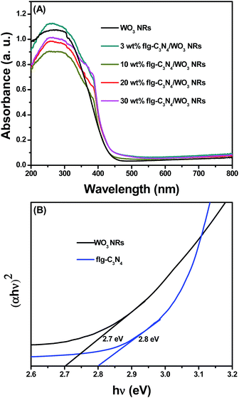 |
| | Fig. 5 (A) The UV-Vis absorption spectra of the as prepared WO3 NRs and the flg-C3N4/WO3 NRs nanocomposites with different contents of flg-C3N4 (B) estimated band gap curves for the as prepared WO3 NRs and the flg-C3N4. | |
3.6 Nitrogen adsorption analysis
The Brunauer–Emmett–Teller (BET) surface areas of WO3 NRs and 20 wt% flg-C3N4/WO3 NRs have also been analyzed. From Fig. 6, the specific surface area of the 20 wt% flg-C3N4/WO3 NRs was 40.6 m2 g−1, which was about 1.7 times as high as that of the WO3 NRs (24.3 m2 g−1), showing that the decoration of the flg-C3N4 could further increase the specific surface areas of the WO3 NRs. The higher surface area values implied that there are more adsorption and active sites for the photocatalytic reaction.
 |
| | Fig. 6 Nitrogen absorption–desorption isotherm of WO3 NRs and the 20 wt% flg-C3N4/WO3 NRs nanocomposites. | |
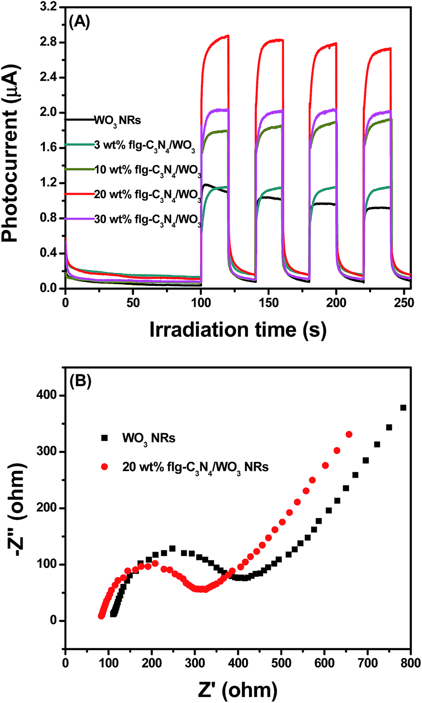
3.7 Electrochemistry analysis
It can be noted that the separation efficiency of electrons and holes plays a critical role for photocatalysis. The separation efficiency of photoinduced-charges was assessed by photocurrent and electrochemical impedance spectroscopy (EIS). From Fig. 7A, in view of photocurrent response, the introduction of flg-C3N4 enhanced the photocurrent density remarkably, indicating boosting separation efficiencies of electrons and holes and longer lifetimes of photogenerated charge carriers.41 In addition, as shown in Fig. 7B, the 20% flg-C3N4/WO3 NRs nanocomposites exhibited a smaller diameter of the Nyquist circle than WO3 NRs, suggesting that 20 wt% flg-C3N4/WO3 NRs nanocomposites had a lower charge transfer resistance and more effective electron–hole pair separation efficiency.42 Thus, these results further supported that decorating with flg-C3N4 facilitated the charge separation.
 |
| | Fig. 7 (A) Transient photocurrent response for the as prepared WO3 NRs and the flg-C3N4/WO3 NRs nanocomposites with different contents of flg-C3N4; (B) electrochemical impedance spectroscopy (EIS) Nyquist plots of the WO3 NRs and the 20 wt% flg-C3N4/WO3 NRs nanocomposites. | |
3.8 Photocatalytic performance
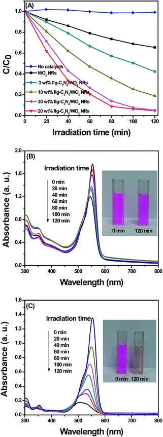
As observed in Fig. 8A, the photoactivities of WO3 NRs and flg-C3N4/WO3 NRs nanocomposites were investigated by degrading RhB. The results indicated that RhB was not degraded obviously without catalysts, indicating that RhB self-degradation could be ignored. The photocatalytic activity of WO3 NRs was improved gradually with the increasing proportion of flg-C3N4. When the contents of flg-C3N4 reached 20%, the highest photocatalytic performance was obtained, and 96% of RhB was degraded after 120 min irradiation. Nevertheless, further increasing the contents of flg-C3N4 in the nanocomposites up to 30% caused decreased degradation efficiency. This result could be attributed to that excessive flg-C3N4 might cover part of the active site of WO3 NRs. Based on the abovementioned results, there might be some synergistic interaction between WO3 NRs and flg-C3N4, which plays a vital role in enhancing the photocatalytic performance. From Fig. 8B and C, the intensity of the characteristic absorption peak dropped as the time increased. In addition, the maximum absorption peak was left-shifted, which was related to the step-by-step de-ethylation process.43 The COD values for the degradation of RhB (Table 1) show that the COD values decreased by about 64% by the 20 wt% flg-C3N4/WO3 NRs. In addition, the corresponding total organic carbon (TOC) values decreased by about 46.2% (Fig. S3†). The rate constant of the 20 wt% flg-C3N4/WO3 NRs nanocomposites was about 7.5 times as high as that of the WO3 NRs (the kinetics were fitted by a first-order model as shown in Table S1†).
 |
| | Fig. 8 (A) Photodegradation of RhB by the as-prepared WO3 NRs and the flg-C3N4/WO3 NRs nanocomposites with different contents of flg-C3N4; (B) time-dependent UV-Vis absorption spectra of the as-prepared WO3 NRs; (C) time-dependent UV-Vis absorption spectra of the 20 wt% flg-C3N4/WO3 NRs. | |
Table 1 The changes of COD values in the degradation processes for RhB under visible light irradiation
| Time (min) |
0 |
60 |
120 |
| COD values by WO3 NRs (mg mL−1) |
213.6 |
169.7 |
151.5 |
| COD values by 20 wt% flg-C3N4/WO3 NRs (mg mL−1) |
150.0 |
75.7 |
54.5 |
3.9 Stability evaluation
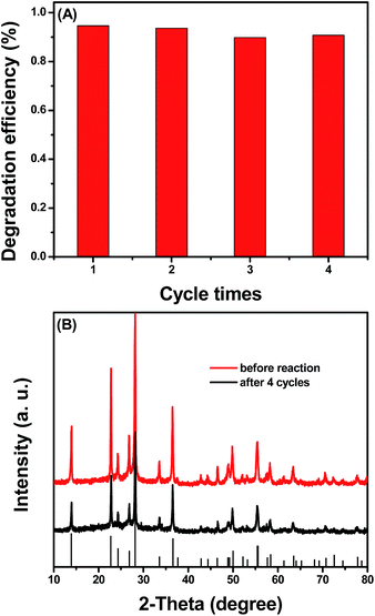
To test the practical reusability of the flg-C3N4/WO3 NRs nanocomposites, the circulating tests were investigated under the same conditions. As revealed in Fig. 9A, the photocatalytic activity of 20 wt% flg-C3N4/WO3 NRs nanocomposites exhibited no obvious distinction after four recycles. Notably, from Fig. 9B, no distinction was found between the XRD results before and after four consecutive tests, suggesting that the nanocomposites were stable during the degradation process of the pollutants.
 |
| | Fig. 9 (A) Cycling runs of 20 wt% flg-C3N4/WO3 NRs nanocomposites; (B) XRD patterns of 20 wt% flg-C3N4/WO3 NRs nanocomposites before and after the photocatalytic reactions. | |
3.10 Proposed photocatalytic mechanism
According to previous reports, the CB and VB positions of WO3 NRs are +0.74 V and +3.44 V, respectively.44 The CB and VB positions of flg-C3N4 are −0.97 eV and 1.83 eV, respectively.22 For the general electron–hole separation process, the electrons in the CB of flg-C3N4 will transfer to the WO3 CB, and holes in the VB of WO3 NRs will transfer to the flg-C3N4 VB. These accumulated electrons lying in WO3 CB cannot be utilized sufficiently via the oxygen reduction process (O2 + e− → ˙O2− (aq), −0.33 V vs. NHE). Likewise, the holes lying in the flg-C3N4 VB cannot have enough potential to effectively oxidize OH− (OH− + h+ → ˙OH (aq), −2.4 V vs. NHE). However, according to ESR analysis, the characteristic peaks of the DMPO- ˙O2− and ˙OH radicals could be observed (Fig. 10A and B), which showed that the ˙O2− and ˙OH radical species were produced under a visible light irradiation process. Therefore, the transfer process of photoinduced charge carriers is not in accordance with the traditional model in this system. Namely, the separation of charge carriers may conform to the Z-scheme.44 As shown in Fig. 11, the photoexcited electrons in the WO3 CB and photoexcited holes in the flg-C3N4 VB combine rapidly. Moreover, not only the molecular oxygen is reduced into ˙O2− by the electrons in the flg-C3N4 CB, which has more negative potential, but also the water molecule is oxidized into ˙OH radicals by the holes in the WO3 VB, which has more positive potential. Therefore, a typical Z-scheme is favorable for the flg-C3N4/WO3 NRs photocatalyst. In addition, the effect of photogenerated holes was investigated by a trapping experiment (as shown in Fig. 10C). After triethanolamine was added, the photocatalytic activity significantly decreased, which showed that the holes might play an important role.
 |
| | Fig. 10 ESR spectra of (A) DMPO-˙O2− and (B) DMPO-˙OH adducts in 20 wt% flg-C3N4/WO3 NRs nanocomposites aqueous dispersion systems before and after visible light irradiation; (C) comparison of the photocatalytic activities of 20 wt% flg-C3N4/WO3 NRs nanocomposites for the degradation of RhB with or without adding triethanolamine. | |
 |
| | Fig. 11 Schematic of the proposed mechanism for the degradation of pollutants. | |
Thus, based on the abovementioned results, the photoexcited holes, ˙O2− and ˙OH radicals act as the major reactive species during the photodegradation process. The transport process of photogenerated electrons and holes is consistent with the Z-scheme in this system.
4. Conclusions
The novel flg-C3N4/WO3 NRs nanocomposites were prepared via a facile hydrothermal process. The 20 wt% flg-C3N4/WO3 NRs nanocomposites showed the highest degradation efficiency for RhB. The improved photocatalytic activity might be attributed to more active sites, the formation of a heterojunction and the high separation of electrons and holes. In addition, the flg-C3N4/WO3 NRs nanocomposites were typical Z-scheme photocatalysts, and a possible mechanism for the improved photocatalytic performance was also proposed. Furthermore, from the perspectives of the decreased COD values and the stability of nanocomposites, this work is also meaningful in designing and fabricating the flg-C3N4-based nanocomposites.
Acknowledgements
The authors genuinely appreciate the financial support for this study by the National Nature Science Foundation of China (21476097) and the Six talent peaks project in Jiangsu Province (2014-JNHB-014).
Notes and references
- S. Cao, J. Low, J. Yu and M. Jaroniec, Adv. Mater., 2015, 27, 2150–2176 CrossRef CAS PubMed.
- M. Guan, C. Xiao, J. Zhang, S. Fan, R. An, Q. Cheng, J. Xie, M. Zhou, B. Ye and Y. Xie, J. Am. Chem. Soc., 2013, 135, 10411–10417 CrossRef CAS PubMed.
- J. Tian, R. Ning, Q. Liu, A. Asiri, A. Al-Youbi and X. Sun, ACS Appl. Mater. Interfaces, 2014, 6, 1011–1017 CAS.
- J. Mao, T. Y. Peng, X. H. Zhang, K. Li, L. Q. Ye and L. Zan, Catal. Sci. Technol., 2013, 3, 1253–1260 CAS.
- S. Han, L. Hu, Z. Liang, S. Wageh, A. Al-Ghamdi, Y. Chen and X. Fang, Adv. Funct. Mater., 2014, 24, 5719–5727 CrossRef CAS.
- R. Pawar and C. Lee, Appl. Catal., B, 2014, 144, 57–65 CrossRef CAS.
- D. Chen, T. Li, Q. Chen, J. Gao, B. Fan, J. Li, X. Li and R. Zhang, Nanoscale, 2012, 4, 5431–5439 RSC.
- J. Xu and X. Cao, Chem. Eng. J., 2015, 260, 642–648 CrossRef CAS.
- M. Miyauchi, M. Shibuya, Z. Zhao and Z. Liu, J. Phys. Chem. C, 2009, 113, 10642 CAS.
- B. Weng, J. Wu, N. Zhang and Y. Xu, Langmuir, 2014, 30, 5574–5584 CrossRef CAS PubMed.
- P. Wang, Y. Bai, P. Luo and J. Liu, Catal. Commun., 2013, 38, 82–85 CrossRef CAS.
- Y. Xie, G. Liu, L. Yin and H. Cheng, J. Mater. Chem., 2012, 22, 6746–6751 RSC.
- Z. Sun, T. Liao, Y. Dou, S. Hwang, M. Park, L. Jiang, J. Kim and S. Dou, Nat. Commun., 2014, 5, 3813 CAS.
- J. Liu, O. Margeat, W. Dachraoui, X. Liu, M. Fahlman and J. Ackermann, Adv. Funct. Mater., 2014, 24, 6029–6037 CrossRef CAS.
- F. Zheng, H. Lu, M. Guo, M. Zhang and Q. Zhen, J. Mater. Chem. C, 2015, 3, 7612–7620 RSC.
- M. Park, J. Seo, H. Song and K. Nam, J. Phys. Chem. C, 2016, 120, 9192–9199 CAS.
- G. Liu, J. Han, X. Zhou, L. Huang, F. Zhang, X. Wang, C. Ding, X. Zheng, H. Han and C. Li, J. Catal., 2013, 307, 148–152 CrossRef CAS.
- Z. Liu, Z. Zhao and M. Miyauchi, J. Phys. Chem. C, 2009, 113, 17132–17137 CAS.
- Y. Liu, Y. Ohko, R. Zhang, Y. Yang and Z. Zhang, J. Hazard. Mater., 2010, 184, 386–391 CrossRef CAS PubMed.
- Y. Nosaka, S. Takahashi, H. Sakamoto and A. Nosaka, J. Phys. Chem. C, 2011, 115, 21283–21290 CAS.
- J. Zhao, Z. Jia, X. Shen, H. Zhou and L. Ma, Ceram. Int., 2015, 41, 5600–5606 CrossRef CAS.
- Y. Ide, F. Liu, J. Zhang, N. Kawamoto, K. Komaguchi, Y. Bando and D. Golberg, J. Mater. Chem. A, 2014, 2, 4150–4156 CAS.
- S. Yang, Y. Gong, J. Zhang, L. Zhan, L. Ma, Z. Fang, R. Vajtai, X. Wang and P. Ajayan, Adv. Mater., 2013, 25, 2452–2456 CrossRef CAS PubMed.
- Z. Zhang, J. Huang, M. Zhang, Q. Yuan and B. Dong, Appl. Catal., B, 2015, 163, 298–305 CrossRef CAS.
- C. Liu, L. Wang, Y. Tang, S. Luo, Y. Liu, S. Zhang, Y. Zeng and Y. Xu, Appl. Catal., B, 2015, 164, 1–9 CrossRef CAS.
- J. Choi, D. Reddy and T. Kim, Ceram. Int., 2015, 41, 13793–13803 CrossRef CAS.
- X. Wang, K. Maeda, A. Thomas, K. Takanabe, G. Xin, J. Carlsson, K. Domen and M. Antonietti, Nat. Mater., 2009, 8, 76–80 CrossRef CAS PubMed.
- F. Dong, Z. Zhao, T. Xiong, Z. Ni, W. Zhang, Y. Sun and W. Ho, ACS Appl. Mater. Interfaces, 2013, 5, 11392–11401 CAS.
- M. Groenewolt and M. Antonietti, Adv. Mater., 2005, 17, 1789–1792 CrossRef CAS.
- Y. Li, L. Fang, R. Jin, Y. Yang, X. Fang, Y. Xing and S. Song, Nanoscale, 2015, 7, 758–764 RSC.
- W. Chen, T. Liu, T. Huang, X. Liu, J. Zhu, G. Duan and X. Yang, Appl. Surf. Sci., 2015, 355, 379–387 CrossRef CAS.
- N. Cheng, J. Tian, Q. Liu, C. Ge, A. Qusti, A. Asiri, A. Al-Youbi and X. Sun, ACS Appl. Mater. Interfaces, 2013, 5, 6815–6819 CAS.
- H. Xu, J. Yan, Y. G. Xu, Y. Song, H. Li, J. Xia, C. Huang and H. Wan, Appl. Catal., B, 2013, 129, 182–193 CrossRef CAS.
- H. Xu, J. Yan, X. She, L. Xu, J. Xia, Y. Xu, Y. Song, L. Huang and H. Li, Nanoscale, 2014, 6, 1406–1415 RSC.
- Y. Lv, Y. Zhu and Y. Zhu, J. Phys. Chem. C, 2013, 117, 18520–18528 CAS.
- X. Bai, L. Wang, R. Zong and Y. Zhu, J. Phys. Chem. C, 2013, 117, 9952–9961 CAS.
- J. Holst and E. Gillan, J. Am. Chem. Soc., 2008, 130, 7373–7379 CrossRef CAS PubMed.
- X. She, L. Liu, H. Ji, Z. Mo, Y. Li, L. Huang, D. Du, H. Xu and H. Li, Appl. Catal., B, 2016, 22, 144–153 CrossRef.
- H. Xu, L. Liu, Y. Song, L. Huang, Y. Li, Z. Chen, Q. Zhang and H. Li, J. Alloys Compd., 2016, 660, 48–54 CrossRef CAS.
- X. She, J. Wu, J. Zhong, H. Xu, Y. Yang, R. Vajtai, J. Lou, Y. Liu, D. Du, H. Li and P. Ajayan, Nano Energy, 2016, 27, 138–146 CrossRef CAS.
- S. Chen, Y. Hu, S. Meng and X. Fu, Appl. Catal., B, 2014, 150, 564–573 CrossRef.
- M. Zhang, X. J. Bai, D. Liu, J. Wang and Y. Zhu, Appl. Catal., B, 2015, 164, 77–81 CrossRef CAS.
- J. Di, J. Xia, S. Yin, H. Xu, L. Xu, Y. Xu, M. He and H. Li, J. Mater. Chem. A, 2014, 2, 5340–5351 CAS.
- P. Zhou, J. Yu and M. Jaroniec, Adv. Mater., 2014, 26, 4920–4935 CrossRef CAS PubMed.
Footnote |
| † Electronic supplementary information (ESI) available. See DOI: 10.1039/c6ra12861k |
|
| This journal is © The Royal Society of Chemistry 2016 |
Click here to see how this site uses Cookies. View our privacy policy here.