Implications of designing clarithromycin loaded solid lipid nanoparticles on their pharmacokinetics, antibacterial activity and safety
Manu Sharma*a,
Namita Guptaa and
Sumeet Guptab
aDepartment of Pharmacy, Banasthali University, Banasthali, Rajasthan-304022, India. E-mail: sharmamanu10@gmail.com; Tel: +91-9694881221
bDepartment of Pharmacology, M M College of Pharmacy, Maharishi Markandeshwar University, Mullana, Ambala-133207, Haryana, India
First published on 4th August 2016
Abstract
The major obstacles for treatment of intracellular infections with clarithromycin are poor gastrointestinal solubility, short half-life (3–4 h), low oral bioavailability and hepatotoxicity. Therefore, clarithromycin loaded solid lipid nanocarriers (CLA-SLNs) were engineered by an emulsification solvent evaporation method to improve CLA in vivo pharmacokinetics and safety. Various formulation variables such as type of surfactant, concentration of surfactant, binary lipid ratio, continuous phase volume and sonication time were optimized considering particle size and entrapment efficiency as quality parameters. The optimized CLA-SLN formulation exhibited a maximum entrapment efficiency of 84 ± 9% and smallest particle size of 307 ± 23 nm with a zeta potential of −29 mV. Physicochemical characterization of CLA-SLNs by differential scanning calorimetry and powder X-ray diffraction confirmed entrapment of CLA and its phase transformation from crystalline to amorphous in the SLN formulation. Morphological characterization revealed a smooth and spherical shape of the optimized CLA-SLNs which prolonged the controlled diffusion of the drug upto 24 h. In vitro antibiofilm activity disclosed the higher potential of CLA-SLNs in biofilm eradication at lower drug concentration (40 μg ml−1) compared to free CLA (140 μg ml−1), which was confirmed by SEM images. A pharmacokinetic study in wistar rats depicted higher Cmax (2.3-fold), Tmax (2-fold), MRT (1.4-fold) and almost 5-fold improvement in relative oral bioavailability of CLA on oral administration of CLA-SLNs. Furthermore, cytotoxicity studies in macrophage cell line and repeated oral dose toxicity study confirmed the safety of the CLA-SLN formulation. In conclusion, CLA-SLNs can serve as a superior carrier system with high oral bioavailability of CLA and can improve the therapeutic potential of CLA in treatment of intracellular infections.
1 Introduction
During the last few decades several new antibiotics have been developed for treatment of a variety of infections. However, most of the infections caused by biofilm (an extracellular polymeric matrix) forming microorganisms are difficult to treat. Pathogens like E. coli, P. aeruginosa, K. pneumonia, S. aureus, S. pneumonia, C. albicans protect themselves from opsonization by antibiotics and phagocytosis by forming a biofilm.1 A biofilm also limits the diffusion of antibiotics into bacterial cells and facilitates the development of tolerance to antibiotics.2 Macrolides are commonly used to treat chronic infections. Among macrolides, clarithromycin (CLA) has higher stability under acidic environments making it suitable for oral delivery.3 It is effective against C. pneumonia, M. pneumomiae, P. aeruginosa, K. pneumonia, S. pneumonia, S. aureus, H. influenza, B. pertussis, Legionella and Campylobacter responsible for respiratory tract infections as well as H. pylori responsible for peptic ulcer.4 Therefore, it is indicated in chronic obstructive pulmonary disorders, skin and soft tissue infections. CLA is a Biopharmaceutical Classification System (BCS) class II drug having dissolution controlled slow absorption due to its insolubility in water leading to its low systemic oral bioavailability.5 It also undergoes extensive hepatic first pass metabolism.6 Thus, short half-life (3–4 h) and poor systemic bioavailability of CLA limits the therapeutic efficacy of CLA in intracellular infections caused by biofilm forming micro-organisms.7,8 Subsequently, higher dose of CLA for longer period need to be administered for achieving therapeutic effect which may lead to hepatotoxicity and elevated serum level of drug. Since CLA inhibits hepatic microsomal cytochrome CYP3A4 involved in its metabolism.9 In view of above facts, continuous efforts have been made to improve its bioavailability, safety and efficacy to treat the infection caused by biofilm forming pathogens. So far, several studies have been done to improve the efficacy of CLA for parenteral and pulmonary delivery such as production of CLA loaded human serum albumin microspheres, polymeric nano-micro carrier system of CLA and liposomal CLA. However, developed formulations have bestowed confined successs.9–13 This constrains on the investigation of novel formulation strategies for improving the oral bioavailability of CLA.Solid lipid nanoparticles (SLNs) offer numerous benefits as carrier over their conventional colloidal predecessors like polymeric nanoparticles, emulsions and liposomes. SLNs provide a tremendous potential of improving bioavailability of BCS class II drugs with minimal expected cytotoxic potential compared to polymeric nanoparticles due to physiological resemblance of lipid excipients with cellular lipids.14,15 Earlier researchers have reported improvement in the bioavailability of variety of drugs like amphotericin B (4.96-fold),14 quercetin (5-fold),16 nitrendipine (3.98-fold),17 rifampicin (10-fold), isoniazid (29-fold),18 fenofibrate (2-fold),19 lopinavir (2.13-fold)20 etc. after oral administration of SLNs. The enhanced bioavailability of drugs loaded in SLNs was attributed to their lymphatic absorption via paracellular and transcellular pathways through enterocytes and endocytosis by phagocytic cells.14 However, particle size as well as physicochemical composition affected their ultimate absorption into lymphatic system. SLNs having particle size in submicron range exhibited higher uptake and easily released from Peyer's patches to lymphatic system.21 Along with small particle size, SLNs with high hydrophobicity and with either negative or neutral charge showed higher accumulation and uptake by M cells.22 So far to our knowledge no work has been reported on oral delivery of CLA loaded solid lipid nanoparticles in literature. Therefore, abovementioned paradigm opens an avenue for development of CLA loaded SLNs for oral delivery to improve CLA bioavailability, therapeutic efficacy and safety.
SLNs are commonly fabricated by employing lipids belonging to class of fatty acids, waxes or glycerides.23 Among the lipids, fatty acids or waxes form highly ordered crystal packing in single lipid matrix system leading to expulsion of drug with superior physical stability. On the other hand glycerides form less ordered crystal lattice with higher drug loading and poor physical stability. Thus, selection of suitable lipid for SLNs fabrication depends upon the degree of crystallinity and polymorphic behavior of lipid which strongly affects drug loading capacity, physical stability and release rate.24,25 Binary lipid matrix composed of fatty acids (like stearic acid) and glycerides (tristearin) may improve drug loading and physical stability compared to single constituent due to amalgamation of properties of different constituents.25 However, use of binary lipid matrix in design of SLNs has not yet been recognized. Stability as well as drug release issue from SLNs is also governed by charge developed on the surface by surfactant.17 Therefore, proper selection of type and concentration of emulsifiers (like soy lecithin, Poloxamer F-68, Poloxamer 188, Brij 78, polysorbates etc.) required for production of SLNs with high entrapment efficiency and smaller submicron particle size (<500 nm) to facilitate uptake of SLNs by M cells.21,22
Keeping in mind above discussed perspectives of solid lipid nanocarriers, the goal of present study was to prepare, characterize and evaluate CLA loaded solid binary lipid nanoparticulate formulation (CLA-SLNs) for their antimicrobial activity against biofilm forming pathogen S. aureus. Furthermore, CLA-SLNs were evaluated for their pharmacokinetics and safety.
2 Results and discussion
2.1 Formulation optimization
Different batches of CLA-SLNs were prepared using stearic acid alone as lipid to select a suitable surfactant and its optimum concentration, volume of continuous phase and sonication time. The single lipid CLA-SLNs exhibited particle size in the range of 348 ± 44 nm to 876 ± 26 nm and polydispersity index 0.2 to 0.6. Among the surfactants (PVA, Pluronic F-68, tween 80), pluronic F-68 resulted in formulation of smaller particles (411 ± 19 nm) with higher entrapment efficiency (67 ± 8%) (Table 1). Significant difference in particle size was observed at a fixed concentration (0.5% w/v) of different stabilizers indicated that type of stabilizer influences the particle size. The results of our study also confirmed that Pluronic F-68 provides stable mechanical and thermodynamic barrier at interface preventing coalescence of particles compared to Tween 80 and PVA similar to earlier reported results.26,27 Particle size increased whereas entrapment efficiency decreased on reducing surfactant concentration (0.25% w/v). This indicated that minimum amount of Pluronic F-68 was required to prepare a stable emulsion.28 Lower entrapment efficiency at higher concentration of Pluronic F-68 might be due to solubilizing property of surfactant. However, smaller particle size at higher Pluronic F-68 concentration is the result of greater reduction in interfacial tension at the interface of lipid and aqueous phase.29| Formulation code | Particle size (nm ± SD) | Polydispersity index ± SD | Zeta potential (mV) | Entrapment efficiency (%) | Drug loading (%) | Yield (%) |
|---|---|---|---|---|---|---|
| A1 | 592 ± 39 | 0.28 ± 0.05 | −18 ± 2 | 59 ± 9 | 4.5 ± 1.0 | 58 ± 8 |
| A2 | 711 ± 21 | 0.37 ± 0.03 | −17 ± 3 | 62 ± 7 | 4.8 ± 0.8 | 53 ± 10 |
| A3 | 411 ± 19 | 0.29 ± 0.04 | −18 ± 3 | 67 ± 8 | 5.2 ± 0.9 | 59 ± 9 |
| A4 | 639 ± 25 | 0.40 ± 0.05 | −17 ± 2 | 58 ± 9 | 4.5 ± 0.9 | 57 ± 10 |
| A5 | 529 ± 23 | 0.20 ± 0.04 | −18 ± 4 | 61 ± 8 | 4.7 ± 0.9 | 58 ± 7 |
| A6 | 876 ± 26 | 0.32 ± 0.05 | −18 ± 3 | 54 ± 7 | 4.2 ± 0.8 | 60 ± 11 |
| A7 | 348 ± 44 | 0.47 ± 0.09 | −19 ± 3 | 58 ± 9 | 4.5 ± 1.0 | 55 ± 9 |
| A8 | 601 ± 19 | 0.57 ± 0.12 | −20 ± 3 | 63 ± 6 | 4.9 ± 0.7 | 57 ± 12 |
| A9 | 801 ± 28 | 0.60 ± 0.14 | −18 ± 4 | 46 ± 11 | 3.5 ± 1.0 | 53 ± 13 |
| A10 | 258 ± 25 | 0.26 ± 0.04 | −21 ± 3 | 74 ± 9 | 5.7 ± 0.9 | 60 ± 9 |
| A11 | 277 ± 57 | 0.38 ± 0.07 | −27 ± 3 | 76 ± 13 | 5.8 ± 1.0 | 58 ± 11 |
| A12 | 289 ± 41 | 0.17 ± 0.05 | −24 ± 3 | 79 ± 15 | 6.1 ± 1.2 | 61 ± 14 |
| A13 | 307 ± 23 | 0.21 ± 0.04 | −29 ± 5 | 84 ± 9 | 6.5 ± 0.9 | 63 ± 11 |
| A14 | 345 ± 26 | 0.21 ± 0.04 | −28 ± 2 | 80 ± 17 | 6.2 ± 1.4 | 61 ± 9 |
| A15 | 362 ± 35 | 0.31 ± 0.08 | −28 ± 4 | 78 ± 16 | 6.0 ± 1.3 | 62 ± 10 |
| A16 | 392 ± 48 | 0.38 ± 0.09 | −27 ± 3 | 73 ± 16 | 5.6 ± 1.2 | 59 ± 9 |
In addition to this, increase in sonication time from 2.5 to 5 min reduced the size of SLNs significantly and increased drug loading due to formation of uniform micelles. However further increase in sonication time (7.5 min) reduced drug loading and increased particle size. This might be due to breakage of surfactant barrier layer or increased particle surface area with increased sonication time making the system thermodynamically unstable facilitating agglomeration of lipid phase.30
Increase in continuous phase volume from 20 to 50 ml decreased the entrapment efficiency whereas increase in particle size was observed (Table 1). This might be due to lower magnitude of shear stress experienced in higher volume of continuous phase compared to lower volume. The maximum encapsulation efficiency (67 ± 8%) and drug loading (5.2 ± 0.9%) was found for batch A3 with smallest particle size (411 ± 19 nm). Thus, optimized variables for preparation of CLA-SLNs were 0.5% w/v pluronic F-68 as surfactant, 20 ml continuous phase volume and 5 min sonication time. However, the entrapment efficiency of optimized formulation was poor. Therefore, stearic acid along with tristearin was used to enhance entrapment efficiency using optimized formulation process variables.31 The particle size and zeta potential of different formulations prepared using binary lipids ranged from 258–392 nm and −21 to −29 mV respectively (Table 1). The results also indicated that formulations prepared with binary lipids (stearic acid and tristearin) in 1![[thin space (1/6-em)]](https://www.rsc.org/images/entities/char_2009.gif) :
:![[thin space (1/6-em)]](https://www.rsc.org/images/entities/char_2009.gif) 1 ratio showed higher entrapment efficiency (84 ± 9%) and actual drug loading (6.5 ± 0.9%) in comparison to stearic acid alone (67 ± 8%) (Table 1). Smaller particle size and higher drug loading in formulations containing binary lipids might be the result of increased disturbances in crystal lattice of stearic acid due to presence of tristearin.32 However, further increase in tristearin content reduced entrapment efficiency along with increased particle size. Higher zeta potential of optimized formulation near −29 mV indicated higher stability of CLA-SLNs. The yield of different batches of SLNs fabricated with stearic acid alone and in binary mixture with tristearin as lipid varied from 53 ± 10% to 63 ± 11%. Low yield might be attributed by losses occurring during formulation due to adherence of particles to glassware, probe as well as during centrifugation.33
1 ratio showed higher entrapment efficiency (84 ± 9%) and actual drug loading (6.5 ± 0.9%) in comparison to stearic acid alone (67 ± 8%) (Table 1). Smaller particle size and higher drug loading in formulations containing binary lipids might be the result of increased disturbances in crystal lattice of stearic acid due to presence of tristearin.32 However, further increase in tristearin content reduced entrapment efficiency along with increased particle size. Higher zeta potential of optimized formulation near −29 mV indicated higher stability of CLA-SLNs. The yield of different batches of SLNs fabricated with stearic acid alone and in binary mixture with tristearin as lipid varied from 53 ± 10% to 63 ± 11%. Low yield might be attributed by losses occurring during formulation due to adherence of particles to glassware, probe as well as during centrifugation.33
2.2 Characterization studies
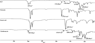
![[double bond, length as m-dash]](https://www.rsc.org/images/entities/char_e001.gif) O stretching vibration of carboxyl group. CLA spectra showed peak for C–H stretching vibration at 2927.90 cm−1; predominant peak of –O–C
O stretching vibration of carboxyl group. CLA spectra showed peak for C–H stretching vibration at 2927.90 cm−1; predominant peak of –O–C![[double bond, length as m-dash]](https://www.rsc.org/images/entities/char_e001.gif) O stretching vibration in lactone ring and –C
O stretching vibration in lactone ring and –C![[double bond, length as m-dash]](https://www.rsc.org/images/entities/char_e001.gif) O stretching vibration of ketone group at 1728.76 cm−1 and 1689.33 cm−1 respectively; peaks at 1167.84 cm−1, 1104.87 cm−1 and 1045.58 cm−1 refers to –O–ether functional band. The spectra of CLA-SLNs showed typical peaks of lactone carbonyl and ketone carbonyl group at 1731.48 cm−1 and 1702.25 cm−1; peak at 2927 cm−1 due to C–H stretching vibration and peaks at 1172.68 cm−1, 1146.29 cm−1 and 1104.60 cm−1 due to ether functional band confirming the presence of CLA in SLNs without any chemical interaction with excipients during SLNs preparation.
O stretching vibration of ketone group at 1728.76 cm−1 and 1689.33 cm−1 respectively; peaks at 1167.84 cm−1, 1104.87 cm−1 and 1045.58 cm−1 refers to –O–ether functional band. The spectra of CLA-SLNs showed typical peaks of lactone carbonyl and ketone carbonyl group at 1731.48 cm−1 and 1702.25 cm−1; peak at 2927 cm−1 due to C–H stretching vibration and peaks at 1172.68 cm−1, 1146.29 cm−1 and 1104.60 cm−1 due to ether functional band confirming the presence of CLA in SLNs without any chemical interaction with excipients during SLNs preparation.
 | ||
| Fig. 5 In vitro release of drug from free CLA and optimized CLA-SLNs in pH progressive dissolution media. | ||
In vitro release data upto 12 h was fitted into different release kinetic equations. The release data showed best fit to Korseymeyer Peppas equation which was confirmed by comparing regression coefficients (R2) of zero order (0.985), first order (0.885), Higuchi model (0.975) and Korseymeyer Peppas equation (0.987). The diffusion exponent (n) of Korseymeyer Peppas equation has value 0.43 < n < 0.85 indicating anomalous behavior of drug release, i.e. drug release was contributed by dissolution and diffusion of CLA from lipid matrix.33,36 Our results are analogous to earlier reported studies indicating that drug loaded SLNs bestowed the controlled drug release pattern.17,37
| S. No. | Time (month) | Storage conditions | Release in 10 h (%) | Entrapment efficiency (%) | Particle size (nm) | PDI | Zeta potential (mV) |
|---|---|---|---|---|---|---|---|
| a Represents significant difference of parameter at the specific condition compared to control at P < 0.05. | |||||||
| 1 | 0 | — | 77 ± 4 | 84 ± 9 | 307 ± 23 | 0.20 ± 0.04 | −29 ± 5 |
| 2 | 1 | Room temperature | 74 ± 5 | 82 ± 8 | 331 ± 40 | 0.27 ± 0.06 | −26 ± 3 |
| Accelerated temperature | 70 ± 4 | 80 ± 7 | 364 ± 29 | 0.20 ± 0.03 | −24 ± 3 | ||
| Cool temperature | 75 ± 6 | 84 ± 4 | 326 ± 36 | 0.23 ± 0.04 | −28 ± 2 | ||
| 3 | 3 | Room temperature | 72 ± 5 | 81 ± 5 | 360 ± 31 | 0.20 ± 0.05 | −25 ± 4 |
| Accelerated temperature | 65 ± 5a | 74 ± 6a | 471 ± 31 | 0.30 ± 0.05a | −22 ± 4 | ||
| Cool temperature | 75 ± 4 | 82 ± 6 | 343 ± 42 | 0.20 ± 0.03 | −28 ± 2 | ||
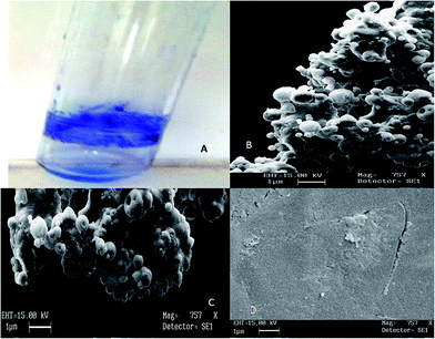
2.2.7.1 In vitro antibiofilm activity. Biofilm formation was confirmed visually by presence of stained film on walls of test tube by crystal violet staining test. Furthermore, SEM images were also taken to confirm the biofilm formation by S. aureus (Fig. 7A and B). Higher bacterial cell damage by optimized A13 formulation at concentration equivalent to 40 μg ml−1 drug was further confirmed by SEM image compared to free drug solution indicates higher inhibitory potential of CLA-SLN (Fig. 7C and D).
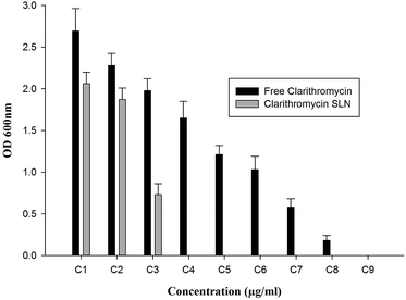
All the concentrations used during MBIC assay had inhibitory effect on the growth of viable organisms compared to control (Fig. 8). However, biofilms treated with A13 formulation having CLA equivalent to 40 μg ml−1 showed complete eradication of bacteria whereas MBIC of CLA was 140 μg ml−1. Even though lower concentration of drug inhibited the bacteria in biofilm but not completely killed the bacteria. Optimized CLA-SLNs had showed almost 3.5 times higher efficacy in destroying the bacteria in biofilm.
Lower MIC (12-fold) and MBIC (3.5-fold) of A13 formulation compared to free drug explains the improved antibacterial activity of nanosized carriers.39 The improved antibacterial activity of CLA-SLN formulation might be due to adherence/adsorption of SLN on cell wall of microorganism facilitating sustained diffusion of drug working as depot for drug.40 Moreover, physiological resemblance of CLA-SLNs to membrane lipids further improves the uptake mechanism of drug by fusion of CLA-SLNs with cell wall or cell membrane. These series of events might have sustained bactericidal activity of drug against target organism.11
| Parameters | CLA suspension | A13 formulation |
|---|---|---|
| a *p < 0.05 level of significant difference. **p < 0.001 level of significant difference. | ||
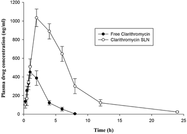
| Tmax (h)** | 1.0 ± 0.2 | 2.0 ± 0.3 |
| Cmax (ng ml−1)** | 451 ± 69 | 1034 ± 90 |
| t1/2 (h)** | 1.1 ± 0.3 | 3.8 ± 0.7 |
| ke (h−1)** | 0.6 ± 0.1 | 0.18 ± 0.01 |
| AUC(0–24) (ng h ml−1)** | 1381 ± 112 | 7094 ± 332 |
| AUMC (ng h2 ml−1)** | 2539 ± 140 | 18![[thin space (1/6-em)]](https://www.rsc.org/images/entities/char_2009.gif) 670 ± 1446 670 ± 1446 |
| MRT (h)* | 1.8 ± 0.3 | 2.6 ± 0.4 |
| Relative bioavailability (%) | — | 514 |
3 Experimental procedures
3.1 Materials
Clarithromycin (CLA) was generously gifted by Ranbaxy laboratories Ltd, New Delhi, India. Tristearin, Pluronic F-68 and nutrient broth medium was procured from Himedia Laboratories Pvt. Ltd, Mumbai. Dulbecco's modified Eagle's medium, fetal bovine serum, penicillin, streptomycin and 3-[4,5-dimethyl thiazolyl]-2,5-diphenyl tetrazolium bromide (MTT reagent) was purchased from Sigma chemicals (St. Louis, MO). Stearic acid (octadecanoic acid), soya lecithin, sodium lauryl sulphate, poly vinyl alcohol (PVA), disodium hydrogen orthophosphate, sodium dihydrogen orthophosphate and sodium hydroxide were purchased from CDH (Cental drug house laboratory). Tween 80 (polysorbate 80) and dichloromethane was procured from Merck (Mumbai, India). Lyophilized bacterial strain of S. aureus (MTCC no. 86) was purchased from Institute of Microbial Technology, Chandigarh, India. Chemicals and solvents employed for experimental work were of analytical grade. Distilled water was used during the study.3.2 Fabrication of clarithromycin loaded solid lipid nanocarriers
CLA-SLNs were fabricated by emulsification solvent evaporation technique.41 An accurately weighed amount of CLA (20 mg), soya lecithin (40 mg) and lipid (200 mg of stearic acid alone or in combination with tristearin) dissolved in dichloromethane (5 ml) constituted lipid phase. Subsequently, lipid phase was introduced at constant rate (1 ml min−1) into aqueous phase containing 0.5% w/v surfactant (Tween 80/PVA/Pluronic F-68) under continuous homogenization for 20 min at 10![[thin space (1/6-em)]](https://www.rsc.org/images/entities/char_2009.gif) 000 rpm. Emulsification was continued using probe sonicator (40 W output power, 60% amplitude, Branson Sonifier) for 5 min at 4 °C. The resulting o/w emulsion was kept on stirring (75 rpm) for 14 h at room temperature to remove organic solvent. CLA-SLNs were separated by centrifuging the dispersion at 30
000 rpm. Emulsification was continued using probe sonicator (40 W output power, 60% amplitude, Branson Sonifier) for 5 min at 4 °C. The resulting o/w emulsion was kept on stirring (75 rpm) for 14 h at room temperature to remove organic solvent. CLA-SLNs were separated by centrifuging the dispersion at 30![[thin space (1/6-em)]](https://www.rsc.org/images/entities/char_2009.gif) 000 rpm, 5 °C for 25 min. CLA-SLNs were re-suspended in approximately 0.5 ml distilled water and lyophilized using mannitol (5% w/v) as cryoprotectant in a lyophilizer (CleanVac Freeze dryer, South Korea) at −50 °C and 0.05 mbar pressure for 48 h. Lyophilized CLA-SLNs were stored in desiccator at 25 °C. Different batches of CLA-SLNs prepared to optimize the formulation variables like type and concentration of surfactant, sonication time, volume of continuous phase as well as stearic acid
000 rpm, 5 °C for 25 min. CLA-SLNs were re-suspended in approximately 0.5 ml distilled water and lyophilized using mannitol (5% w/v) as cryoprotectant in a lyophilizer (CleanVac Freeze dryer, South Korea) at −50 °C and 0.05 mbar pressure for 48 h. Lyophilized CLA-SLNs were stored in desiccator at 25 °C. Different batches of CLA-SLNs prepared to optimize the formulation variables like type and concentration of surfactant, sonication time, volume of continuous phase as well as stearic acid![[thin space (1/6-em)]](https://www.rsc.org/images/entities/char_2009.gif) :
:![[thin space (1/6-em)]](https://www.rsc.org/images/entities/char_2009.gif) tristearin ratio are coded in Table 4.
tristearin ratio are coded in Table 4.
| S. No. | Formulation code | Lipid | Surfactant | Sonication time (min) | Volume of continuous phase (ml) | |||
|---|---|---|---|---|---|---|---|---|
| Stearic acid (mg) | Tristearin (mg) | Tween 80 (% w/v) | Pluronic F-68 (% w/v) | PVA (% w/v) | ||||
| 1 | A1 | 200 | — | 0.50 | — | — | 5 | 20 |
| 2 | A2 | 200 | — | — | — | 0.5 | 5 | 20 |
| 3 | A3 | 200 | — | — | 0.5 | — | 5 | 20 |
| 4 | A4 | 200 | — | — | 0.5 | — | 2.5 | 20 |
| 5 | A5 | 200 | — | — | 0.5 | — | 7.5 | 20 |
| 6 | A6 | 200 | — | — | 0.25 | — | 5 | 20 |
| 7 | A7 | 200 | — | — | 1.0 | — | 5 | 20 |
| 8 | A8 | 200 | — | — | 0.5 | — | 5 | 30 |
| 9 | A9 | 200 | — | — | 0.5 | — | 5 | 50 |
| 10 | A10 | 50 | 150 | — | 0.5 | — | 5 | 20 |
| 11 | A11 | 60 | 140 | — | 0.5 | — | 5 | 20 |
| 12 | A12 | 70 | 130 | — | 0.5 | — | 5 | 20 |
| 13 | A13 | 100 | 100 | — | 0.5 | — | 5 | 20 |
| 14 | A14 | 130 | 70 | — | 0.5 | — | 5 | 20 |
| 15 | A15 | 140 | 60 | — | 0.5 | — | 5 | 20 |
| 16 | A16 | 150 | 50 | — | 0.5 | — | 5 | 20 |
3.3 Characterization of CLA-SLNs
![[thin space (1/6-em)]](https://www.rsc.org/images/entities/char_2009.gif) :
:![[thin space (1/6-em)]](https://www.rsc.org/images/entities/char_2009.gif) 2 (v/v). The resultant samples were screened through 0.45 μm nylon membrane filters and analyzed using UV/Visible spectrophotometer (LabIndia 3000+, India) at 206 nm. The actual drug loading (ADL) and entrapment efficiency (EE) of CLA-SLNs was calculated by formula
2 (v/v). The resultant samples were screened through 0.45 μm nylon membrane filters and analyzed using UV/Visible spectrophotometer (LabIndia 3000+, India) at 206 nm. The actual drug loading (ADL) and entrapment efficiency (EE) of CLA-SLNs was calculated by formula
 | (1) |
 | (2) |
 | (3) |
The results were expressed as mean ± SD of three independent experiments.
![[thin space (1/6-em)]](https://www.rsc.org/images/entities/char_2009.gif) 000 Da) immersed in dissolution medium (500 ml). Aliquots (3 ml) were withdrawn upto 24 h and analyzed for CLA content using UV/Visible spectrophotometer at 206 nm. Each volume withdrawn during sampling was replaced with fresh buffer. The results of dissolution study were expressed as mean ± SD of three experiments. Release data was plotted for different release kinetic models like zero order, first order, Higuchi model and Korsermeyer–Peppas model to predict the mechanism and kinetics of drug release. The linearity of graph determined by calculating regression coefficient was used to determine best fit model.
000 Da) immersed in dissolution medium (500 ml). Aliquots (3 ml) were withdrawn upto 24 h and analyzed for CLA content using UV/Visible spectrophotometer at 206 nm. Each volume withdrawn during sampling was replaced with fresh buffer. The results of dissolution study were expressed as mean ± SD of three experiments. Release data was plotted for different release kinetic models like zero order, first order, Higuchi model and Korsermeyer–Peppas model to predict the mechanism and kinetics of drug release. The linearity of graph determined by calculating regression coefficient was used to determine best fit model.Serial dilutions of optimized CLA-SLNs and CLA solution equivalent to different concentrations i.e. 0.125, 0.25, 0.5, 1, 2, 4, 8, 10 and 12 μg ml−1 of CLA were evaluated for determining minimum bacterial growth inhibitory concentration. Briefly, sterile nutrient broth in culture tubes was inoculated with 100 μl of bacterial suspension (equivalent to 0.5 McFarland standard) followed by addition of different concentrations of test samples in culture tubes respectively. Tubes were incubated for 24 h at 37 °C and scrutinized for any evidence of bacterial growth by measuring turbidity of samples at 600 nm. Sterile PBS was used as control during MIC determination.
3.3.9.1 Determination of minimum biofilm inhibitory concentration (MBIC). Before determining antibiofilm activity of CLA and optimized CLA-SLNs, biofilm formation by S. aureus was confirmed by crystal violet dye method and SEM. Briefly, S. aureus inoculum adjusted to 0.5 McFarland standards was inoculated in nutrient broth followed by incubation for 96 h at 37 °C to facilitate biofilm formation. Broth was replenished with fresh media to remove non-adherent bacteria from the surface of test tube after every 24 h. Broth was decanted and biofilm formed on the walls and bottom of culture tube after 96 h was washed with sterile distill water thrice to remove all non adherent bacteria from culture tube. The formation of biofilm was detected by adding 0.1% w/v crystal violet dye solution (0.5 ml) in culture tubes and swirled uniformly. The excess of stain was rinsed off twice with sterile distilled water. The presence of stain on wall and bottom of test tube indicated the formation of biofilm by S. aureus which was further confirmed by taking SEM images of biofilm formed.
SEM images of biofilm formed by S. aureus were taken under optimized conditions explained by Ceri et al. with slight modification.47 Biofilm was rinsed off twice with sterile phosphate buffer saline pH 7.4. Biofilm from culture tube surface was removed by sonication for 1 min with sterile PBS (1 ml). Bacterial biofilms were fixed with Karnovsky's fixative for 1 h at room temperature followed by treatment for 1 h with 1% w/v osmium tetraoxide. Biofilm samples were rinsed twice with sterile distilled water and subjected to stepwise dehydration with ethanol (50, 60, 70, 80, 90, 95 and 100%). Sputter coating of dehydrated samples was done with gold palladium and analyzed at appropriate magnification by SEM (LEO435VP). Antibiofilm activity of CLA and optimized CLA-SLNs was also confirmed by SEM images. Biofilms were treated with different concentration of CLA or optimized CLA-SLNs equivalent to 20, 40 and 80 μg ml−1 CLA respectively for 24 h at 37 °C and treated similarly as explained above before capturing images by scanning electron microscope. Untreated biofilm images worked as control.
MBIC of CLA and optimized CLA-SLNs was determined according to following procedure. Culture tubes having biofilms on walls and bottom were washed twice with sterile distilled water to remove unattached bacteria from surface of tubes. Sterile PBS (1 ml) was added in each tube and sonicated for 1 min to detach the bacteria from the surface of test tube. Bacterial biofilm was treated with different concentrations of CLA or CLA-SLNs (equivalent to 5, 10, 20, 40, 80, 100, 120 and 140 μg ml−1 of drug) respectively for 24 h at 37 °C. Sample (100 μl) was taken from each culture tube and incubated in fresh nutrient broth respectively. Turbidity in culture tubes indicating the presence of viable organisms was quantified at 600 nm. Biofilm assay for each sample was performed in triplicate.
 | (4) |
3.3.11.1 Pharmacokinetic evaluation. Eighteen male wistar rats were fasted for 24 h prior to start of experiment with free access to water ad libitum. Fasted male rats were randomly divided into three groups (n = 6). Group I (control) received saline solution orally; group II was orally treated with CLA suspension (5 mg kg−1) and group III received optimized CLA-SLN (A13) formulation (dose equivalent to 5 mg kg−1 CLA). Blood samples (250 μl) were serially withdrawn into heparinized tubes from retro-orbital plexus at an interval of 0.25, 0.5, 0.75, 1, 2, 4, 8, 12 and 24 h. Normal sterile saline solution equivalent to blood sample withdrawn was injected to rats to compensate for blood loss. Blood samples were centrifuged at 5000 rpm for 10 min at 4 °C to separate plasma. Thereafter, collected plasma samples were stored at −20 °C prior to analysis for drug content by reverse phase high performance liquid chromatography (RP-HPLC). The amount of drug in plasma was determined by HPLC (LC-2010CHT; Shimadzu, Japan) equipped with C18 column (Phenonmex column, 250 mm × 4.6 mm, 5 μm), UV-Vis detector (Shimadzu Co., Kyoto, Japan), an auto injector (Shimadzu model SIL-6A) and a system controller (Shimadzu module SCL-6A). HPLC resolution of sample was achieved with mobile phase consisting of a mixture of 0.067 M aqueous solution of monosodium dihydrogen phosphate buffer (adjusted to pH 5.5 with orthophosphoric acid) and acetonitrile (60
![[thin space (1/6-em)]](https://www.rsc.org/images/entities/char_2009.gif) :
:![[thin space (1/6-em)]](https://www.rsc.org/images/entities/char_2009.gif) 40 v/v) at 25 ± 2 °C. Mobile phase before use was filtered with 0.45 μm membrane filter and deaerated for 5 min using bath sonicator. The flow rate being adjusted to 1 ml min−1. Sample injection volume was 20 μl and eluted analytes for drug were analyzed at 206 nm.48
40 v/v) at 25 ± 2 °C. Mobile phase before use was filtered with 0.45 μm membrane filter and deaerated for 5 min using bath sonicator. The flow rate being adjusted to 1 ml min−1. Sample injection volume was 20 μl and eluted analytes for drug were analyzed at 206 nm.48The observed pharmacokinetic data was analyzed by non-compartmental pharmacokinetic analysis using WinNonlin (5.1: Pharsight, Mountain View, CA).
3.3.11.2 In vivo repeated oral dose acute toxicity study. Male wistar rats (180 ± 20 g) obtained from institutional animal house were used for determination of repeated oral dose toxicity of CLA and optimized CLA-SLNs (A13) at a dose equivalent to 10 mg per kg body weight. All animals were kept on normal diet and filtered drinking water throughout study. Animals were divided into three groups (n = 6) i.e. group I [control group (PBS treated)], group II (CLA suspension treated) and group III (optimized CLA-SLNs treated). Male wistar rats in each group were orally administered test sample at dose equivalent to 10 mg per kg body weight once a day for 14 days. Animals were observed for any symptoms of toxicity or mortality during this period. Animals were anaesthetized and sacrificed on day 14. Liver was harvested for histological study from control group and test sample group. Liver was rinsed with PBS and fixed in 10% neutral formalin at pH 7 for 24 h. Liver blocks were embedded in paraffin, sectioned and stained with hematoxylin and eosin. Histological samples were analyzed by capturing images using an optical imaging system (Nikon Eclipse TS 100, Nickon, Toyoto, Japan) equipped with camera.
3.4 Statistical analysis
Results of all the studies are illustrated as mean ± SD. One way ANOVA followed by Holm–Sidak test using Sigma plot 12 software (Systat Software Inc CA, USA) was used for data analysis to ascertain the significant difference at the level of P < 0.05.4 Conclusions
Clarithromycin loaded solid lipid nanocarriers were successfully prepared with high loading capacity by emulsification solvent evaporation method. Sustained drug release characteristics of optimized CLA-SLNs and higher resemblance to cell membrane compared to free drug facilitated higher systemic availability of CLA after oral administration of CLA-SLNs as well as improved their efficacy in eradication of biofilm formed by S. aureus at lower drug concentration. In vitro cytotoxicity study and repeated oral dose toxicity study also confirmed the safety of CLA-SLNs compared to free drug. Thus CLA-SLN formulation possessing high biofilm eradication potential, cellular biocompatibility and low acute toxicity profile offers their candidature for development of clinically relevant targeted therapy against biofilm forming bacteria. Furthermore, studies are needed for industrial scale up and clinical use of CLA-SLNs.Ethical statement
Animal studies were conducted in accordance with legislation of institute on animal experiments. The protocol was approved by the Institutional Animal Ethical Committee (MMCP/IEC/11/20). Animals were housed in plastic cages in climatically controlled rooms (22 ± 2 °C, 55 ± 5% RH on a 12 h dark/light cycle) and fed with standard rodent food pellet (Lipto India Ltd, Bombay) and water ad libitum. All animal experiments were conducted according to guideline established for care and use of laboratory animals by CPCSEA, Ministry of Social Justice and Empowerment, Government of India.Declaration of interest
The authors report no declaration of interest.Acknowledgements
Authors would like to express their gratitude to Ranbaxy laboratories Ltd, New Delhi, India for providing clarithromycin as gift sample. Authors are thankful to MNIT Jaipur for providing PXRD facility.References
- H. Valizadeh, G. Mohammad, R. Ehyaei, M. Milani, M. Azhdarzadeh, P. Zakeri-Milani and F. Lotfipour, Pharmazie, 2012, 67, 63–68 CAS.
- O. Lieleg and K. Ribbeck, Trends Cell Biol., 2011, 21, 543–551 CrossRef CAS PubMed.
- M. N. Mordi, M. D. Pelta, V. Boote, G. A. Morris and J. Barber, J. Med. Chem., 2000, 43, 467–474 CrossRef CAS PubMed.
- T. Nagata, H. Mukae, J. Kadota, T. Hayashi, T. Fujii, M. Kuroki, R. Shirai, K. Yanagihara, K. Tomono, T. Koji and S. Kohno, Antimicrob. Agents Chemother., 2004, 48, 2251–2259 CrossRef CAS PubMed.
- E. Yonemochi, S. Kitahara, S. Maeda, S. Yamamura, T. Oguchi and K. Yamamoto, Eur. J. Pharm. Sci., 1999, 7, 331–338 CrossRef CAS PubMed.
- Y. Inoue, S. Yoshimura, Y. Tozuka, K. Moribe, T. Kumamotoa, T. Ishikawa and K. Yamamotoa, Int. J. Pharm., 2007, 331, 38–45 CrossRef CAS PubMed.
- Y. Nakagawa, S. Itai, T. Yoshida and T. Nagai, Chem. Pharm. Bull., 1992, 40, 725–728 CrossRef CAS PubMed.
- K. A. Rodvold, Clin. Pharmacokinet., 1999, 37, 385–398 CrossRef CAS PubMed.
- M. Alhajlan, M. Alhariri and A. Omri, Antimicrob. Agents Chemother., 2013, 57, 2694–2704 CrossRef CAS PubMed.
- Y. Özkan, N. Dıkmen, A. Işmer, Ö. Günhan and H. Y. Aboul-Enein, Il Farmaco, 2000, 55, 303–307 CrossRef.
- G. Mohammadi, A. Nokhodchi, B. M. Jalali, K. Adibkia, N. Ehyaeif and H. Valizadeh, Colloids Surf., B, 2011, 88, 39–44 CrossRef CAS PubMed.
- P. H. Moghaddam, V. Ramezani, E. Esfandi, A. Vatanara, M. Nabi-Meibodi, M. Darabi, K. Gilani and A. R. Najafabadi, Powder Technol., 2013, 239, 478–483 CrossRef CAS.
- Y. Li, T. Su, Y. Zhang, X. Huang, J. Li and C. Li, Drug Delivery, 2015, 22, 627–637 CrossRef CAS PubMed.
- S. Jain, P. U. Valvi, N. K. Swamakar and K. Thanki, Mol. Pharmaceutics, 2012, 9, 2542–2553 CrossRef CAS PubMed.
- Z. Mei, X. Li, Q. Wu, S. Hu and X. Yang, Pharmacol. Res., 2005, 51, 345–351 CrossRef CAS PubMed.
- H. Li, X. Zhao, Y. Ma, G. Zhai, L. Li and H. Lou, J. Controlled Release, 2009, 133, 238–244 CrossRef CAS PubMed.
- K. Manjunath and V. Venkateswarlu, J. Drug Targeting, 2006, 14, 632–645 CrossRef CAS PubMed.
- R. Pandey, S. Sharma and G. K. Khuller, Tuberculosis, 2005, 85, 415–420 CrossRef CAS PubMed.
- A. Hanafy, H. Spahn-Langguth, G. Vergnault, P. Grenier, M. TubicGrozdanis, T. Lenhardt and P. Langguth, Adv. Drug Delivery Rev., 2007, 59, 419–426 CrossRef CAS PubMed.
- M. R. Aji Alex, A. J. Chacko, S. Jose and E. B. Souto, Eur. J. Pharm. Sci., 2011, 42, 11–18 CrossRef CAS PubMed.
- M. P. Desai, V. Labhasetwar, G. L. Amidon and R. J. Lew, Pharm. Res., 1996, 13, 1838–1845 CrossRef CAS.
- M. A. Jepson, M. A. Clark, N. Foster, C. M. Mason, M. K. Bennett, N. L. Simmons and B. H. Hirst, J. Anat., 1996, 189, 507–516 Search PubMed.
- K. Manjunath, J. S. Reddy and V. Venkateswarlu, Methods Find. Exp. Clin. Pharmacol., 2005, 27, 127–144 CrossRef CAS PubMed.
- M. Windbergs, C. J. Strachan and P. Kleinebudde, Eur. J. Pharm. Biopharm., 2009, 71, 80–87 CrossRef CAS PubMed.
- V. Jenning and S. Gohla, Int. J. Pharm., 2000, 196, 219–222 CrossRef CAS PubMed.
- J. Lee, J. Y. Choi and C. H. Park, Int. J. Pharm., 2008, 355, 328–336 CrossRef CAS PubMed.
- L. Peltonen and J. Hirvonen, J. Pharm. Pharmacol., 2010, 62, 1569–1579 CrossRef CAS PubMed.
- Q. Y. Xieng, M. T. Wang, F. Chen, T. Gong, Y. L. Jien, Z. R. Zhang and Y. Huang, Arch. Pharmacal Res., 2007, 30, 519–525 CrossRef.
- I. Ghosh, S. Bose, R. Vippagunta and F. Harmon, Int. J. Pharm., 2011, 409, 260–268 CrossRef CAS PubMed.
- A. M. Cerdeira, M. Mazzotti and B. Gander, Int. J. Pharm., 2010, 396, 210–218 CrossRef CAS PubMed.
- H. Harde, M. Das and S. Jain, Expert Opin. Drug Delivery, 2011, 8, 1407–1424 CrossRef CAS PubMed.
- B. Singh, P. R. Vuddanda, M. R. V. Kumar, V. Kumar, P. S. Saxena and S. Singh, Colloids Surf., B, 2014, 12, 92–98 CrossRef PubMed.
- M. Sharma, V. Sharma, A. K. Panda and D. K. Majumdar, Int. J. Nanomed., 2011, 6, 2097–2111 CAS.
- B. Morakul, J. Suksiriworapong, J. Leanpolchareanchai and V. B. Junvaprasert, Int. J. Pharm., 2013, 457, 187–196 CrossRef CAS PubMed.
- M. Uner, Pharmazie, 2006, 61, 375–386 CAS.
- S. Das, P. N. Murthy, L. Nath and P. Chowdhury, Acta Pol. Pharm., 2010, 67, 217–223 Search PubMed.
- M. Grassi, D. Voinovich, E. Franceschinis, B. Perissutti and J. Filipovic-Grcic, J. Controlled Release, 2003, 92, 275–289 CrossRef CAS PubMed.
- S. Ghaffari, J. Varshosaz, A. Saadat and F. Atyabi, Int. J. Nanomed., 2011, 6, 35–43 CAS.
- S. M. Abdelghany, D. J. Quinn, R. J. Ingram, B. F. Gilmore, R. F. Donnelly, C. C. Taggart and C. J. Scott, Int. J. Nanomed., 2012, 7, 4053–4063 CAS.
- M. Nabi-Meibodi, A. Vatanara, A. R. Najafabadi, M. R. Rouini, V. Ramezani, K. Gilani, S. M. H. Etemadzadeh and K. Azadmanesh, Colloids Surf., B, 2013, 112, 408–414 CrossRef CAS PubMed.
- M. K. Rawat, A. Jain and S. Singh, J. Pharm. Sci., 2011, 100, 2406–2417 CrossRef CAS PubMed.
- L. Shargel, B. Andrew and S. Wu-Pon, Applied Biopharmaceutics and Pharmacokinetics, Mc-Graw Hill, New York, 2005 Search PubMed.
- C. M. Mansbach and P. Nevi, J. Lipid Res., 1998, 39, 963–968 CAS.
- R. Cavalli, A. Bargoni, V. Podio and E. Muntoni, J. Pharm. Sci., 2003, 92, 1085–1094 CrossRef CAS PubMed.
- B. Sanjula, J. Drug Targeting, 2009, 17, 249–256 CrossRef CAS PubMed.
- K. A. Keller and C. Banks, in Toxicological testing handbook principles, applications and data interpretation, ed. D. Jacobson-Kram and K. A. Keller, Informa healthcare, USA, New York, 2006, p. 499 Search PubMed.
- H. Ceri, M. E. Olson, C. Stremick, R. R. Read, D. Morck and A. Buret, J. Clin. Microbiol., 1999, 37, 1771–1776 CAS.
- J. Li, S. Nie, X. Yang, C. Wang, S. Cui and W. Pan, Int. J. Pharm., 2008, 356, 282–290 CrossRef CAS PubMed.
| This journal is © The Royal Society of Chemistry 2016 |


