Hydrolysis-condensation reactions of diethylphosphato-ethyltriethoxysilane involved in organic–inorganic talc-like hybrid synthesis: liquid and solid-state NMR investigations

A. Garniera, F. Da Cruz-Boissonb, S. Rigoleta, J. Brendlé*a and V. Bounor-Legaréc
aAxe Matériaux à Porosité Contrôlée, Institut de Science des Matériaux de Mulhouse CNRS UMR 7361, Université de Haute-Alsace, Université de Strasbourg, 3b rue Alfred Werner, 68093 Mulhouse Cedex, France. E-mail: Jocelyne.Brendle@uha.fr
bUniv Lyon, INSA de Lyon, CNRS, Laboratoire Ingénierie des Matériaux Polymères, UMR 5223, F-69621 Villeurbanne, France
cIngénierie des Matériaux Polymères, Univ Lyon, CNRS-UMR 5223 IMP@Lyon1, 15 Bd. A. Latarjet, F-69622 Villeurbanne cedex, France

Received 16th May 2016 , Accepted 1st August 2016

First published on 1st August 2016


Abstract

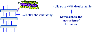
Hydrolysis and condensation reactions involved in the synthesis of organic–inorganic talc like hybrids (TLH) starting from diethylphosphatoethyltriethoxysilane (SiP), magnesium nitrate (Mg(NO3)2), ethanol and sodium hydroxide have been studied. The influence of magnesium nitrate, concentration of NaOH aqueous solution and pH value of the reaction media was investigated by high resolution 1H, 29Si and 31P liquid Nuclear Magnetic Resonance (NMR) and 29Si and 31P solid state NMR. It was shown that Mg(NO3)2 has a catalytic effect on the hydrolysis-condensation reaction rates of SiP and that high concentrations of NaOH lead to a scission of siloxane bonds as well as a partial hydrolysis of ethoxy groups linked to phosphorous. The final TLH products have been characterized by solid state 29Si NMR.


1. Introduction

2[thin space (1/6-em)]:[thin space (1/6-em)]1 phyllosilicates have a lamellar structure in which an octahedral sheet (O) containing one or more hexacoordinated element (aluminium, magnesium, …) is sandwiched between two tetrahedral (T) sheets containing one or more tetracoordinated elements (silicon, aluminium). Depending on the occupancy of the different sheets, the framework can be neutral, like in talc or pyrophyllite, or negatively charged due to isomorphic substitutions in the tetrahedral and/or octahedral sheets like in montmorillonite, hectorite or beidellite. In this case, interlayer cations balance the negative charges. Two post-treatments can be used to obtain organic–inorganic compounds starting from 2[thin space (1/6-em)]:[thin space (1/6-em)]1 phyllosilicates: by ion exchange between the inorganic interlayer cations and organic cations1 or by grafting. In this later case, an organoalkoxysilane is grafting directly on the OH group present at the edges of the inorganic frameworks.2,3 In 1995, a one-step synthesis involving a sol–gel process, was proposed by Fukushima and Tani4 to obtain an organic–inorganic talc like hybrid (TLH) with organic moieties covalently linked to the silicon atoms of the tetrahedral sheets and pending in the interlayer space. The chemical formula of these new compounds is: Mg3(RSi)4O8 (OH)2, with R being an organic moiety. To prepare TLH, a magnesium source such as magnesium nitrate5 or magnesium chloride4,6–10 is necessary to build the octahedral sheet. This salt is dissolved in water,5,11 ethanol9,10,12 or methanol4,7 prior addition of an organoalkoxysilane as silicon source. Many organoalkoxysilane were used, depending of the desired final properties. For example for adsorption of heavy metal ions, Lagadic et al.13 used a mercaptoorganoalkoxysilane. Later, Badshah et al.14 used this type of organoalkoxysilane for the adsorption of cations at liquid/solid interface. For the same properties Moscofian et al.15 used an organoalkoxysilane with an urea group. Patel et al.16 focused on tetraorthoethoxysilane (TEOS) and aminopropyltriethoxysilane (APTES) for the synthesis of TLH used as catalysts for condensation of aldehyde and ketone. After the silicon source addition, an aqueous solution of sodium hydroxide is added to allow the formation of the inorganic structure. Generally the concentration of basic solution is around 0.05 M.7–9 However in the presence of aminopropyltriethoxysilane (APTES), Lebeau et al.10 showed that the basic character of APTES is sufficient to form the TLH structure. The synthesis of TLH can be carried out at room temperature5,7,8,10,12 between 40 °C and 50 °C or under hydrothermal treatment.9–17 The synthesis duration goes from 5 days for Da-Fonseca et al.17 in 1999 to 4 hours for Ferreira et al.18 depending on the synthesis conditions and on the type of organoalkoxysilane. Usually the synthesis is performed during 24 hours.5,8,10,12

Fukushima et al.11 synthesized the first organic–inorganic talc like hybrid with 3-methacryloxypropyltriethoxysilane, magnesium chloride and sodium hydroxide at room temperature. They followed the procedure proposed by Mizutani et al.6 who studied the formation of Ni-phyllosilicate and Mg-phyllosilicate having a talc like structure. The synthesis of these compounds were carried out by mixing a silicic acidic solution with magnesium chloride and a solution of sodium hydroxide at room temperature and under hydrothermal condition (200 °C). By X-ray diffraction (XRD) and chemical analysis, it was shown that at room temperature the formation of the Mg-phyllosilicate was very slow. Mizutani et al.6 explained that that Ni or Mg phyllosilicates can selectively be prepared by controlling the copolymerization reaction of silicic acid and Ni or Mg salts.

Ukrainzyck et al.19 investigated the synthesis and characterization of Al and Mg phyllosilicate. The synthesis of these compounds were performed with several organoalkoxysilane (dodecyltriethoxysilane, penthyltriethoxysilane, phenyltrimethoxysilane, buthyltrimethoxysilane and 3-methacryloyloxypropyltrimethoxysilane) which were mixed with alcoholic solution of magnesium chloride and sodium hydroxide (0.5 M). It was suggested that the formation of lamellar structure is directed by the hydrolysis reactions of the alkoxide groups. This later acts as a surfactant organized as lamellar micelles that form a template which in presence of basic medium and metal species allow the formation of the phyllosilicate-like structure. However, this mechanism of formation is only observed in the case of Al phyllosilicates and is not elucidated to explain the formation of Mg phyllosilicates. They assume that this is due to the presence of a large micelles sterically hindering hydrolysis reactions. So they join to the Mizutani et al.6 proposal for the formation of the phyllosilicate like structure for some alkoxysilane. All the mechanisms were proposed according to X-ray diffraction (XRD), solid state nuclear magnetic resonance (ss-NMR), infrared spectroscopy (FTIR) and chemical analysis of the final solids, but no evidence of the kinetics and formation mechanism was demonstrated in the liquid phase.

In that frame, the present study focus on the mechanism of formation of talc like structure (TLH) prepared by sol–gel process. Diethylphosphatoethyltriethoxysilane (SiP) is used as silicon source and magnesium nitrate as magnesium source.20–23 The hydrolysis–condensation reactions of SiP in ethanol/water solutions were studied in 1996 by Cardenas et al.24 and more recently in 2008 by Van Nieuwenhuyse et al.25 They identified the different species present in solution and the kinetics of the SiP hydrolysis–condensation reactions. Here the formation of TLH will be followed by liquid 29Si and 31P NMR as well as by 29Si and 31P solid state NMR. The influence of magnesium nitrate in the hydrolysis and condensation reactions of SiP in ethanol will be first investigated, then the influence of the pH, the volume of sodium hydroxide as well as its concentration will be studied. All the data will contribute to gather information on the initial steps of TLH formation mechanism.

2. Experimentals

2.1. Materials

Magnesium nitrate hexahydrate (Fluka, 99.9 wt%), diethyphosphatoethyltriethoxysilane (ABCR, 92 wt%), EtOH (Analpur, 99.9 wt%), sodium hydroxide (Carlo Erba, 99 wt%), chromium(III) acetylacetonate (Cr(acac)3, Sigma Aldrich, 97 wt%), hexamethyldisilane (HDMS, Sigma Aldrich, 98 wt%), phosphoric acid-d3 (Sigma Aldrich, 85 wt% in D2O), ethanol-d6 (Euriso-Top) and D2O (Euriso-Top) were used without further purification.

2.2. TLH preparation for liquid state NMR kinetics studies

In a first step, magnesium nitrate hexahydrate (0.03 mol), ethanol (EtOH) (16 mL) and chromium acetylacetonate (2 × 10⁻² M) were mixed at room temperature until complete dissolution of magnesium and chromium salts. Then diethylphosphatoethyltriethoxysilane (SiP) (0.04 mol) was added. To reach a pH value of 12, an aqueous solution of NaOH (10 M or 1 M) was added dropwise. Two base concentrations were used in order to observe the dilution effect. The NMR analysis were performed prior the addition of NaOH solution and after each addition.

2.3. TLH preparation for solid state NMR kinetics studies

Magnesium nitrate hexahydrate (0.03 mol) and EtOH (16 mL) were first mixed at room temperature until complete dissolution of magnesium salts. Then diethylphosphatoethyltriethoxysilane (SiP) (0.04 mol) was added. To reach a pH value of 12, an aqueous solution of NaOH (10 M or 1 M) was added dropwise. After each addition of base, a fraction of the precipitate was collected by centrifugation (10[thin space (1/6-em)]000 rpm, 5 min) and dried during 24 h at 70 °C. Only two samples at pH 9 and pH 12 could be analyzed because for previous pH values, the amount of precipitate was too small.

The different steps of the formation of the TLH and the specific studies carried out are summarized in Fig. 1.


image file: c6ra12719c-f1.tif
Fig. 1 Scheme for the steps of the organic–inorganic talc like synthesis.

2.4. Characterization

2.4.1. Liquid-state NMR. 29Si NMR experiments were recorded at 300 K on a Bruker Avance II 400 spectrometer (79.5 MHz for 29Si) using a 10 mm 29Si selective probe with a z-gradient coil. A co-axial tube filled with a deuterated ethanolic (EtOH-d6) solution of HMDS (0.10 M) and Cr(acac)3 (2 × 10⁻² M) was used for calibration and lock. Chemical shifts are referenced to external HMDS (δ = −19.8 ppm) which is also used as a standard for quantification. An inverse gate decoupling pulse sequence was used with a 70° pulse angle and a recycle delay of 11 s.

1H and 31P NMR spectra were recorded at 300 K on a Bruker Avance III 400 spectrometer (400.13 MHz for 1H and 162.0 MHz for 31P) using a 5 mm BBFO + probe with a z-gradient coil. A co-axial tube filled with a deuterated water (D2O) solution of D3PO4 (2.85 mol L−1) was used for calibration and lock. A single pulse sequence with a 30° pulse angle and a recycle delay of 6 s and a power gate decoupling pulse sequence with a 30° pulse angle and a recycle delay of 5.5 s were used for 1H and 31P experiments respectively. 1H chemical shifts are referenced to external HDO signal set to 4.7 ppm and 31P chemical shifts are referenced to external D3PO4 signal set to 0 ppm.

2.4.2. Solid state NMR (ss-NMR). 29Si and 31P ss-NMR was performed on Bruker Advance II 300 MHz and 400 MHz spectrometers respectively. Main conditions are summarized in Table 1.
Table 1 Analysis recording conditions for 29Si and 31P solid state NMR experiments
  29Si 31P
CPMAS MAS + DEC MAS
a The measurement of the recycle time was made by inversion-recuperation method.
Spectrometer Bruker advance II 300 MHz Bruker advance II 400 MHz
Reference TMS H3PO4
Frequency (MHz) 59.6 162.0
Spinning rate (MHz) 4 12
Probe (mm) 7 4
Pulse (μs) 6.5 6.5 3.5
Contact time (ms) 4
Recycle time (s) 10a 80 25a
Flip angle π/2 π/6 π/2


3. Results and discussion

In 29Si NMR studies, the classical notation for silicon environments is T for silicon with three oxygen bridging atoms (Table 2). T notation is usually completed by two letters, i and j (Tij i, j = 0, 1, 2 or 3) where i is the number of oxo bridges Si–O–Si and j is the number of hydroxy groups.
Table 2 Assignment of 29Si NMR signals 7 for hydrolysis and condensation of SiP in EtOH/Mg (NO3)2,6H2O/NaOH according to Van Nieuwenhuyse et al.25 Signal labels used in Fig. 2, 4, 5, 7 and 9 are also indicated
Zones Chemical shift (ppm) Assignment Signal label Formula
a Can be T1 and/or T2.
T0 −48.1 T00 a RSi(OEt)3
−45.7 T01 b RSi(OH)(OEt)2
−43.9 T02 c RSi(OH)2(OEt)
−42.1 T03 d RSi (OH)3
T1 T12 −51.0 T12T12 e R(OH)2SiOSi(OH)2R
−51.3 image file: c6ra12719c-t1.tif f R(OH)(OEt)SiOSi(OH)2R
−51.1 image file: c6ra12719c-t2.tif i R(OH) (OH) SiOSi(OH) ROSi(OH)2R
T11 −53.0 image file: c6ra12719c-t3.tif g R(OH)(OEt)SiOSi(OH)2R
−53.1 T11T11 h R(OH)(OEt)SiOSi(OEt)R
T10 −55.2 T10T10 k R(OEt)2SiOSi(OEt)2R
T2 T21 −57 to −65 image file: c6ra12719c-t4.tif j R(OH)(OH)SiOSi(OH) ROSi(OH)2R
T20 image file: c6ra12719c-t5.tif m RSi(OH) (OSia)2
T3 T30 −65 to −70 image file: c6ra12719c-t6.tif l RSi(OSia)3


3.1. Influence of the presence of magnesium nitrate

Fig. 2 shows the evolution of 29Si NMR spectra of SiP/Mg (NO3)2·6H2O/EtOH mixture with reaction time.
image file: c6ra12719c-f2.tif
Fig. 2 29Si NMR spectra of mixture SiP/Mg(NO3)2·6H2O/EtOH with Cr(acac)3 at 0 min (1), 31 min (2), 1 h 46 min (3), 7 h 30 min (4) and 40 h (5) of reaction, where 0 min correspond to the SiP incorporation (labelling of the signals according to Table 2).

In the early stage of the reaction, four signals were observed at −48.3 ppm (a), −45.9 ppm (b), −44.1 ppm (c) and −42.3 ppm (d), corresponding to the SiP monomer (T00), and the hydrolyzed species (T01, T02 and T03) respectively (see Table 2). Multiple resonances were also observed between −50 ppm and −56 ppm corresponding to T10/T11/T12 condensed species. These later are present whatever the reaction time with slight differences in their relative intensity. T0 species slowly disappear with increasing reaction time and only traces are observed after 7 h 30 min (Fig. 2(4)). T2 species are already formed after 31 min (Fig. 2(2)) while T3 species are not yet detectable after 7 h 30 min. Broad signals from T3 species come out of the background noise only after 20 h of reaction (not shown), their presence is confirmed on the spectrum recorded after 40 h (Fig. 2(5)).

The spectra show fast hydrolysis and condensation reactions of SiP with a water content of 4.5 mol for 1 mol of SiP. It is worthy to note that previous studies on SiP hydrolysis–condensation reactions made by Cardenas et al.24 and Van Nieuwenhuyse et al.25 demonstrated that more than 4 hours are necessary at room temperature to have a complete hydrolysis of SiP in EtOH/water solutions with 5 mol of water for 1 mol of SiP and that even more time is needed to observe the first condensed species.

As a partial conclusion, for the same amount of water, the presence of magnesium nitrate seems to lead to a fastening of the hydrolysis–condensation reactions.

To confirm these observations, the evolution of the amounts of hydrolyzed and condensed species versus reaction time is depicted in Fig. 3 and compared to the study made by Van Nieuwenhuyse et al.25 with SiP in EtOH/water solutions (molar ratio water/SiP = 5).


image file: c6ra12719c-f3.tif
Fig. 3 Evolution over time of the different species contents (from integration of 29Si NMR signals) in SiP/Mg(NO3)2/EtOH and SiP/EtOH in mixtures prepared by Van Nieuwenhuyse et al.25

For the kinetics of SiP/EtOH with magnesium nitrate, three steps can be distinguished (Fig. 3). The first one occurs between 0 min and 10 min. At the beginning of the experiment, T0 hydrolysed species and T1 are formed very rapidly, whereas the formation of T2 is slower and no T3 environment is noticed. Between 10 min and 90 min, part of the T0 species are transformed into T1 and T1 are transformed into T2 species. Between 90 and 465 min, T0 and T1 environments contributions decreased and T2 environment increased which is consistent with the formation of the structure. After 465 min, it is worthy to note that T3 species started growing, T1 are still decreasing, T2 species increased and T0 species are almost totally consumed. This results can be compared to the one obtained by Van Nieuwenhuyse et al.25 in the presence of SiP/EtOH and H2O mixture. Indeed, the trend of kinetic curves is similar but the rate is extremely slow in their case which confirm that the presence of magnesium salt increase the condensation rate of the SiP. This is also proven by comparing the consumption of T0 species (50% of T0 are consumed in 15 min for the mixture with magnesium nitrate while 510 min are needed to consume them without magnesium ions).

Fig. 4 focus on the −49 ppm to −63 ppm 29Si NMR spectrum region for the mixture after 7 h 30 min of reaction. According to literature,24,25 the T1 region can be divided in three main areas: [−50/−52], [−52/−54] and [−54/−56] ppm which are assigned to T10, T11 and T12 moieties respectively. In the T2 region, two main areas [−60/−61.5] and [−61.5/−63.5] ppm can also be assigned to T20 and T21 moieties respectively. All those condensed compounds involve Si–O–Si bonds. New signals from −58.5 ppm to −60 ppm not described in the case of hydrolysis and condensation reactions of alkoxysilanes alone were are also observed and assumed to belong to T2 silicon atoms involved in Si–O–Mg bonds. Indeed, similar shifts were observed by Chabrol et al.3 for the grafting of talc with N-(3-triethoxysilylpropyl)-4,5-dihydroimidazole and 2-hydroxy-4-(3-triethoxysilylpropoxy)-diphenylketone. The talc formed before grafting, show in 29Si solid state NMR, for the Q2 environment, one resonance at −91.8 ppm corresponding to the (Si*(OSi)2(OH)2) groups and two shifted resonances at −87.5 ppm and −85.2 ppm assigned to the (Si*(OSi)2(OMg) (OH)) or (Si*(OSi)2(O□) (OH)) (were □ stands for Mg2+ missing in the octahedral sheet) respectively. However, in this study no equivalent T1 Si–O–Mg species are observed.


image file: c6ra12719c-f4.tif
Fig. 4 Zoom of the T1 and T2 29Si NMR spectrum region for SiP/Mg (NO3)2·6H2O/EtOH mixture at 7 h 30 min of reaction (labelling of the signals according to Table 2).

3.2. Influence of the aqueous solution of NaOH

To enlighten the further discussion, Table 3 first gathered the SiP to water ratio for all the different reactional medium studied.
Table 3 SiP to water ratio for the different reactional mixtures
  SiP/EtOH25 SiP/EtOH/Mg(NO3)2·6H2O SiP/EtOH/Mg(NO3)2·6H2O, NaOH 10 M SiP/EtOH, NaOH 10 M
pH 9 pH 12 pH 10 pH 12
SiP (mole) 1 1 1 1 1 1
Water (mole) 5 4.5 6.2 8.7 0.3 1.7


3.2.1. Influence on hydrolysis and condensation reactions.
Mixture of SiP/EtOH and NaOH 10 M. The evolution of the reactional solution mixture SiP/EtOH in basic medium was monitored by 29Si NMR after addition of sodium hydroxide solution to reach pH 12 (Fig. 5).
image file: c6ra12719c-f5.tif
Fig. 5 29Si NMR spectra of mixture SiP/EtOH and NaOH 10 M for pH 5 (1), pH 8 (2), pH 10 (3), pH 12 (4) and pH 12 after 2 h (5) (labelling of the signals according to Table 2).

In this case the water/SiP ratio increases with addition of NaOH solution but remain below the value used by Van Nieuwenhuyse et al. so it is difficult to compare the kinetics. From these spectra, it is interesting to note that whatever the volume of NaOH added to the solution and the reaction time, no hydrolysed species are observed in the reaction media (even for the water/SiP ratio of 1/1.7 at pH = 12). Moreover it appears clearly that for all the studied pH values, the condensation rate is very fast and quite instantaneous in presence of NaOH.

Fig. 6 displays the evolution of SiP T0 species during sodium hydroxide addition, the comparison was made with the same mixture without base but in presence of water.25


image file: c6ra12719c-f6.tif
Fig. 6 Evolution of the SiP hydrolyzed T0 species during the sodium hydroxide addition monitored by 29Si liquid state NMR.

Additions of NaOH allow to drastically fasten the consumption of T0 species, when compared to the study by Van Nieuwenhuyse et al.25 When reaching pH 12, T0 species are fully consumed. This observation confirms the fast condensation of the SiP species in the presence of NaOH.

In frame on the classical conditions used for the TLH synthesis; evolution of the mixture when pH 10 and pH 12 are reached is more specifically studied (Fig. 7 and 8).


image file: c6ra12719c-f7.tif
Fig. 7 Evolution of the 29Si NMR spectra of the mixture SiP/EtOH and NaOH 10 M at pH 10: (1) 10 min after SiP addition, (2) 45 min, (3) 2 h and (4) 4 h (labelling of the signals according to Table 2).

image file: c6ra12719c-f8.tif
Fig. 8 Different environments obtained at pH 12 for the mixture SiP/EtOH and NaOH 10 M where (1) corresponds to 10 min after SiP addition, (2) 5 h, (3) 18 h and (4) 91 h.

As previously observed, at pH 10 only 0.3 mole of water is present in the solution for 1 mole of the SiP. This rate is not sufficient to hydrolyze all the ethoxy group present in the SiP based solution. This is evidenced in Fig. 7 where the evolution of the species during time for a given pH is displayed.

At pH 12 (Fig. 8) hydrolyzed species are not present, only T2 and T3 condensed species are observables. As the amount of water is 1.67 mole for 1 mole of SiP, it is sufficient to hydrolyze all the ethoxy functions. Among the broad peaks, the presence of thin resonances in the T2 and T3 environment suggests the presence of silsesquioxane structures. In 1999 Fasce et al.26 studies the synthesis of polyhedral silsesquioxanes (POSS) and characterized the obtaining structure by 29Si NMR. They showed the presence of broader peaks due to the presence of different organic group but they observed the presence of a thin peaks at −66 ppm and they supposed the presence of a Tx structure, were x stands for the number of silicon atoms. Many other studies dedicated to POSS structures confirmed that point.27–30 Beside some studies focusing on the influence of the basic medium as the work of Rikowski et al.27 showed, that Tx cubes can be formed under basic medium. In our study thin 29Si peaks suggest therefore the formation of cages in which the phosphorus group is present as doublet signals are observed.


Mixture of SiP/EtOH/Mg(NO3)2·6H2O and NaOH 10 M. Fig. 9 shows the NMR spectra of the SiP/Mg(NO3)2/EtOH reactional medium with addition of an aqueous solution of NaOH at different pH. As the reaction proceeds with the addition of NaOH, a solid precipitate in the solution. NMR analysis were made only on the soluble part. After addition of 1 drop of 10 M aqueous solution of NaOH, a pH value of 5 is obtained and only three of the hydrolyzed species (T00, T01, T02), remain in the mixture whereas T03 species (d) is no more present (Fig. 9(1)). This underlines the influence of NaOH on the kinetic of formation of the different species. As previously mentioned the signal attributed to the T2 Si–O–Mg environments is observed. After addition of 7 drops (pH = 8, t = 1 h 29 min) Fig. 9(2) of NaOH 10 M solution, hydrolyzed species T00 and T01 are still present. Concerning the condensed species broader and broader signals are observed even between the usual spectral widths of Si–O–Si bonds suggesting the contribution of the magnesium inside the framework. When reaching pH 12 (Fig. 9(4)), thin signals appear again in the T1, T2 and T3 region but no noteworthy evolution is observed after pH 12.
image file: c6ra12719c-f9.tif
Fig. 9 29Si NMR spectra of mixture SiP/Mg (NO3)2·6H2O/EtOH and NaOH 10 M for pH 5 (t = 33 min) (1), pH 8 (t = 1 h 29 min) (2), pH 10 (t = 2 h 33) (3), pH 12 (t = 3 h 52) (4) and pH 12 + 1 h 30 min (t = 5 h 29) (5) (t = 0 correspond to the SiP incorporation) (labelling of the signals according to Table 2).

As underlined previously for TLH synthesis a basic medium is usually needed. However the obtained results suggest that when the pH value equals 12, a scission of Si–O–Si bonds (or Si–O–Mg) occurs. In regards to the conventional preparative method, the NaOH concentration is here ten times higher (i.e. 10 M instead of 1 M). In order to check the influence of the relative concentration of NaOH, experiments were carried out using NaOH 1 M solution and same pH conditions. The kinetics data recorded by liquid 29Si NMR (not shown here) are similar in both case even if the dilution of the medium induces a lower signal to noise ratio for the detection of silicon signals. These analysis confirmed that at high pH values, chains scissions occurred.

To complete this hypothesis, Fig. 10 displays the 29Si CP-MAS spectra of the precipitate formed when synthesis are carried out in the same conditions as for 29Si liquid NMR kinetics. That will gather information in order to go deeper in the understanding of TLH formation mechanism. Table 4 summarizes the range of chemical shift known for the different expected silicon environments.9,10,12


image file: c6ra12719c-f10.tif
Fig. 10 Comparison of 29Si CP-MAS NMR spectra of mixture SiP/Mg(NO3)2·6H2O/EtOH in presence of NaOH 10 M and NaOH 1 M for different pH; (1-a) NaOH 1 M pH 9; (1-b) NaOH 1 M pH 12; (2-a) NaOH 10 M pH 9; (2-b) NaOH 10 M pH 12.
Table 4 29Si ss-NMR signals assignment for hydrolysis and condensation of silicone with magnesium. (M = Si or Mg)9,10,12
Environments Assignments (M = Si or Mg) Chemical shift (ppm)
T1 RSi(OH)2(OM) −45 to −55
T2 RSi(OSi)(OH)(OM) −55 to −65
T3 RSi(OSi)2(OM) −65 to −80


In these spectra, regardless the concentrations of base, the results were similar. The resonances at −51 ppm, −57 ppm indicate the presence of T1 and T2 environment for all the samples. One supplementary resonance around −69 ppm corresponding to T3 species is observed on the spectra of the samples prepared at pH 9 starting from NaOH 1 M and 10 M. At higher pH, the formation of fully condensed species is not observed; this can be correlated to the liquid NMR results which showed that at high pH soluble condensed species undergo chemical bonds scission. The presence of higher amount of T3 species for synthesis made with NaOH 1 M suggests an effect of the dilution on the condensation rate. The reaction time should have also an effect on the Si–O–Si or Si–O–Mg cleavage. As mentioned in the literature,31 long reaction times are expected to promote the cleavage at high pH values. In our case this is confirmed by the disappearance of T3 environment in solid phase and the appearance of hydrolyzed species in the liquid phase. MAS-DEC NMR spectra were also recorded in order to quantify the proportions of different silicon environments in the precipitates. The results obtained after deconvolution (Table 5) confirm the observations made in Fig. 10. Between pH 9 and pH 12 whatever the NaOH concentration (1 M and 10 M) T3 environment disappear (16–0% and 8–0%) and T1 environment became more significant (20–60% and 40–50%). As the signal to noise ratio is quite low owing to the very low amount of precipitate, the values obtained by the signal decomposition are not very accurate. It has to be underlined that whatever the NaOH concentration, there is an increase of the T1 and T2 species and a decrease of T3 species as the pH increases.

Table 5 Percentage of the different environment from MAS + DEC 29Si NMR for the precipitates synthesized with different pH and NaOH concentrations
pH 9 pH 12
Environment Chemical shift ppm Percentage Environment Chemical shift ppm Percentage
1 M
T0 T0
T1 −49.9 25 T1 −51.2 51
−53.8 9
T2 −57.3 56 T2 −56.9 40
−64.1 3
T3 −70.1 16 T3
[thin space (1/6-em)]
10 M
T0 T0
T1 −50.5 39 T1 −50.3 42
T2 −56.5 50 T2 −56.4 56
−61.8 3 −61.7 2
T3 −69.6 8 T3


3.2.2. Diethylphosphato group reactivity. As the synthesis of TLH proceeds in basic conditions with magnesium salts, it is important to check if there is a modification of the phosphorous environment during the synthesis.

The 31P NMR spectra of the of SiP/Mg(NO3)2/EtOH reactional medium before and after addition of NaOH (10 M) were recorded versus reaction time. They are shown in Fig. 11–13.


image file: c6ra12719c-f11.tif
Fig. 11 31P liquid NMR spectra of the mixture SiP/EtOH (1) and the mixture SiP/Mg(NO3)2/EtOH versus time: (2) 22 min after SiP addition, (3) 1 h, (4) 5 h, (5) 10 h 30 min, and (6) 41 h.

image file: c6ra12719c-f12.tif
Fig. 12 31P liquid NMR spectra of the mixture SiP/Mg(NO3)2/EtOH and NaOH 10 M at pH 10 during time: (1) 22 min after NaOH 10 M addition, (2) 1 h, (3) 2 h, (4) 4 h, (5) 25 h, (6) 48 h.

image file: c6ra12719c-f13.tif
Fig. 13 31P liquid NMR spectra of the mixture SiP/Mg(NO3)2/EtOH and NaOH 10 M at pH 12 during time: (1) 22 min after NaOH 10 M addition, (2) 1 h 30 min, (3) 7 h, and (4) 95 h.

The first spectrum obtained after addition of SiP exhibits broad and split signals between 32 and 33.5 ppm close to SiP phosphorous group chemical shift in ethanolic solvent. Those signals become broader and broader with reaction times while the corresponding 1H spectra show only a decrease of the signals of ethoxy group linked to the silicon atom. No modification of proton ethoxy signals linked to phosphorous was detected. As the corresponding liquid 29Si NMR spectra (Fig. 2) also disclose a fast hydrolysis and condensation of SiP at those reaction times, this splitting and broadening the phosphorus signal in presence of magnesium salts was only attributed to those first mentioned reactions.

After addition of NaOH to reach pH 10, the signals of phosphorous are slightly shifted [32.5 to 34 ppm] compared to the ones observed with only magnesium salts in the mixture and a broader signal is observed at 35 ppm. As the hydrolysis–condensation reactions proceeds, according to 29Si NMR kinetics, the phosphorous signals become broader and broader but the corresponding spectra obtained by 1H NMR do not show any modification of the proportion of ethoxy groups linked to the phosphorus. This points out that at pH = 10, the chemical shift and shape of the phosphorus signal, is only impacted by the condensation of SiP and slightly by the change in pH conditions.

Then, Fig. 13 displays the 31P liquid NMR spectra of the SiP/EtOH/Mg(NO3)2 mixture with NaOH 10 M at pH 12 versus time, corresponding to the last step of the talc like hybrid synthesis.

The signals of phosphorous are slightly shifted [34 to 38 ppm] and after 22 min of reaction the signal becomes broader and extends toward 33 ppm. The presence of a broadening of the peaks can, as previously mentioned, be attributed to the formation of the condensed species at pH 12 flowed by subsequent scission as observed by 29Si NMR analysis. 1H NMR analysis was also performed (not shown here) and depicted the disappearance of ethoxy groups linked to silicon while no significant change was evidenced in the signals of ethoxy groups linked to phosphorus, except a shift and a broadening of the CH2–P signal.

Lastly Fig. 14 exhibit the 31P MAS NMR spectra of precipitates from SiP/EtOH/Mg (NO3)2 mixture with NaOH 10 M at pH 9 and pH 12.


image file: c6ra12719c-f14.tif
Fig. 14 31P MAS NMR spectra of the precipitate from SiP/Mg(NO3)2, 6H2O/EtOH in presence of NaOH 10 M for different pH; (1) pH 9 and (2) pH 12.

As previously shown on the soluble species, the 31P signals of the precipitates are slightly different depending on the pH of synthesis. At pH 9 one environment seems present at 35 ppm, but after decomposition two environments are detected at 33 ppm and 36 ppm. For the precipitate obtained at pH 12, two environment are clearly identified at 37 ppm and 30 ppm. Actually even if no reaction of phosphorous was evidenced by liquid state NMR, these observations in the solid state tend to demonstrate that in the TLH structure organic moieties are not all located in the same sites. We are still investigating this point.

4. Conclusion

The present study was dedicated to the elucidation of talc like structure (TLH) formation mechanism. Compared to the hydrolysis–condensation reactions of the diethylphosphatoethyltriethoxysilane (SiP) as a silicon source, it was shown that the presence of the magnesium nitrate hexahydrate enhanced the sol–gel reactions. From the multinuclear NMR analysis, the presence of Si–O–Mg bonds were evidenced participating to the TLH building. Similarly, deeper NMR analysis depicted the role of the addition of the base on the condensed species observation. Moreover, the impact of the pH on the final structure allowed to determine that for a pH value over 9, siloxane bonds scissions are observed limiting the growth of the inorganic material. Finally, 31P NMR analysis shown that the diethylphosphato group is located in different environments according to the growth of the TLH but keep its initial chemical structure. It was also evidenced that the formation of Si–O–Mg is observed with or without addition of NaOH and that this later allows reducing the reaction time of the synthesis. The formation of Si–O–Mg bonds suggests the formation of the octahedral sheet prior the formation of the tetrahedral ones.

Acknowledgements

The authors thank the ANR for the funding of this study through the MATETPRO Project ANR12-RMNP 0017.

References

  1. S. Williams-Daryn and R. K. Thomas, J. Colloid Interface Sci., 2002, 255, 303–311 CrossRef CAS PubMed.
  2. K. A. Carrado, Appl. Clay Sci., 2000, 17, 1–23 CrossRef CAS.
  3. K. Chabrol, M. Gressier, N. Pebere, M.-J. Menu, F. Martin, J.-P. Bonino, C. Marichal and J. Brendle, J. Mater. Chem., 2010, 20, 9695–9706 RSC.
  4. Y. Fukushima and M. Tani, J. Chem. Soc., Chem. Commun., 1995, 241–242 RSC.
  5. J.-C. Gallégo, M. Jaber, J. Miehé-Brendlé and C. Marichal, New J. Chem., 2008, 32, 407–412 RSC.
  6. T. Mizutani, Y. Fukushima, A. Okada and O. Kamigaito, Bull. Chem. Soc. Jpn., 1990, 63, 2094–2098 CrossRef CAS.
  7. S. L. Burkett, A. Press and S. Mann, Chem. Mater., 1997, 9, 1071–1073 CrossRef CAS.
  8. N. T. Whilton, S. L. Burkett and S. Mann, J. Mater. Chem., 1998, 8, 1927–1932 RSC.
  9. C. R. Silva, M. G. Fonseca, J. S. Barone and C. Airoldi, Chem. Mater., 2002, 14, 175–179 CrossRef CAS.
  10. B. Lebeau, J. Brendle, C. Marichal, A. J. Patil, E. Muthusamy and S. Mann, J. Nanosci. Nanotechnol., 2006, 6, 1–8 CrossRef.
  11. Y. Fukushima and M. Tani, Bull. Chem. Soc. Jpn., 1996, 69, 3667–3671 CrossRef CAS.
  12. G. Kumaraswamy, Y. Deshmukh, V. V. Agrawal and P. Rajmohanan, J. Phys. Chem. B, 2005, 109, 16034–16039 CrossRef CAS PubMed.
  13. I. L. Lagadic, M. K. Mitchell and B. D. Payne, Environ. Sci. Technol., 2001, 35, 984–990 CrossRef CAS PubMed.
  14. S. Badshah and C. Airoldi, Thermochim. Acta, 2013, 552, 28–36 CrossRef CAS.
  15. A. S. O. Moscofian, C. R. Silva and C. Airoldi, Microporous Mesoporous Mater., 2008, 107, 113–120 CrossRef CAS.
  16. H. A. Patel, S. K. Sharma and R. V. Jasra, J. Mol. Catal. A: Chem., 2008, 286, 31–40 CrossRef CAS.
  17. M. G. Da Fonseca, C. R. Silva and C. Airoldi, Langmuir, 1999, 15, 5048–5055 CrossRef CAS.
  18. R. B. Ferreira, C. R. da Silva and H. O. Pastore, Langmuir, 2008, 24, 14215–14221 CrossRef CAS PubMed.
  19. L. Ukrainzyck, R. A. Bellman and A. B. Anderson, J. Phys. Chem. B, 1997, 101, 531–539 CrossRef.
  20. J. Alongi, C. Colleoni, G. Rosace and G. Malucelli, J. Therm. Anal. Calorim., 2011, 110, 1207–1216 CrossRef.
  21. J. Alongi, C. Colleoni, G. Rosace and G. Malucelli, Polym. Degrad. Stab., 2013, 98, 579–589 CrossRef CAS.
  22. P. Theil-Van Nieuwenhuyse, V. Bounor-Legaré, P. Bardollet, P. Cassagnau, A. Michel, L. David, F. Babonneau and G. Camino, Polym. Degrad. Stab., 2013, 98, 2635–2644 CrossRef CAS.
  23. J. Bonnet, V. Bounor-Legaré, F. Boisson, F. Melis, G. Camino and P. Cassagnau, Polym. Degrad. Stab., 2012, 97, 513–522 CrossRef CAS.
  24. A. Cardenas, N. Hovnanian and M. Smaihi, J. Appl. Polym. Sci., 1996, 60, 2279–2288 CrossRef CAS.
  25. P. Van Nieuwenhuyse, V. Bounor-Legaré, F. Boisson, P. Cassagnau and A. Michel, J. Non-Cryst. Solids, 2008, 354, 1654–1663 CrossRef CAS.
  26. D. P. Fasce, R. J. J. Williams, F. Méchin, J. P. Pascault, M. F. Llauro and R. Pétiaud, Macromolecules, 1999, 32, 4757–4763 CrossRef CAS.
  27. E. Rikowski and H. C. Marsmann, Polyhedron, 1997, 16, 3357–3361 CrossRef CAS.
  28. H. Cai, X. Zhang, K. Xu, H. Liu, J. Su, X. Liu, Z. Fu, Y. Guo and M. Chen, Polym. Adv. Technol., 2012, 23, 765–775 CrossRef CAS.
  29. S. Chimjarn, R. Kunthom, P. Chancharone, R. Sodkhomkhum, P. Sangtrirutnugul and V. Ervithayasuporn, Dalton Trans., 2015, 44, 916–919 RSC.
  30. M. G. Voronkov and V. I. Lavrentev, Top. Curr. Chem., 1982, 102, 199–236 CrossRef CAS.
  31. W. Prince and R. Perami, Cem. Concr. Res., 1993, 23, 1121–1129 CrossRef CAS.

This journal is © The Royal Society of Chemistry 2016
Click here to see how this site uses Cookies. View our privacy policy here.