DOI:
10.1039/C6RA12189F
(Paper)
RSC Adv., 2016,
6, 66394-66406
Assessment on the antibacterial activity of nanosized silica derived from hypercoordinated silicon(IV) precursors
Received
10th May 2016
, Accepted 2nd July 2016
First published on 6th July 2016
Abstract
Silica nanoparticles were synthesized through a versatile sol–gel combustion method from hydrazide based hypercoordinated silicon complexes derived from the reaction of silicon tetrachloride with O-silylated hydrazide derivatives. The complexes were characterized by 1H, 13C, 29Si NMR and ESI-mass spectrometric techniques. A refined morphology was observed in the product after sintering i.e. from spherical to rod shaped nanoparticles. The powder X-ray diffraction patterns and the TEM images of silica show the formation of silica nanoparticles. The IR spectra show Si–O linkages and DLS studies indicate the particle size distribution to be between 20 and 100 nm for the material before sintering and 70–120 nm after sintering at 1000 °C. A TEM image of the decomposed gel indicates the formation of crystalline silica rods. This work also demonstrates the influence of nano-sized silica particles on antibacterial activity (DIZ, MIC and MBC) i.e. better activity was shown for nano-rods derived from the hypercoordinated silicon complexes than the conventional TEOS (sol–gel) method. Experiments on the generation of reactive oxygen species (ROS) through oxidative stress demonstrate the toxicity of nanosilica particles.
1. Introduction
Porous silica and silica nanoparticles are well-known industrial materials due to their widespread applications in sorption, separation, sensing and catalysis because of their large surface area.1 The physicochemical properties of nanomaterials strongly depend on the size, shape, surface area and structure of particles.2 Apart from this, the recent focus has been centered on biomedical applications of porous silica in controlled drug delivery systems and bio-sensing.3 The major applications of nano-sized silica in biology are used in gene transfer, drug delivery and bio-imaging.4 Various approaches have been developed to synthesize porous silica and silica nanoparticles using tetraethyl orthosilicate (TEOS) as the silicon source.5–8 Also, the research on silica nanoparticles and frameworks derived from photosynthetic organisms and biomass has been growing due to the flexibility and ease of scaling-up.9 Although several processes such as sol–gel synthesis,10 combustion,11 flame synthesis12 and micro-emulsion methods13 are used in the synthesis of silica nanoparticles, the sol–gel process among them involving hydrolysis and polycondensation of the respective silica precursor is the unique and highly used method.14 Interestingly, silica is also used by some living organisms like diatom algae (known as silicon-containing organisms because of their high ecological importance) as the construction material for elements of the skeleton.15
Tetraethoxysilane (TEOS) is most commonly used precursor for the preparation of spherical nanosilica particles selectively.16 Considering the ever increasing demand on porous silica and nanosilica materials for new applications,1–4 it is highly desirable to seek an alternative approach that can provide controlled morphology and porosity for future prospects. In this regard, it seems that organometallic approach is a valuable alternative for the synthesis of nano-sized materials as the displacement of organic ligands under mild conditions should afford a clean surface without any contaminants detrimental for the chemical or physical properties of the resulting nanomaterials.17 Also, this is one of the best methods to synthesize alloys of metals having varied reduction potentials.18
In order to obtain a better control of the size and size distribution of the particles as well as to obtain clean surfaces, we have used an alternative approach using hypercoordinated silicon(IV) complexes as precursors by controlling the pretreatment and reaction conditions. In general, these hypercoordinated silicon(IV) complexes show flexibility towards bond breaking and bond forming reactions as well as facile ligand exchangeability depending on the nature of solvent, temperature and substituents on silicon and the ligand systems.19
2. Materials and methods
All the reactions were carried out under dry nitrogen using Schlenk techniques. Solvents were dried and purified by standard methods. Reagents (including polyhalosilanes and TEOS (99%), triethylamine (99%), trimethylsilylchloride (98%)) were purchased from Sigma-Aldrich. Halosilanes were distilled and kept in sealed ampules prior to use. Throughout the procedures double de-ionized (DI) water (with a measured resistivity of 18.2 MΩ cm−1) was used. Nutrient agar media (Himedia Lab Ltd., Bangalore, India) was used for evaluation of bacterial growth in liquid broth culture and was supplemented with a 2% bacterial agar (Himedia Lab. Ltd., Banglore, India) to prepare the solid media used in plate culture studies.
NMR spectra were recorded on a Bruker Avance DMX-400 spectrometer operating at 500.13, 125.76, and 99.36 MHz, respectively, for 1H, 13C, and 29Si spectra. For identification of functional group by Shimadzu instrument for IR analysis with KBr pellet over the range to 4000 cm−1. The phase purity was investigated by Bruker D8 Advance XRD, at the step intervals of 0.02 and using copper Kα (1.5406 Å) and Ni filtered radiation. The decomposition of the complex was characterized by thermogravimetric analysis (TGA) coupled with differential thermal analysis (DTA). The surface morphology of calcined silica product was analyzed by scanning electron microscopy (SEM, S4800 Hitachi). The SiNP powders (precursor and calcinated one) were suspended uniformly in ethanol and sonication for 5 min. Grids for the high resolution tunneling election microscopy (HR-TEM) analysis were made and analyzed by HR-TEM (JEOL JEM 2000 Fx II, Japan, at 200 kV).
The particle size distribution was analyzed by Dynamic Light Scattering (DLS, HORIBA Instruments Pvt Ltd., Singapore) by preparing a clear and uniform suspension using a sonicator. The scattering angle was kept at 173 °C with holder temperature of 25.2 °C, mono-dispersion medium viscosity of 1.000 mPa s, and count ratio of 1247 kcps. The analyzer used was HORIBA SZ-100 for windows [Z Type] version 2.0.
2.1. Reactive oxygen species (ROS) assay
The oxidant sensitive dye DCFH2DA was used for the detection of reactive oxygen species (ROS). When the cells are exposed to the respective silica nanoparticles (SiC and SiT), they induce the oxidative stress over E. coli cells and subsequently the release of reactive oxygen species happens. This involves further in the rupture of cells and leading to death of the cells.
2.2. Methodology for ROS assay
The cultured E. coli cells were treated with different types of silica nanoparticles (SiC and SiT) for next 48 hours and one negative control was kept to analyze comparatively. The cultured cells were centrifuged at 4000 rpm at 4 °C for 10 minutes to aspirate out the medium and cells were then re-suspended in phosphate buffer at pH 7.0. It can be noted that 10 mM DCFH2DA (2′,7′-dichlorodihydrofluorescein diacetate, a weak fluorescent dye) in methanol was diluted in culture medium to get 100 μM solution. The treated samples as well as negative control were treated with 100 μM DCFH2DA for 60 min. Next, centrifugation of the cellular suspension followed by removal of extracellular media to remove any excess DCFH2-DA was carried out and further washed three times to have clear readings. The fluorescence of oxidized green fluorescent 2′,7′-dichlorofluorescein (DCF) was measured at excitation wavelength 492 nm and emission at 523 nm using HITACHI F-700 fluorescent spectrophotometer. It can be noted that, the same has been confirmed with fluorescent microscope.
3. Results and discussion
3.1. Synthesis and characterization of hypercoordinated silicon(IV) complexes
Hypercoordinated silicon complexes 1–3 were synthesized by general silylation and transsilylation of N-(cyclohexylideneimino)-O-(trimethylsilyl) isonicotinimidate derivatives as shown in Scheme 1.20 The purity and structure of synthesized complexes were confirmed by UV-Vis, FT-IR, NMR and ESI MS.
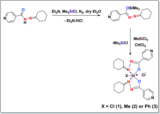 |
| | Scheme 1 General route for the synthesis of hydrazide based hypercoordinated silicon(IV) complexes.12 | |
The ESI mass spectrum of 3 exhibited directly [M − Cl]+ at m/z 537.2 without the molecular ion peak (Fig. 1 and Scheme 2). In general, ion formation arises from charge exchange between the fast-moving incident particles and the neutral molecules in solution to generate M± or M+ and ions.21 Complexes with halide ligands often lose HX, concomitant with oxidative addition of a carbon–hydrogen bond of a ligand to the metal center, or alternatively, formation of an [M − X]+ ion is a common ionization route. Further the peak at m/z 298.3 could be due to the formation of a cyclized product, 4. The same product was also obtained during thermolysis of 1–3.
 |
| | Fig. 1 ESI-mass spectrum of 3. | |
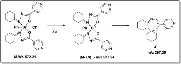 |
| | Scheme 2 Possible mass fragmentation pattern of 3. | |
3.2. Synthesis of nanosilica particles
The silica nanomaterials were synthesized through sol–gel combustion method from the hypercoordinated silicon complexes as described in Fig. 2.
 |
| | Fig. 2 Synthesis of nanoparticles from the hydrazide based hypercoordinated silicon complexes. | |
Moreover, the hydrazide ligands which are coordinated to the hyper-coordinated silicon complex act as a fuel during combustion process and nitric acid acts as an oxidant. The reaction is highly exothermic and the large volume of gases evolved during combustion process leads to the high porosity of the particles.
3.3. Characterization of nanosilica particles
3.3.1. IR spectral analysis. The stretching modes corresponding to azomethine group (1629 cm−1), –NCO (1672 cm−1, 1652 cm−1) and Si–O (760 cm−1) in silicon complexes (Fig. 3) were completely disappeared in the precursor obtained from combustion as well as in the calcined product. The appearance of Si–O–Si bonding vibrations (1068 cm−1, 796 cm−1 and 457 cm−1) in both precursor and calcined products shows the formation of silica nanoparticles as shown in Table 1.
 |
| | Fig. 3 FT-IR spectral comparison for the sintered silica and the precursor. | |
Table 1 Comparative infrared spectral data of the nanosilica particles obtained from hypercoordinated silicon complexes (1–3)
| Complex |
Infrared absorptions |
| νSi–O (cm−1) |
νNCO (cm−1) |
νC![[double bond, length as m-dash]](https://www.rsc.org/images/entities/char_e001.gif) N (cm−1) N (cm−1) |
νSi–O–Si (cm−1) (symmetric stretching) |
νSi–O–Si (cm−1) (symmetric bending) |
νSi–O–Si (cm−1) (asymmetric bending) |
| 1–3 |
740–750 |
1672 and 1652 |
1629 |
Absent |
Absent |
Absent |
| Precursor |
Absent |
Absent |
Absent |
1068 |
796.60 |
457 |
| Calcinated product (1000 °C for 4 h) |
Absent |
Absent |
Absent |
1068 |
792.74 |
457 |
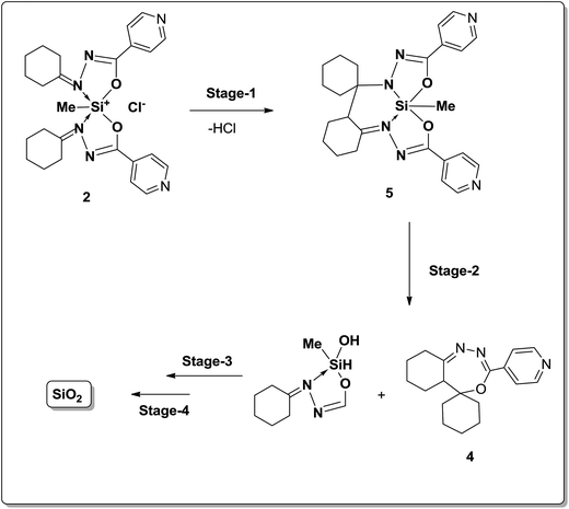
3.3.2. TGA analysis of complex 2. The thermal degradation of 2 was monitored with a thermogravimetric analyzer (TGA) (Fig. 4). Samples were heated from ambient temperature to 900 °C at a heating rate of 5 °C min−1. Before 200 °C, a weight loss of 7.6% is observed, and this can be attributed to the loss of HCl to form a neutral trischelate 3 as described previously.22 The next weight loss (54%) occurred between 200 and 300 °C, which may correspond to the elimination of pyridyl groups and a cyclized product (4) without silicon. Further weight loss could be due to remainder of the organic ligand; the losses that follow may correspond to the formation of residual silica.23 Further the formation of silica was observed from the proposed decomposition pathways as shown in Scheme 3. The TGA analysis data is given in Table 2.
 |
| | Fig. 4 Thermal analysis of the hypercoordinated silicon(IV) complex studied in the present work. | |
 |
| | Scheme 3 Proposed mechanistic pathway for the formation of nanosilica particles. | |
Table 2 Thermogravimetric analysis data of complex 2
| Decomposition stages |
Temperature range (°C) |
Weight loss (%) |
Calculated weight loss (%) |
| Stage-1 |
28–200 |
7.6 |
7.1 |
| Stage-2 |
200–300 |
53.0 |
54.7 |
| Stage-3 |
300–400 |
27.3 |
26.0 |
| Stage-4 |
400–600 |
3.5 |
3.4 |
3.3.3. XRD analysis. XRD pattern (Fig. 5a) of the precursor obtained from combustion indicates a broad pattern i.e. the amorphous nature of the particles formed at the time combustion. The increase in particle size is due to the aggregation of spherical particles during calcination and this is confirmed by the powder XRD analysis. Moreover, the XRD pattern of calcined product shows sharp peaks which indicates the product formed is well crystalline. Calcination of the sample at 1000 °C shows a single phasic well crystalline crystobalite phase which matches with the JCPDS file no. 39-1425. The XRD pattern indicates a diffracted broad peak at 2θ = 22.51, which reveals the formation of amorphous silica without any other ordered crystalline structures. The XRD pattern (Fig. 5b) of the precursor calcined at 1000 °C for 4 h shows a crystalline pattern with a sharp peak at 2θ = 21.28 (101) and 35.56 (200), which confirms the presence of cristobalite. The average crystalline size was calculated by using Scherer's formula as 82 nm. The sintered product containing nano-silica represents cubic crystal symmetry.
 |
| | Fig. 5 Powder XRD patterns of (a) precursor synthesized from sol–gel method at 80 °C (b) precursor calcinated at 1000 °C for 4 h. | |
The high intensity peaks were considered for calculating the average particle size by using Debye–Scherrer formula (eqn (1)).
| |
D = Kλ/β![[thin space (1/6-em)]](https://www.rsc.org/images/entities/char_2009.gif) cosθ cosθ
| (1) |
K is the constant approximate value of 0.9,
λ is Cu-Kα wavelength 1.5406 Å and
β are the full width half maximum.
3.3.4. DLS studies. Average particle size of prepared nanosilica particles was estimated by DLS (Fig. 6). According to these results, the particle size of silica particles before sintering was in the range of 20–100 nm and after calcinating at 1000 °C the particle size was found to be in the range of 70–120 nm. The formation of nano-sized silica particles is also confirmed by TEM analysis (Fig. 6b and c) where the particles formed are found to be in nano regime.
 |
| | Fig. 6 (a) and (d) – Particle size distribution (by DLS); (b) and (c) – TEM images of precursor and calcinated products respectively. | |
3.3.5. TEM analysis. The TEM micrographs of silica nanoparticles obtained before and after calcination show that with change in calcinations temperature, the dispersion states and sizes of silica nanoparticles also changes (Fig. 6b and c). Fig. 6b shows the precursor sample obtained at 80 °C in which the particles are highly agglomerated. The TEM micrograph of silica nanoparticles obtained by calcinating the precursor at 1000 °C in which the organic moieties and carbon particles were removed by oxidation, reveals the rod shaped morphology as shown in Fig. 6c. The product shows a refined surface morphology i.e. from spherical to rod shaped nanoparticles after sintering.
3.4. Bacterial toxicological evaluation of silica nanoparticles
The antibacterial activity of various materials like graphene,24 chitin, oligosaccharides, chitosan,25 metal nanoparticles,26 metal-carbon coatings,27 and metal-doped hydroxyapatite28 has been reported. Among them, chitosan derivatives29 have been extensively studied using various modified surfaces.
In the present work, a comparative antibacterial activity profile has been determined for silica nanoparticles. Silica nanoparticles prepared from the silicon complex is denoted as SiC whereas the silicon nanoparticles prepared from TEOS is denoted as SiT. The antibacterial activity of the silicon nanoparticles prepared from two different sources were analyzed for various Gram negative (E. coli) and Gram positive (S. aureus and B. subtilis) bacterial strains by different factors – DIZ, MIC, MBC and growth kinetics. The results have been compared with the control drug, gentamicin and the antibacterial activity is found to be significantly high for the silica nanoparticles.
The magnitude of susceptibility of microorganisms is reflected by DIZ; the more susceptible microbial strains exhibit more DIZ and vice versa. The disks impregnated with SiC were surrounded by larger DIZ compared to SiT for B. subtilis while the E. coli strain has lesser susceptibility followed by S. aureus. It can be noted that, although the difference in DIZ and MIC is there but the growth kinetics (GrK) is almost similar for B. subtilis and S. aureus. The representative image showing the DIZ for SiC and SiT impregnated disks for microbial strains (Fig. 7a and b) indicates the evidence for the more activity of SiC than SiT. The DIZ and AI are 10–30% more for SiC than SiT which may be due to the change in morphology or change in particle size.
 |
| | Fig. 7 (a) Zone of Inhibition in S. aureus with nano-sized silica derived from TEOS (SiT) and hydrazide based complex (SiC); (b) zone of inhibition in E. coli with nanosilica derived from TEOS (SiT) and hydrazide based complex (SiC). | |
To demonstrate the antibacterial activity of nanosilica particles different bacterial strains have been selected (one Gram negative and two Gram positive strains). Table 3 summarizes the DIZ, MIC and MBC of SiC and SiT; which represents the antibacterial activities of nanosilica particles dispersed in batch culture. With the initial bacterial concentration of 103 to 104 CFU mL−1, MIC and MBC represents the bacteriostatic and bactericidal effects of nanosilica particles respectively. To the best of our acquaintance, this is the first study providing a systematic and detailed study regarding, the complete antimicrobial profiling for nanosilica particles synthesized by sol–gel approach which also includes MIC, MBC and GrK analysis. The MIC ranges from 40 to 60 μg mL−1 and 30 to 50 μg mL−1 for SiT and SiC respectively against different Gram positive and Gram negative bacterial strains. Similarly, MBC ranges from 60 to 80 μg mL−1 and 40 to 60 μg mL−1 for SiT and SiC respectively against different Gram positive and Gram negative bacterial strains. In order to optimize the concentration, various amounts of silica nanoparticles were used in the experiments. From these results, it can be concluded that silica nanoparticles are having broad-spectrum of bactericidal activities which was found to be concentration dependent, it can be noted that for comparative analysis, the concentration of SiC and SiT was considered at their respective MBC concentration for a particular bacterial strains, from which bacterial growth curve was inhibited. The above mentioned initial concentration was adjusted in 50 mL nutrient broth and the antibacterial activity was evaluated up to 3 hours, since the minimum time to achieve bacteriostatic and bactericidal effects is likely to fall within said period.
Table 3 Summary on antibacterial activity of SiC and SiT
| Antimicrobial characteristics |
SiTa |
SiCb |
| SiT-silica nano derived from TEOS. SiC-silica nano derived from hydrazide based hypercoordinated complexes. |
| DIZ in cm (E. coli) |
2.3 |
2.9 |
| MIC in μg mL−1 (E. coli) |
40 |
30 |
| MBC in μg mL−1 (E. coli) |
60 |
40 |
| DIZ in cm (B. subtilis) |
2.7 |
3.3 |
| MIC in μg mL−1 (B. subtilis) |
60 |
50 |
| MBC in μg mL−1 (B. subtilis) |
80 |
60 |
| DIZ in cm (S. aureus) |
2.4 |
3.0 |
| MIC in μg mL−1 (S. aureus) |
50 |
40 |
| MBC in μg mL−1 (S. aureus) |
70 |
50 |
The growth profile of bacterial strains with varying concentration of SiC and SiT are depicted in Fig. 8a–c. In batch culture studies, as the concentration of silica nanoparticles increases, the lower maximum absorbance (at 600 nm) and greater lag phase was observed. Notably, for B. subtilis and S. aureus strains, both the nanosilica particles show maximum bacteriostatic effect as there is sharp decrease in absorbance for particular concentration of nanosilica particles also, the increase in greater lag phase. Additionally, the graphs represent SiC has more bacteriostatic activity which establishes the fact for its higher radius values of DIZ and lower MIC and MBC. Similar reports have been found with nanosilver and nano cupric oxide on E. coli and other microbial strains as reported earlier and they used higher concentrations of silver nanoparticles (107 and 105 to 106 CFU mL−1).30–32 As per our earlier studies, the degree of antibacterial activity of polyelectrolyte stabilized Ag nanoparticles varies with their particle size.33 Moreover in this study we used (103 to 104 CFU mL−1) initial bacterial concentration has been taken irrespective of microbial strains and nanosilica concentration. No growth was observed in the flask as the concentration of silica nanoparticles (SiC and SiT) increased to MIC.
 |
| | Fig. 8 (a–c) Growth kinetics of (a) Gram negative (E. coli) and Gram positive (S. aureus) (b) and B. subtilis (c) bacterial strains. | |
The mechanistic pathways involved in antibacterial activity of nanoparticles are not fully understood and the complexity of nanoparticle-related cytotoxicity emphasizes the need for more detailed studies.34 It is speculated that the internalization of the nanosilica particles cause the death of the cell by inhibiting the DNA replication and blocking cellular respiration (Fig. 9).
 |
| | Fig. 9 Fluorescence image and emission spectrum of dead E. coli cells of a control. | |
3.5. Mechanism of action of cytotoxic activity of nanosilica particles on bacterial cells
It is well known that the cells under stress generate reactive oxygen species i.e. oxygen free radical. Notably, this free radical reacts with the cellular proteins, DNA, membrane etc., which may further leads to death of cells.34–37 Even though few reports have discussed the possible electrostatic interaction between other nanoparticles and bacteria, but the exact fact behind the mechanism of action of silica nanoparticles towards the death of bacterial cells has not been established.34–40 In this context, the exact mechanism for bacterial cell death was sought. Reactive oxygen species generation was induced in the cells by creating the oxidative stress with two different types of nanosilica particles (SiC and SiT) as mentioned above and the intensity of reactive oxygen has been measured by the fluorescent color developed after reaction between ROS and DCFH2-DA. It can be noted that, the more fluorescence depicts the more generation of ROS, which ultimately depicts more bacterial cell death. This fluorescence has been observed by fluorescent spectrophotometer and fluorescent microscopy.
The results on fluorescent microscopic images and emission spectra confirm the involvement of ROS in the cell damage. The nanosilica particles (SiC and SiT) showed higher emissions as well as higher number of fluorescent cells (Fig. 10 and 11) over the control (Fig. 9). This could be due to the generation of ROS that is responsible for cell damage under over stressed conditions. Fig. 12 represents the schematic representation of bacterial cell death due to ROS generation in the presence of silica nanoparticles. It can be noted that, the concentrations of both the nanomaterials were used as 70 ppm.
 |
| | Fig. 10 Fluorescence image and emission spectrum of dead E. coli cells of the SiC culture. | |
 |
| | Fig. 11 Fluorescence image and emission spectrum of dead E. coli cells of the SiT culture. | |
 |
| | Fig. 12 Schematic representation of cell death due to ROS generation under stressed conditions of nanosilica particles.41 | |
4. Conclusion
Hydrazide based hypercoordinated silicon complexes were prepared and used as a precursor to synthesize silica nanoparticles through sol–gel combustion method. Calcination at 1000 °C provided a refined morphology of the product from spherical to rod-shaped nanoparticles. The nano-size was confirmed by powder X-ray diffraction patterns and the TEM images. The IR spectral and DLS studies clearly shows the Si–O linkages and size distribution level from 20–100 nm. The influence of nanosized silica particles showed better antibacterial activity (DIZ, MIC and MBC) for nano-rods derived from the hypercoordinated silicon complexes than conventional TEOS method. Nanosilica-induced oxidative stress is responsible for the damage of cells and better antibacterial activity.
Acknowledgements
Support for this work from DST-SERB, New Delhi, India (Ref. No. SR/Si/IC-38/2011) is gratefully acknowledged. Mr Janardan Sannapaneni thanks DST for the fellowship. ASRK and his group members thank DST-VIT-FIST for NMR and XRD spectrometers and SIF-VIT University for other instrumentation facilities.
References
-
(a) F. Glasser, Silicates, Encyclopedia of Materials Science and Engineering, Pergamon Press Ltd, 1986, vol. 6, p. 4393 Search PubMed;
(b) S. Uhrlandt, in Kirk–Othmer Encyclopaedia of Chemical Technology, ed. A. Seidel, John Wiley & Sons, Hoboken, NJ, 2006, p. 365 Search PubMed;
(c) P. Liu, C. Gao, C. Han, H. Tang, F. Wang, Y. Ding, S. Zhang and M. Yang, Polym. Int., 2015, 64, 1053–1059 CrossRef CAS;
(d) J. L. Gurav, I. K. Jung, H. H. Park, E. S. Kang and D. Y. Nadargi, J. Nanomater., 2010, 11 Search PubMed;
(e) G. M. Pajonk, Colloid Polym. Sci., 2003, 281, 637–651 CrossRef CAS;
(f) G. Jia-Hu, L. Yu-Cun, C. Tao, J. Su-Ming, M. Hui, Q. Ning, Z. Hua, Y. Tao and H. Wei-Ming, RSC Adv., 2015, 5, 44990–44997 RSC.
- F. Tang, L. Li and D. Chen, Adv. Mater., 2012, 24, 1504–1534 CrossRef CAS PubMed.
-
(a) B. G. Trewyn, S. Giri, I. I. Slowing and V. S. Y. Lin, Chem. Commun., 2007, 31, 3236–3245 RSC;
(b) I. I. Slowing, B. G. Trewyn, S. Giri and V. S. Y. Lin, Adv. Funct. Mater., 2007, 17, 1225–1236 CrossRef CAS.
-
(a) C. Barbe, J. Bartlett, L. Kong, K. Finnie, H. Q. Lin, M. Larkin, S. Calleja, A. Bush and G. Calleja, Adv. Mater., 2004, 16, 1959–1966 CrossRef CAS;
(b) J. Athinarayanan, V. S. Periasamy and A. A. Alshatwi, J. Mater. Sci.: Mater. Med., 2014, 25, 1637–1644 CrossRef CAS PubMed.
- A. S. Dorcheh and M. Abbasi, J. Mater. Process. Technol., 2008, 199, 10–26 CrossRef.
- N. J. Halas, ACS Nano, 2008, 2, 179–183 CrossRef CAS PubMed.
- J. L. V. Escoto, I. I. Slowing, B. G. Trewyn and V. S. Lin, Small, 2010, 6, 1952–1967 CrossRef PubMed.
- N. Baccile, F. Babonneau, B. Thomas and T. Coradin, J. Mater. Chem., 2009, 19, 8537–8559 RSC.
-
(a) V. Bansal, P. Poddar, A. Ahmad and M. Sastry, J. Am. Chem. Soc., 2006, 128, 11958–11963 CrossRef CAS PubMed;
(b) H. Ehrlich, K. D. Demadis, O. S. Pokrovsky and P. G. Koutsoukos, Chem. Rev., 2010, 110, 4656–4689 CrossRef CAS PubMed;
(c) S. Mann, Nature, 1993, 365, 499–505 CrossRef CAS;
(d) W. Wang, J. C. Martin, X. Fan, A. Han, Z. Luo and L. Sun, ACS Appl. Mater. Interfaces, 2012, 4, 977–981 CrossRef CAS PubMed;
(e) A. A. Alshatwi, J. Athinarayanan and V. S. Periasamy, Mater. Sci. Eng., C, 2015, 47, 8–16 CrossRef CAS PubMed.
- H. Du, P. D. Hamilton, M. A. Reilly, A. Avignon, P. Biswas and N. Ravi, J. Colloid Interface Sci., 2009, 340, 202–208 CrossRef CAS PubMed.
- M. Wooldridge, P. Torek, M. Donovan, D. Hall, T. Miller, T. Palmer and C. Schrock, Combust. Flame, 2002, 131, 98–109 CrossRef CAS.
- H. Chang, J.-H. Park and H.
D. Jang, Colloids Surf., A, 2008, 313–314, 140–144 CrossRef.
- W. Wang, X. Fu, J. Tang and L. Jiang, Colloids Surf., A, 1993, 81, 177–180 CrossRef CAS.
- T. Coradin and P. J. Lopez, ChemBioChem, 2003, 4, 251–259 CrossRef CAS PubMed.
-
(a) P. Treguer, D. M. Nelson, A. J. Van Bennekom, D. J. Demaster, A. Leynaert and B. Queguiner, Science, 1995, 268, 375 CAS;
(b) R. Gordon, D. Losic, M. A. Tiffany, S. S. Nagy and F. A. S. Sterrenburg, Trends Biotechnol., 2009, 27, 116–127 CrossRef CAS PubMed;
(c) V. V. Annenkov, V. A. Pal'shin, O. N. Verkhozina, L. I. Larina and E. N. Danilovtseva, Mater. Chem. Phys., 2015, 165, 227–234 CrossRef CAS.
- W. Stöber, A. Fink and E. Bohn, J. Colloid Interface Sci., 1968, 26, 62–69 CrossRef.
-
(a) K. Philippot and B. Chaudret, C. R. Chim., 2003, 6, 1019–1034 CrossRef CAS;
(b) B. Chaudret and K. Philippot, Oil Gas Sci. Technol., 2007, 62, 799–817 CrossRef CAS;
(c) B. Chaudret, C. R. Phys., 2005, 6, 117–131 CrossRef CAS;
(d) M. Green, Chem. Commun., 2005, 24, 3002–3011 RSC.
- C. N. R. Rao, A. Müller and A. K. Cheetham, Nanomaterials Chemistry: Recent Developments and New Directions, Wiley-VCH, Weinheim, 2007, p. 5 Search PubMed.
-
(a) S. Janardan, P. Suman, K. Vijayakrishna and A. Sivaramakrishna, Synth. React. Inorg., Met.-Org., Nano-Met. Chem., 2016, 46, 311–316 CrossRef CAS;
(b) I. Kalikhman, B. Gostevskii, A. Sivaramakrishna, D. Kost, N. Kocher and D. Stalke, Steric Effect on the Formation, Structure and Reactions of Pentacoordinate Siliconium-Ion Salts, in Organosilicon Chemistry VI, ed. N. Auner and J. Weis, VCH-Wiley, Weinheim, 2008 Search PubMed;
(c) B. Gostevskii, G. Silbert, K. Adear, A. Sivaramakrishna, D. Stalke, S. Deuerlein, N. Kocher, M. G. Voronkov, I. Kalikhman and D. Kost, Organometallics, 2005, 24, 2913–2920 CrossRef CAS;
(d) B. Gostevskii, K. Adear, A. Sivaramakrishna, G. Silbert, D. Stalke, N. Kocher, I. Kalikhman and D. Kost, Chem. Commun., 2004, 14, 1644–1645 RSC;
(e) B. Gostevskii, V. Pestunovich, I. Kalikhman, A. Sivaramakrishna, N. Kocher, S. Deuerlein, D. Leusser, D. Stalke and D. Kost, Organometallics, 2004, 23, 4346–4348 CrossRef CAS;
(f) P. Suman, S. Janardan, K. Vijayakrishna and A. Sivaramakrishna, Polyhedron, 2015, 99, 266–274 CrossRef CAS;
(g) I. Kalikhman, B. Gostevskii, O. Girshberg, A. Sivaramakrishna, N. Kocher, D. Stalke and D. Kost, J. Organomet. Chem., 2003, 686, 202–2014 CrossRef CAS.
- S. Janardan, P. Suman, A. Sivaramakrishna and K. Vijayakrishna, Polyhedron, 2015, 85, 34–40 CrossRef CAS.
- W. Henderson and J. S. McIndoe, A Wiley Text book on Mass spectrometry of inorganic and organometallic compounds, John Wiley & Sons, Ltd, 2005, p. 62 Search PubMed.
- B. Gostevskii, V. Pestunovich, I. Kalikhman, A. Sivaramakrishna, N. Kocher, S. Deuerlein, D. Leusser, D. Stalke and D. Kost, Organometallics, 2004, 23, 4346–4348 CrossRef CAS.
-
(a) F. Mucha, J. Haberecht, U. Böhme and G. Roewer, Monatsh. Chem., 1999, 130, 117–132 CAS;
(b) K. W. Terry, P. K. Ganzel and T. D. Tilley, Chem. Mater., 1992, 4, 1290–1295 CrossRef CAS.
- H. M. Hegab, A. E. Mekawy, L. Zou, D. Mulcahy, C. P. Saint and M. Ginic-Markovic, Carbon, 2016, 105, 362–376 CrossRef CAS.
- J. Dutta, S. Tripathi and P. K. Dutta, Food Sci. Technol. Int., 2012, 18, 3–34 CrossRef CAS PubMed.
-
(a) M. S. Rubina, E. E. Kamitov, Y. V. Zubavichus, G. S. Peters, A. V. Naumkin, S. Suzer and A. Y. Vasil'kov, Appl. Surf. Sci., 2016, 366, 365–371 CrossRef CAS;
(b) H. Li, Q. Cui, B. Feng, J. Wang, X. Lu and J. Weng, Appl. Surf. Sci., 2013, 284, 179–183 CrossRef CAS.
- H. Huang, Y.-Y. Chang, J.-X. Liu, M.-T. Tsai and C.-H. Lai, Thin Solid Films, 2015, 596, 111–117 CrossRef CAS.
- M. Koizhaiganova, I. Yasa and G. Gulumser, Int. Biodeterior. Biodegrad., 2015, 105, 262–267 CrossRef CAS.
-
(a) S. Kumar, V. Deepak, M. Kumari and P. K. Dutta, Int. J. Biol. Macromol., 2016, 84, 349–353 CrossRef CAS PubMed;
(b) P. Tripathi, A. K. Gupta and P. K. Dutta, Asian Chitin J., 2013, 9, 7 Search PubMed;
(c) S. Tripathi, G. K. Mehrotra and P. K. Dutta, Bull. Mater. Sci., 2011, 34, 29–35 CrossRef CAS;
(d) K. Rinki, P. K. Dutta, A. J. Hunt, D. J. Macquarrie and J. H. Clark, Int. J. Polym. Mater., 2011, 60, 988–999 CrossRef CAS;
(e) J. Singh and P. K. Dutta, Int. J. Polym. Mater., 2010, 59, 793–807 CrossRef CAS.
- I. Sondi and B. Salopek-Sondi, J. Colloid Interface Sci., 2004, 275, 177–182 CrossRef CAS PubMed.
- J. P. Ruparelia, A. K. Chatterjee, S. P. Duttagupta and S. Mukherji, Acta Biomater., 2008, 4, 707–716 CrossRef CAS PubMed.
- S. Agnihotri, S. Mukherji and S. Mukherji, RSC Adv., 2014, 4, 3974–3983 RSC.
- K. T. Prabhucharan, N. Pothanagandhi, K. Vijayakrishna, A. Sivaramakrishna, D. Mecerreyes and B. Sreedhar, Eur. Polym. J., 2014, 60, 114–122 CrossRef CAS.
- T. J. Brunner, P. Wick, P. Manser, P. Spohn, R. N. Grass, L. K. Limbach, A. Bruinink and W. J. Stark, Environ. Sci. Technol., 2006, 40, 4374–4381 CrossRef CAS PubMed.
-
(a) A. Nel, T. Xia, L. Mädler and N. Li, Science, 2006, 311, 622–627 CrossRef CAS PubMed;
(b) N. Dasgupta, S. Ranjan, B. Rajendran, V. Manickam, C. Ramalingam, G. S. Avadhani and A. Kumar, Environ. Sci. Pollut. Res. Int., 2016, 23, 4149–4163 CrossRef CAS PubMed;
(c) H. J. Johnston, G. Hutchison, F. M. Christensen, S. Peters, S. Hankin and V. Stone, Crit. Rev. Toxicol., 2010, 40, 328–346 CrossRef CAS PubMed.
- T. G. Dong, S. Dong, C. Catalano, R. Moore, X. Liang and J. J. Mekalanos, Proc. Natl. Acad. Sci. U. S. A., 2015, 112, 2181–2186 CrossRef CAS PubMed.
- A. Jain, R. Shivendu, D. Nandita and C. Ramalingam, Crit. Rev. Food Sci. Nutr., 2016 DOI:10.1080/10408398.2016.1160363.
- W. Jiang, H. Mashayekhi and B. Xing, Environ. Pollut., 2009, 157, 1619–1625 CrossRef CAS PubMed.
- J. T. Seil and T. J. Webster, Int. J. Nanomed., 2012, 7, 2767–2781 CAS.
- N. Dasgupta, S. Ranjan, B. Rajendran, V. Manickam, C. Ramalingam, G. S. Avadhani and A. Kumar, Environ. Sci. Pollut. Res. Int., 2016, 23, 4149–4163 CrossRef CAS PubMed.
- S. Ranjan, N. Dasgupta, S. Chinnappan, C. Ramalingam and A. Kumar, Proc. Natl. Acad. Sci., India, Sect. B, 2016 DOI:10.1007/s40011-015-0673-z.
|
| This journal is © The Royal Society of Chemistry 2016 |
Click here to see how this site uses Cookies. View our privacy policy here. 


