DOI:
10.1039/C6RA11658B
(Paper)
RSC Adv., 2016,
6, 64531-64543
Induction of intrinsic apoptotic pathway and cell cycle arrest via baicalein loaded iron oxide nanoparticles as a competent nano-mediated system for triple negative breast cancer therapy
Received
5th May 2016
, Accepted 23rd June 2016
First published on 27th June 2016
Abstract
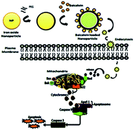
Magnetic nanoparticles have shown an increasing number of applications in the field of molecular medicine. In the present study we investigated the controlled synthesis of biocompatible polymer coated iron oxide nanoparticles (Fe2O3) loaded with baicalein and evaluated these for their drug loading and release behaviour. Moreover, the loaded particles were evaluated for their anti-proliferation, cell cycle arrest and apoptosis activation properties in triple negative breast cancer cells. The structure, morphology and magnetic properties of the prepared materials were studied by using scanning electronic microscopy, transmission electronic microscopy, Fourier transform infrared spectroscopy, X-ray diffraction analysis, thermogravimetric analysis, dynamic light scattering analysis and zeta potential analysis. DLS and TEM analysis confirms that the size of the synthesized iron oxide nanoparticles are about 90–100 nm and that they are extremely crystalline and spherical in nature. FTIR analysis confirms that the baicalein molecules were conjugated with PEG-coated iron oxide nanoparticles. X-ray diffraction patterns indicate that the magnetic nanoparticles were highly pure with a spinel structure. Baicalein loaded nanoparticles show a controlled release profile in response to various pH levels. We found significant cell cycle arrest at various phases in the treated group and subsequent apoptotic cell death to be evidenced by AO/EtBr, DAPI and PI of fluorescence microscopy analysis. The TUNEL assay evidences that DNA damage occurs in the treated cells. Further baicalein loaded iron oxide nanoparticles exhibit significant down-regulation of the anti-apoptotic protein Bcl-2 and up-regulation of pro-apoptotic proteins evidenced by Western blotting. Our findings clearly demonstrate that baicalein loaded iron oxide nanoparticles could efficiently deliver the drug of interest, and initiate and execute the apoptotic process in triple negative breast cancer cells. This could open a new window in polymer surface chemistry to develop metal oxide based drug delivery systems for all kinds of cancer therapeutics.
Introduction
Cancer is one of the most fatal diseases in the world, and the number of new cases increases each year. Despite the fact that chemotherapeutic and radiotherapeutic options are available for cancer, the critical challenge is to deliver them specifically to cancer cells with the additional concern of minimizing toxic effects to normal cells. Triple negative breast cancer cells (TNBC) lack ER− (estrogen), PR− (progesterone) and HER2− (Herceptin) receptors. Nowadays such receptors are targets in cancer treatment, and also are responsible for triple negative breast cancer being faster and more aggressive compared to other types of breast cancer.1 There are many treatment options for other breast cancers but the absence of receptors in TNBC is the main drawback in the treatment of this type of cancer. When exploring new strategies for treatment, one possibility is the use of nanocomposites to overcome this problems, to minimize the side effects of drugs and also to use a targeted drug delivery system.2,3
Baicalein, a flavonoid obtained from the roots of the traditional Chinese herbal medicine Huangqin, Scutellaria baicalensis Georgi, has been widely used for treatment of inflammation, hypertension, cardiovascular disease, bacterial infection and cancer. Baicalein has been found to selectively induce apoptosis in human cancer cell lines with minimal toxicity to normal cells.4
Modern developments in nanotechnology mean that a variety of superparamagnetic and ferromagnetic iron oxide (FeO/Fe2/Fe3) nanoparticles of different surface chemistry have been widely studied for several biological applications such as drug delivery, diagnostics, hyperthermic treatment and magnetic resonance imaging.5,6 These wide applications of Fe/Fe3O4 nanoparticles are possible since they exhibit favorable properties such as high magnetization ability, they are smaller than 100 nm in size, they can be encrusted with several ligands which allow drug delivery at a specific site and are biocompatible. Through using Fe/Fe2O3, the costs and side effects of using Fe3O4 nanoparticles as drug delivery agents can be reduced, meanwhile therapeutic efficacy can be increased.7
Due to their unique physicochemical properties and ability to function at the cellular and molecular level of biological interactions, iron nanoparticles have been actively investigated as next generation targeted drug delivery agents for over thirty years. The importance of targeted drug delivery is to transport a drug directly to the site of the disease under various conditions and thereby treat it consciously, without disturbing normal tissues.8,9
In drug delivery, drug uptake by the targeted cancer cells generally relies on specificity and dispersion process. Biocompatible nanocarriers like Fe2O3 may help in achieving reasonably greater specificities of particles loaded with anticancer drugs, whereas this parameter may be influenced by an additional surface coating. This form of passive drug delivery of anticancer agents, adsorbed on the surface of nanoparticles, is a promising alternative to conventional chemotherapy to increase drug-targeting probability and reduce undesirable side effects.10 A very critical parameter needed to be known in this case is the amount of drug loaded on to the nanoparticles behaving as nanocarriers. This loaded drug is then required to be released at the targeted inflammation side to have its therapeutic effect. No such detailed investigation is available into how the loaded drug is delivered to the targeted site.11
As far as this is concerned, the aspiration of the present study was to develop iron oxide nanoparticles by a co-precipitation method with controllable parameters and enhanced biocompatible properties. They were also evaluated for their standard drug encapsulation and release properties which respond to various pHs in the system. Cellular internalization of the nanoparticles was also observed using cancer cells. In addition, baicalein loaded iron oxide nanoparticles were synthesized and characterized and were evaluated to explore their potential anticancer properties in triple negative breast cancer cells (Schemes 1 and 2).
 |
| Scheme 1 The mechanism of baicalein linked to the surface iron oxide nanoparticles with activated PEG. The PEGylation process activated with NHS forms the efficient linkage between baicalein and iron surface. | |
 |
| Scheme 2 The preparation of baicalein loaded iron oxide nanoparticles for a controlled drug delivery system and activation of the intrinsic apoptotic signalling pathway in MDA-MB-231 breast cancer cells. The intrinsic pathway of caspase activation is initiated by events such as DNA damage and growth factor deficiency. These events ultimately lead to changes in the integrity of the mitochondrial membrane, which is regulated by Bcl-2 family proteins. The balance between pro- and anti-apoptotic Bcl-2 family members determines whether or not a cell will undergo apoptosis. In healthy cells, phosphorylated Bad is sequestered in the cytoplasm by the 14-3-3 protein, and Bcl-2 and Bcl-xL bind to the pro-apoptotic BAX and BAK proteins to inhibit apoptosis. Loss of mitochondrial integrity results in the release of pro-apoptotic proteins including cytochrome c and apoptosis-inducing factor (AIF). Cytochrome c interacts with APAF-1, which recruits pro-caspase-9 to caspase 3 and executes programmed cell death. | |
Materials and methods
Chemicals and reagents
Iron(III) chloride hexahydrate (FeCl3·6H2O) pure granulated (99%) and baicalein (98%) were purchased from Sigma India. Urea (CH4N2O), KOH, ethylene glycol and butanol were purchased from Himedia. All other chemicals were of reagent grade and used without further purification.
Cell line and culture conditions
Triple negative breast cancer MDA-MB-231 cells and HBL-100 human normal breast epithelial cells were purchased from the National Centre for Cell Science (Pune, India). The cells were maintained in Dulbecco’s Modified Eagle’s medium supplemented with 2 mM L-glutamine and Earle’s BSS adjusted to contain 1.5 g l−1 sodium bicarbonate, 0.1 mM nonessential amino acids, and 1.0 mM of sodium pyruvate in a humidified atmosphere containing 5% CO2 at 37 °C.
Synthesis of Fe2O3 nanoparticles
The Fe2O3 magnetic nanoparticles were synthesized by a simple co-precipitation method. In this experiment 2 mM of ferric chloride hexahydrate (FeCl3·6H2O) was used as source. In addition, 15 mM of sodium hydroxide (NaOH) was used as a basic medium and 40 ml of diethylene glycol and 0.5 mM hexamine were added to the reaction mixture. The solution was allowed to stir continuously for 45 minutes at room temperature to form a homogeneous solution. Then, this homogeneous solution was transferred to a 50 ml capacity Teflon lined stainless steel vessel and kept in a hot air oven at 180 °C for 12 h. After the reaction was completed, the solution was allowed to cool at room temperature and the precipitate was washed repeatedly more than five times with distilled water and three times with ethanol under centrifugation at 8000 rpm. The precipitate was separated and dried at 65 °C overnight to get the final product Fe2O3 nanoparticles.
Synthesis of PEG coated Fe2O3
Polyethylene glycol-coated nanoparticles were synthesized using a similar synthesis procedure by the addition of PEG. The detailed experimental procedure is as follows; 5 ml of PEG in a 1 mM concentration was added to a 10 ml solution of Fe2O3 nanoparticles (1 mg ml−1 concentration). In addition 1 mM of N-hydroxy succinimide (NHS) was added to activate the PEGylation process. NHS-activation for efficient PEGylation of primary amines occurs at pH 7–9, reaction of the NHS-ester group results in the formation of stable, irreversible amide bonds. The amide bond formed is highly physiologically stable. The PEG spacer provides unique advantages to the materials, including increased stability, reduced tendency toward aggregation and reduced immunogenicity. The solution was then sonicated for 3 h at 30 °C for uniform dispersion and disaggregation of particles. The solution was centrifuged and washed several times with ethanol and dried overnight under vacuum. The sample thus prepared was heated for 24 h at 200 °C.
Preparation of baicalein loaded Fe2O3 nanoparticles
Baicalein conjugated iron oxide nanoparticles were prepared by a co-precipitation method with minor modifications. In general, baicalein is virtually insoluble in water but it is soluble in ethanol. The required amount of baicalein was dissolved in 3 ml of ethanol. Then, 100 mg of PEG coated iron oxide nanoparticles were dissolved in 30 ml of dimethyl sulfoxide (DMSO) and deionized water under ultrasonication. Subsequently, the baicalein solution was added to the above mixture and the ultrasonication was continued and allowed to stir for 24 h at room temperature. The pH of the solution was adjusted to 10 by adding KOH solution. The resulting black-coloured precipitate was washed with distilled water to remove any unbound drug or any other organic impurities. The resultant drug loaded iron oxide nanoparticles were collected using a lyophilizer.
FT-IR characterization
Fourier transform infrared (FT-IR) spectroscopy of Fe2O3 was performed by using a Nicolet 5700 instrument (Nicolet Instrument, Thermo Company, USA) with a KBr pellet method. Each KBr disk was scanned over a wavenumber region of 500–4000 cm−1.
X-ray diffraction analysis
An X-ray diffraction (XRD) method was used to investigate the crystalline structure of the Fe2O3 nanoparticles. XRD analysis was conducted using a Philips PW 17291 powder X-ray diffractometer with a voltage of 40 kV, 25 mA. The scanning rate employed was 10 min−1 over a 2θ range of 10–80°.
Zeta potential analysis
The zeta potential of the synthesized nanoparticles was determined by means of a zeta potential analyzer (90 Plus Particle Size Analyzer, Brookhaven Instruments Corporation, using Zeta plus software). The measurement of zeta potential is based on the direction and velocity of particles under the influence of a known electric field.
Particle size analysis
The particle size range of the nanoparticles along with their polydispersity was determined using a particle size analyzer (90 Plus Particle Size Analyzer, Brookhaven Instruments Corporation). Particle size was derived based on measuring the time dependent fluctuation of laser light scattering by nanoparticles undergoing Brownian motion.
VSM analysis
The saturation magnetization (Ms) and coercive force (Hc) of the samples were measured using a vibrating sample magnetometer (VSM, Dexing, Model: 250) under magnetic fields of up to 10 kOe.
TGA analysis
TGA analysis was performed on a TGA Q50. Samples were placed in platinum sample pans and heated under an argon atmosphere at a rate of 20 °C min−1 to 100 °C and held for 30 min to completely remove residual solvent. Samples were then heated to 600 °C at a rate of 20 °C min−1.
Scanning electron microscopy (SEM) analysis
The samples were placed on a polycarbonate substrate and the excess water was left to dry at room temperature. They were then dried in a critical point dryer using carbon dioxide, and sputter coated with gold in a metallizer, and examined under a scanning electron microscope (JSM5600LV, JEOL, Japan) operated at an accelerating voltage of 20 kV.
Transmission electron microscope (TEM) analysis
The particle size and morphology of Fe2O3 nanoparticles were examined using a Philips CM120 transmission electron microscope at a voltage of 80 kV. An aqueous dispersion of the particles was drop-cast onto a carbon-coated copper grid and the grid was air dried at room temperature before viewing under the microscope.
Baicalein loading and encapsulation efficiency
Baicalein loaded Fe2O3 nanoparticles (3 mg) were dispersed into 6 ml of phosphate buffer solution (PBS) and centrifuged at 12
000 rpm for 30 min. The supernatant was collected to measure the ultraviolet absorption at 280 nm. The loading efficiency and encapsulation efficiency of the baicalein loaded Fe2O3 nanoparticles were calculated as follows.
Loading efficiency = W0/W × 100% |
Encapsulation efficiency = W0/W1 × 100% |
where, W0 is the amount of baicalein contained in the Fe2O3 nanoparticles, W is the amount of Fe2O3 nanoparticles, and W1 is the amount of baicalein added into the system.
In vitro baicalein release studies
Release properties of baicalein from iron oxide nanoparticles was investigated at different pH levels, 7.4, 6.8 and 5.5. The baicalein loaded iron oxide nanoparticles were dispersed in PBS (pH 7.4) and transferred into a dialysis bag. The dialysis bag was immersed in 95 ml of PBS at pH 5.5, 6.8 and 7.4 under shaking. The drug release profile was performed at 37 °C. At constant time intervals, 5 ml of the aqueous solution was withdrawn and replaced with 5 ml of fresh PBS. The amount of drug release was estimated by measuring the absorbance at 280 nm for baicalein using UV-visible spectrophotometry.
Cell viability assay
Cell viability was determined by measuring the ability of cells to transform MTT to a purple formazan dye. Cells were seeded in 96-well tissue culture plates at 2.5 × 10³ cells per well for 24 h. The cells were then incubated with baicalein, loaded and unloaded nanoparticles at different concentrations for different time periods. After treatment, 200 μl of MTT solution was added per well and incubated for another 5 h. To dissolve the formazan salt formed, the medium was aspirated and replaced with 150 μl of DMSO per well. The cell growth conditions were reflected by the color intensity of the formazan solution. Absorbance at 570 nm was recorded on a 96 well microplate reader.
Morphological study
The triple negative breast cancer cells (TNBC) that were grown on cover slips (1 × 10⁵ cells per cover slip) were incubated for 6–24 h with compounds at the IC50 concentration, and they were then fixed in an ethanol
:
acetic acid solution (3
:
1; v/v). The cover slips were gently mounted on glass slides for morphometric analysis. Three monolayers per experimental group were photomicrographed. The morphological changes of the TNBC cells were analyzed using a Nikon (Japan) bright field inverted light microscope at 40× magnification.
Fluorescence microscopic analysis of cell death
Approximately 1 μl of a dye mixture, 100 mg ml−1 acridine orange (AO) and 100 mg ml−1 ethidium bromide (EtBr) in distilled water, was mixed with 9 ml of cell suspension (1 × 10⁵ cells per ml) on clean microscope cover slips. The cells were collected, washed with phosphate buffered saline (PBS) (pH 7.2) and stained with 1 ml of AO/EtBr. After incubation for 2 min, the cells were washed twice with PBS (5 min each) and visualized under a fluorescence microscope (Nikon Eclipse, Inc, Japan) at 400× magnification with an excitation filter at 480 nm. The same procedure was also followed for DAPI, PI and annexin V/FITC.
Terminal deoxynucleotidyl transferase dUTP nick end labeling (TUNEL) assay
Baicalein loaded iron oxide nanoparticle induced DNA damage was determined using a terminal deoxynucleotidyl transferase dUTP nick end labeling (TUNEL) assay using the DNA Fragmentation Imaging Kit (Roche) following the manufacturers’ instructions. Briefly, MDA-MB-231 and HBL-100 cells were seeded in six-well plates (1.5 × 10⁶ cells per well) for 24 h, and then treated with 20 μg ml−1 loaded NPs for another 24 h. The cells were then detached with trypsin–EDTA, placed on 0.01% polylysine-coated slides, fixed with 4% methanol-free formaldehyde solution, and stained using terminal deoxynucleotidyltransferase and fluorescein-labeled dUTP for fluorescence-based detection of cells containing DNA breaks.
Apoptotic analysis of MDA-MB-231 cells
The apoptotic effects of synthesized nanoparticles on MDA-MB-231 cells were determined by the annexin V-FITC and propidium iodide double staining flow cytometric method. Cells (1 × 10⁵ cells per ml) were treated with loaded and unloaded nanoparticle concentrations and incubated for 6 h. The treated cells were harvested, washed with PBS, and then treated with trypsin/EDTA solution. The suspended cells were centrifuged at 200 × g for 10 min. To the cell pellet was added 100 μl of annexin V-FITC staining solution (Strong Biotech Co., Taipei, Taiwan) and the solution incubated for 10–15 min at 25 °C. The cells were then analyzed with a flow cytometer (FACS verse, BD Bioscience, USA).
Cell cycle determination
The MDA-MB-231 cells were seeded in six-well plates at a density of 2 × 10⁶ cells per well. The cells were treated with baicalein and baicalein loaded iron nanoparticles for 48 h. Thereafter, cells were collected and fixed in ice-cold 70% ethanol and stored at 4 °C overnight. Prior to analysis, the cells were washed twice with phosphate buffered saline (PBS) (0.01 M, pH 7.4), and suspended in 0.5 ml of cold propidium iodide (PI) solution containing 10 μl RNase A (25 μg ml−1) and 10 μl PI (50 μg ml−1). Then cells were incubated at 37 °C for an additional 30 minutes in the dark, and determined by analyzing 15
000 ungated cells using a FACSverse (BD Bioscience, USA) and FACSuite software.
Western blotting analysis
Western blotting was performed to detect the regulation of apoptotic and anti-apoptotic proteins in treated cells. MDA-MB-231 cells (1.5 × 10⁶) were seeded onto 100 mm culture dishes in the presence or absence of the nanoparticles and were treated for 12 h. The medium was removed and the cells were washed with PBS (0.01 M, pH 7.2) several times. Following removal of the supernatant solution, the cells were lysed with lysis buffer (0.1 ml of lysis buffer each plate) for 20 min. The supernatants were collected by centrifugation at 10
000 × g for 5 min at 4 °C, and were used as the cell protein extracts. The harvested protein concentration was measured using a protein assay kit (Bio-Rad). The same amount of protein from each extract was applied to 12% SDS-polyacrylamide gel electrophoresis. Proteins were transferred onto a nitrocellulose membrane (Millipore, Bangalore), and then blocked for 1 h using 10% skimmed milk in water. After washing in PBS containing 0.1% Tween 20 three times, primary antibodies against cytochrome c, caspase-3, PARP, p53, BAX, Bcl-2 and β-actin were added at a v/v ratio of 1
:
1000. After incubation overnight at 4 °C, the primary antibodies were washed away and secondary antibodies were added for 1 h incubation at room temperature.
Statistical analysis
All the experiments were performed in triplicate for each group. The data are expressed as means ± standard deviation. Differences with a p value < 0.05 were considered to be statistically significant.
Results
In this study we used baicalein as drug of interest to deliver into triple negative breast cancer MDA-MB-231 cells. Fig. 1 displays the chemical structure of baicalein. Previous studies have reported that baicalein has a significant role in cancer cell proliferation. In general the functional performance of nanoparticle based delivery systems depends on the physicochemical properties of the nanoparticles, such as size, morphology and physical state.
 |
| Fig. 1 Chemical structure of baicalein. | |
Fig. 2 shows the hydrodynamic size distribution of the pure Fe2O3 nanoparticles, PEG coated and baicalein coated Fe2O3 nanoparticles determined by a dynamic laser scattering (DLS) method. The size distributions of the pure and PEG coated Fe2O3 nanoparticles were found to be 80 and 90 nm (Fig. 2a and b) respectively, whereas the baicalein coated Fe2O3 nanoparticles are about 100 nm in diameter (Fig. 2c). The increase in average size is probably due to the effective coating and entrapment of baicalein on the PEG coated Fe2O3 nanoparticles via the N-hydroxy succinimide (NHS) reaction. The slender increase in the hydrodynamic size confirms the significant colloidal stability of the baicalein conjugated Fe2O3 nanoparticles.
 |
| Fig. 2 DLS analysis of the particle size distribution of the synthesized nanoparticles; (a) iron oxide nanoparticles, (b) PEG coated iron oxide nanoparticles, and (c) baicalein coated iron oxide nanoparticles. | |
FTIR
FTIR in generally used to identify the functional groups found on the nanoparticles. Fig. 3a–d shows the FTIR spectra of pure, PEG coated, baicalein and baicalein conjugated Fe2O3 nanoparticles. The broad absorption peak at 580 cm−1 represents the Fe–O bond in a spinel structure and validates the formation of Fe2O3 nanoparticles, as shown in Fig. 3a. The peak at Fig. 3b for the Fe–O bond is slightly shifted from 580 to 594 cm−1 which may be due to the surface interaction between PEG and Fe2O3 nanoparticles. The absorption bands at 1023 cm−1 represent the characteristic stretching vibration of the alcoholic hydroxyl C–OH bond. These results strongly show that PEG was successfully grafted onto the Fe2O3 nanoparticles. Fig. 3c shows the FTIR spectrum of the pure baicalein molecule and the major peaks are present in the range of 800–1200 cm−1. All the peaks represent the hydroxyl, carboxylic and aromatic groups present in the baicalein molecule. The broad peaks at 3055 and 1230 cm−1 were assigned to stretching and bending vibrations of hydroxyl groups. The peak at 571 cm−1 in Fig. 3d represents the shift in Fe–O bond compared to pure Fe2O3 nanoparticles due to the encapsulation of baicalein molecules and it does not influence significant change in the electronic structure of the Fe2O3 nanoparticles. The peak at 1467 cm−1 attributed to the C–N stretching vibration confirms the amide group to be present on the surface of the Fe2O3 nanoparticles. The linkage between the drug and Fe2O3 nanoparticles is represented by peaks at 945 and 1028 cm−1 in Fig. 3c and d.
 |
| Fig. 3 FTIR spectra of (a) iron oxide nanoparticles, (b) PEG coated iron oxide nanoparticles, (c) baicalein alone, and (d) baicalein coated iron oxide nanoparticles. | |
XRD
Fig. 4a shows the XRD patterns of the Fe2O3 nanoparticles. It indicates that Fe2O3 is the predominant phase in all the particles through a notable broadening of the peaks going from the pure Fe2O3 nanoparticles to the baicalein conjugated Fe2O3 nanoparticles. The crystallization of the Fe2O3 nanoparticles at the most intense peak, corresponding to the (311) intensity in Fe2O3, is related to the mean size of the crystals according to the Scherrer equation.
 |
| Fig. 4 X-ray powder diffraction analysis of the synthesized nanoparticles; (a) iron oxide nanoparticles, (b) PEG coated iron oxide nanoparticles, and (c) baicalein coated iron oxide nanoparticles. | |
The minor decrease in the intensities of the diffraction peak for the PEG coated nanoparticles as shown in Fig. 4b may be due to the addition of PEG on the surface of Fe2O3 nanoparticles and it also significantly reduces the average crystallite size. The intensity of the XRD pattern of baicalein conjugated Fe2O3 nanoparticles also reduces to 50% as compared to PEG coated Fe2O3 nanoparticles as shown in Fig. 4c. This may be due to the reduction in the grain size and amorphous material, probably baicalein, that covers the surface of the Fe2O3 nanoparticles. No other extra peaks were observed in the XRD pattern which confirms the highly pure nature of the Fe2O3 nanoparticles.
Surface charge analysis
Zeta potential measurements were performed to identify the surface charge of the synthesized nanoparticles. Generally, they are used to study the physicochemical stability and interaction of biomolecules with nanoparticles. The surface charges of the pure Fe2O3 nanoparticles, PEG coated, and baicalein conjugated Fe2O3 nanoparticles are shown in Fig. 5a–c.
 |
| Fig. 5 Zeta potential analysis of the synthesized nanoparticles; (a) iron oxide nanoparticles, (b) PEG coated iron oxide nanoparticles, and (c) baicalein coated iron oxide nanoparticles. | |
The zeta values of both pure Fe2O3 nanoparticles and PEG coated Fe2O3 nanoparticles indicate a positive surface charge of 20 mV and 0 mV, respectively, due to the deprotonated surface. The baicalein conjugated Fe2O3 nanoparticles show a negative surface charge of −7.14 mV due to strong electrostatic interaction between drug and Fe2O3 particles and protonation of the surface groups via carboxyl molecules and the presence of anionic ions from the drug molecules. This positively charged surface quickly interacts with the negatively charged cell surface and provides efficient delivery of baicalein. The results confirm the increased efficiency of the anticancer drug by using negatively charged nanoparticles. The cause for the change in surface charge from positive to negative may be because of residual drug binding to the surface of the Fe2O3 nanoparticles. The change in the surface charge of the nanoparticles confirms the successful conjugation of baicalein on the Fe2O3 nanoparticles surface.
Electron microscopy analysis
SEM analysis was used to confirm the morphology of the synthesized particles. The scanning electron microscopy (SEM) micrograph for the synthesized Fe2O3 nanoparticles is shown in Fig. 6a–c. The results obtained from scanning electron microscopy analysis clearly show that the Fe2O3 nanoparticles have a spherical shape. Rather than this shape, the coated particles have a narrow size distribution where most of the Fe2O3 particles are within 80–100 nm diameter. The morphological size variation of the synthesized pure Fe2O3 nanoparticles, PEG coated and baicalein coated Fe2O3 nanoparticles are presented in Fig. 5a–c. The increase in the size of the nanoparticles confirms the encapsulation of PEG and baicalein on the Fe2O3 nanoparticles.
 |
| Fig. 6 Scanning electron micrograph of the synthesized nanoparticles; (a) iron oxide nanoparticles, (b) PEG coated iron oxide nanoparticles, and (c) baicalein coated iron oxide nanoparticles. Transmission electron micrographs of the synthesized nanoparticles; (d) iron oxide nanoparticles, (e) PEG coated iron oxide nanoparticles, and (f) baicalein coated iron oxide nanoparticles. | |
Further, TEM studies were performed to confirm the size and morphology of the synthesized Fe2O3 nanoparticles. TEM micrographs show monodispersed particles with a spherical shape and a size distribution of 100 nm, Fig. 6d–f. These spherical aggregates were obtained due to the absence of a driving force at the surface of the particles or due to inter-particle interaction. This confirms that the nanoparticles alone were prone to aggregation due to strong magnetic interaction without using a surfactant. The PEG coated Fe2O3 nanoparticles (Fig. 6d and e) have a monodisperse sphere-like shape with a size of 90 nm and significantly reduced aggregation whereas, the baicalein conjugated Fe2O3 nanoparticles with size range of 90–100 nm (Fig. 6f). Sodium hydroxide might control the aggregation and also act as a stabilizing agent to generate individual spherical shape and to induce the growth of Fe2O3 nanoparticle formation. The hydrodynamic size of the nanoparticles achieved by DLS analysis as compared to the size obtained by TEM analysis may be contributed by the hydration of the surface associated surfactant and bioactive molecules.
VSM analysis
Hysteresis loops for all the Fe2O3 nanoparticles are shown in Fig. 7a–c. They confirm that the synthesized iron oxide nanoparticles have superparamagnetic behaviour with an insignificant level of hysteresis. This superparamagnetic behaviour which occurs due to thermal energy overcomes the anisotropic barrier and randomizes the magnetic moment. This behaviour is very important for biomedical applications due to the zero magnetization when the applied magnetic field is removed. The saturated magnetization value of pure iron oxide nanoparticles, PEG coated and baicalein conjugated iron oxide nanoparticles were found to be 21, 13 and 4 emu g−1, respectively.
 |
| Fig. 7 Vibrating sample magnetometry analysis of the synthesized nanoparticles; (a) iron oxide nanoparticles, (b) PEG coated iron oxide nanoparticles, and (c) baicalein coated iron oxide nanoparticles. | |
PEG and baicalein adsorption on iron oxide nanoparticles
The amount of PEG and baicalein adsorbed by the iron oxide nanoparticles was studied using thermogravimetric analysis (TGA). Uncoated, and PEG and baicalein coated iron oxide nanoparticles were heated up to 600 °C in air. Fig. 8 shows that pure PEG degrades completely when heated up to 600 °C. Similarly, baicalein completely degrades at 600 °C. There is no significant weight loss found in the TGA curve of the uncoated iron oxide nanoparticles, whereas, there are significant weight losses in the TGA curves of the PEG and baicalein coated iron oxide nanoparticles (Fig. 8). The weight loss due to PEG and baicalein are presented in Table 1.
 |
| Fig. 8 Weight loss vs. temperature TGA curves of the PEG and baicalein coated iron oxide nanoparticles. | |
Table 1 Weight loss due to PEG and baicalein in thermogravimetric analysis (TGA) in air
Sample set |
wt loss (%) |
1 wt% PEG coated |
13 |
4 wt% PEG coated |
16 |
1 wt% PEG coated |
21 |
4 wt% PEG coated |
25 |
Baicalein loading and encapsulation efficiency
The drug loading and encapsulation efficiency of baicalein loaded iron oxide nanoparticles with different baicalein concentrations are shown in Table 2 and release was found to be proportional to baicalein concentration. The nanoparticles with 5 mg ml−1 of baicalein showed highest loading and encapsulation efficiency, 9.3% and 95.3%, respectively.
Table 2 Loading and entrapment efficiency of baicalein loaded iron oxide nanoparticles
Baicalein concentration (mg ml−1) |
1 (mg ml−1) |
3 (mg ml−1) |
5 (mg ml−1) |
Loading efficiency (%) |
2.5 |
6.8 |
9.3 |
Encapsulation efficiency (%) |
22.5 |
85.2 |
95.3 |
In vitro drug release studies
For the in vitro drug release study, 5 mg ml−1 baicalein loaded iron oxide nanoparticles were used. The release behaviour of baicalein from the iron oxide nanoparticles was tested under different pH conditions, namely pH 7.4 (corresponding to the environment of blood), pH 6.8 (the pH of cancer cells), and pH 5.5 (the pH in mature endosomes of tumor cells). In vitro baicalein release profiles of baicalein loaded Fe2O3 nanoparticles are shown in Fig. 9.
 |
| Fig. 9 Drug release profiles of baicalein coated iron oxide nanoparticles at different pHs. | |
The findings clearly demonstrate that the baicalein loaded Fe2O3 nanoparticles exhibit apparent pH-responsive release behaviour. Less than 20 ± 2.2% of the incorporated baicalein was released from the loaded Fe2O3 nanoparticles within 48 h in PBS (pH 7.4). At pH 6.8, the drug release ratio was very slightly higher than that at pH 7.4. However, a rapid release occurred at pH 5.5, and more than 48 ± 1.6% of entrapped baicalein was released after an incubation period of 48 h, which may be attributed to the acidic pH-induced deformation of the linkage between baicalein and Fe2O3 nanoparticles. It is well known that the extracellular pH of tumors is slightly more acidic than that of blood and normal tissue. So far the specific pH 5.5 is shown to facilitate the efficient release of coated baicalein.
Antiproliferative activity
The present study investigates the antiproliferative potential of the synthesized loaded nanoparticles on the viability of MDA-MB-231 breast cancer cells after treatment with various concentrations (10–100 μg) for 48 h. The experimental results indicate that all the nanoparticles and compounds inhibited cell proliferation in a dose and time dependent manner (Fig. 10). The determined IC50 values for the tested nanoparticle groups are 38 μg ml−1 (iron oxide nanoparticles), 36 μg ml−1 (PEG coated iron nanoparticles), 27 μg ml−1 (baicalein), and 22 μg ml−1 (baicalein loaded) against MDA-MB-231 breast cancer cells by MTT assay.
 |
| Fig. 10 Cytotoxic effect of the synthesized nanoparticles and compounds on the viability of MDA-MB-231 cells. | |
Fig. 11 shows the morphological changes in MDA-MB-231 breast cancer cells after treatment with unloaded and loaded nanoparticles for 24 h. Bright field micrographs reveal that the baicalein loaded iron nanoparticle complex induces increased cell shrinkage, membrane blebbing and forms floating cells, unlike the other three groups, in a dose-dependent manner. Cytological investigations elucidate the antiproliferative effect routed through membrane blebbing, membrane instability and disturbing the cytoskeleton of the cells by the nanocomplex. In (Fig. 11e–h), the arrows indicate the appearance of membrane blebbing and formation of floating cells in the treated MDA-MB-231 breast cancer cells, these adverse effects were not found in normal HBL-100 cells (Fig. 11a–d), which implies that the nanoparticle complex induces a significant cytotoxic effect only on triple negative breast cancer cells.
 |
| Fig. 11 Morphometric analysis of treated cells; (a–d) HBL-100 cells: (e–h) MDA-MB-231 cells: (b and f) iron NP treated, (c and g) baicalein treated, and (d and h) baicalein loaded iron oxide treated; the arrows indicate membrane-blebbed cells. | |
Fluorescence microscopy analysis of cell death
Acridine orange/ethidium bromide (AO/EtBr) staining method. Fluorescence microscopy studies reveal that the effects of loaded nanoparticles significantly induce apoptosis in MDA-MB-231 breast cancer cells. The arrows in Fig. 12e–h show that induced apoptosis was observed in MDA-MB-231 cells treated with the synthesized nanoparticles. A control of HBL-100 cells did not show any significant change compared to the treated cells (Fig. 12a–d). The iron oxide and baicalein alone groups required an increased concentration relative to the baicalein loaded iron nanoparticles to induce apoptosis and the nuclear condensation effect on the cells. However, lower to moderate levels of induced apoptosis were observed in cells treated with other groups when compared to loaded ones.
 |
| Fig. 12 AO/EtBr apoptotic analysis of treated cells; (a–d) HBL-100 cells: (e–h) MDA-MB-231 cells: (b and f) iron NP treated, (c and g) baicalein treated, and (d and h) baicalein loaded iron oxide treated; the arrows indicate apoptotic cells. | |
Similarly, DAPI, (Fig. 13a–d normal HBL-100 cells; Fig. 13e–h MDA-MB-231 cancer cells), PI (Fig. 14a–d normal HBL-100 cells; Fig. 14e–h MDA-MB-231 cancer cells) and annexin V/FITC (Fig. 15a–d normal HBL-100 cells; Fig. 15e–h MDA-MB-231 cancer cells) staining also reveals that a significant level of nuclear fragmentation and cell death could be found for the baicalein loaded nanoparticles treated MDA-MB-231 cancer cells when compared to the other groups.
 |
| Fig. 13 DAPI apoptotic analysis of treated cells; (a–d) HBL-100 cells: (e–h) MDA-MB-231 cells: (b and f) iron NP treated, (c and g) baicalein treated, and (d and h) baicalein loaded iron oxide treated; the arrows indicate apoptotic cells. | |
 |
| Fig. 14 PI apoptotic analysis of treated cells; (a–d) HBL-100 cells: (e–h) MDA-MB-231 cells: (b and f) iron NP treated, (c and g) baicalein treated, and (d and h) baicalein loaded iron oxide treated; the arrows indicate apoptotic cells. | |
 |
| Fig. 15 Annexin V/FITC apoptotic analysis of treated cells; (a–d) HBL-100 cells: (e–h) MDA-MB-231 cells: (b and f) iron NP treated, (c and g) baicalein treated, and (d and h) baicalein loaded iron oxide treated; the arrows indicate apoptotic cells. | |
TUNEL assay. DNA fragmentation is a hallmark of apoptosis. Therefore, we sought to determine whether the synthesized nanoparticles are involved in DNA fragmentation in the selected cells. MDA-MB-231 cells were exposed to 20 μg ml−1 of the prepared nanoparticle groups for 24 h and then TUNEL analysis was performed. The results indicate that the treatment with loaded iron oxide nanoparticles caused an appearance of a significant number of TUNEL-positive cells, whereas no apoptotic cells were observed in the untreated controls (Fig. 16a–h). The baicalein and iron oxide alone groups exhibit insignificant levels of positive cells. Moreover there no significant numbers of TUNEL positive cells in human normal breast HBL-100 cells.
 |
| Fig. 16 TUNEL analysis of treated cells; (a–d) HBL-100 cells: (e–h) MDA-MB-231 cells: (b and f) iron NP treated, (c and g) baicalein treated, and (d and h) baicalein loaded iron oxide treated; the arrows indicate TUNEL positive cells. | |
Apoptotic cell death. To further confirm the baicalein loaded iron nanoparticle-induced apoptosis, an annexin V-FITC staining assay was performed with MDA-MB-231 cells. Treatment with baicalein loaded iron nanoparticles shows a significant population of annexin-V positive cells (pro and late apoptotic cells) in the right hand quadrants of flow cytometric graphs in a dose dependent manner (Fig. 17d). Baicalein and iron oxide nanoparticle alone groups exhibited moderate levels of apoptosis but not levels comparable to the activity of baicalein loaded iron nanoparticles (Fig. 17b and c). These results indicate that baicalein loaded iron oxide nanoparticle-induced cell death is mediated by the induction of apoptotic pathways in MDA-MB-231 cells more significantly than the alone groups in triple negative breast cancer cells.
 |
| Fig. 17 Dual parameter flow cytometry analysis of MDA-MB-231 cancer cells. Quadrant 4 represents necrotic cells, Quadrant 3 represents late apoptotic cells, Quadrant 2 represents proapoptotic cells, and Quadrant 1 represents live cells. (a) Control, (b) iron oxide, (c) baicalein, and (d) baicalein loaded iron oxide treated cells. | |
Cell cycle analysis. To explore the mechanisms by which the prepared nanoparticle groups inhibit the growth of MDA-MB-231 cells, cell cycle analysis was employed. After exposure of these cells to the synthesized nanomaterials, the untreated control cells were mainly distributed in the G0/G1 and G2/M phases (that means an actively proliferative stage; Fig. 16). On treatment with 20 μg ml−1 baicalein loaded iron nanoparticles for 24 h, the percentage of S phase cells was significantly reduced (38%) and that of G2/M phase cells also decreased 65% compared to the control (Fig. 18d). Moreover, the percentage of S phase cells significantly decreased (25% compared to the control; Fig. 16c), whereas, the treatment of MDA-MB-231 cells with iron oxide nanoparticles and baicalein group alone for 24 h saw the percentage of S phase cells significantly reduced (only 27% treated group in the case of control 55%; Fig. 18b).
 |
| Fig. 18 Cell cycle analysis of MDA-MB-231 cells after treatment with (a) control, (b) iron, (c) baicalein and (d) baicalein loaded iron oxide nanoparticles. | |
Effect of the synthesized nanoparticles on regulation of apoptotic protein expression. The Bcl-2 gene family has been revealed as a complex network that regulates the apoptotic process. To elucidate the apoptotic pathways activated by the synthesized nanoparticles, Western blot analysis was carried out. Most of the studies reveals that the bcl-2 protein prevents apoptotic mechanism, whereas BAX protein induce the cell death in cancer cells. Downregulation of bcl-2 leads to the release of cytochrome c from mitochondria to the cytosol, resulting in cell death. We examined the cellular levels of cytochrome-c, caspase-3, PARP, p53, BAX, bcl-2 and β-actin expression after treatment with respective IC50 concentrations of nanoparticles and nanocomplexes. We found that the significant downregulation of Bcl-2 (Fig. 19) and upregulation of cytochrome-c, caspase-3, PARP, p53 and BAX in treated MDA-MB-231 cells is comparable to the untreated control. The expression of internal control protein, β-actin, was not affected throughout the experiments. Moreover, apoptotic protein expression was gradually increased with an increased concentration of synthesized nanoparticles.
 |
| Fig. 19 Regulation of apoptotic and anti-apoptotic proteins in MDA-MB-231 cells treatment with iron, baicalein and baicalein loaded iron oxide nanoparticles through Western blot analysis. | |
Discussion
The recent development of functionalized nanoparticles is expected to bring about a breakthrough in their possibilities for several biomedical applications. We have developed an innovative water-dispersible iron oxide nanoparticle-based formulation that can be efficiently loaded with the water-insoluble anticancer agent baicalein. The drug loaded formulations demonstrate sustained drug loading and release behaviour. Magnetic nanoparticles are generally surface modified with hydrophilic polymers such as albumin, dextran and starch to disperse them in an aqueous vehicle. However, nanoparticles stabilized by the existing methods have limited applications in drug delivery primarily because of the difficulty of loading these formulations with high doses of therapeutic agents, and especially with water-insoluble drugs. Magnetite and maghemite have attracted attention for biomedical applications because of their biocompatibility and low toxicity in nature.12 Many drugs, particularly those associated with cancer therapy, cannot be used effectively without the added complications of non-specific toxicity and severe side effects that affect healthy cells.
One common method of increasing biocompatibility is to coat particles with a biocompatible material. Several polymers such as oleic acid,13 Pluronic-127,14 PVA (polyvinyl alcohol)15 and PLGA poly(D,L-lactide-co-glycolide)16 have been used. As well as increasing the compatibility, polymer coatings can serve to increase the hydrophilic nature of the particles in addition to providing an environment for drug loading. The attachment of PEG promotes water solubility, reduces toxicity and decreases enzymatic degradation of the nanoparticles.17 PEG was chosen as the coating material because it has the desired solution properties in water and it contains many isolated hydroxyl functional groups, which can absorb and complex with metal ions and also act as linker molecule for baicalein. In our study we observed particles with uniform shape and narrow particle size distribution in the presence of PEG.
The surfactant molecules in the adsorbed state were subjected to a field at the solid surface. As a result, the characteristic bands shifted to a lower frequency region which indicates that the hydrocarbon chains in the monolayer surrounding the nanoparticles were in a closed-packed, crystalline state.18 Moreover the C
O stretching band of the carboxyl group, which is present at 1710 cm−1 in the spectrum of pure oleic acid, is absent in the spectrum of the coated nanoparticles.19 Similarly the C
O stretching band of the carboxyl group confirms that the baicalein is conjugated to the iron surface. It also shows a pH dependent baicalein release pattern due to the cancer microenvironment.
The effective use of MNPs in biomedical applications such as targeted drug delivery depends on a number of factors related to the size and magnetism of the nanoparticles. In this study we obtained both PEG and drug coated particles of about 90 nm and 100 nm in size, respectively. Increasing the magnetization is advantageous to facilitate manipulation in drug delivery schemes. The nanoparticles must be small so that they can be superparamagnetic in order to avoid agglomeration after stopping the magnetic field and to remain in circulation (Fig. 6).
In situ addition of dextran on the surface of magnetite nanoparticles also significantly reduces the average crystallite size of the particles. The intensity of the XRD profile of quercetin conjugated MNPs was also reduced by 50% compared with pure magnetic nanoparticles.20 As is evident from the findings, our XRD patterns of PEG and baicalein coated iron oxide nanoparticles exhibit uniformly high levels of crystallinity even after baicalein conjugation.
MNPs are proven candidates due to their biocompatibility and wide applications in medical fields. Thus, in order to create a vehicle capable of carrying an anticancer drug for targeted therapeutic purposes, we have developed drug loaded baicalein conjugated magnetic nanoparticles that show efficient drug release properties (Fig. 9). Iron oxide nanoparticles show a spherical shape, evidenced by TEM and SEM analysis, and the particle size is about 19 nm (ref. 21). Doxorubicin coated poly(butylcyanoacrylate) nanoparticles (PBCA NPs) are highly stable spherical particles.22 Our results and findings too are in accordance with these previously reported studies.
Even though MDA-MB-231 cells originate from a metastatic, hormone independent breast cancer type, they were strongly influenced by the synthesized nanomaterials. In addition to a noticeable antiproliferative effect, the nanocomplexes led to normal cells being undisturbed and induced apoptosis in all three receptor negative cells. At a high dose, all Fe3O4 nanoparticles induce G0/G1 arrest and decrease G2/M in HEK293 cells. Among them, nanomaterials with a high level of magnetism show significant effects on cell cycle progression.23 Therefore, the effects of baicalein loaded iron nanoparticles on MDA-MB-231 proliferation (Fig. 10) can be explained by their interference on the cell cycle, and the same significant antiproliferative effect is observed in normal breast epithelial cell line HBL-100. This also indicates that the effect of baicalein loaded iron nanoparticles on the cell cycle is cell type specific in nature.
Oxidative stress is one of several mechanisms that induce cell death through apoptosis. Our findings clearly demonstrate that compared to the control, iron nanoparticles and baicalein alone treated cell groups fail to show significant apoptotic cell death whereas baicalein loaded groups show the same concentration at a higher level of apoptotic activity (Fig. 12–15). Since iron oxide alone has proven to be a potential anticancer agent,24 due to the synergetic effect of baicalein and iron nanoparticles, baicalein shows the maximum level of anticancer activity. Apoptosis is a tightly controlled process in which cell death is executed through the activation of specific signalling pathways. Although it is well established that many organelles contribute to apoptosis, extensive research has shown that nanoparticles induce cell apoptosis via a mitochondria-dependent pathway.25 As an indicator of this, a TUNEL assay (Fig. 16) shows a significant number of positive cells confirming that the nanoparticles could initiate breaking of DNA in MDA-MB-231 cells, leading to initiation of the apoptotic process.
In our study baicalein loaded nanoparticles can also induce ROS-mediated apoptosis in triple negative breast cancer cells. Baicalein is also known to have significant mitochondrial dysfunction activity and to regulate intracellular levels of ROS in human hepatoma cells.26 The present study elucidates that apoptosis is a major mechanism by which the prepared nanoparticles induce cell death in triple negative breast cancer cells (Fig. 17). The concentration of baicalein loaded nanoparticles was low even though cytotoxicity was significantly higher. This also suggests that the cytotoxicity of baicalein loaded nanoparticles was dose-dependent in manner.
ROS could induce apoptosis through the activation of cell cycle checkpoints and mitochondrial apoptotic pathways. In the present study, we noticed a decreased percentage of S phase in triple negative breast cancer cells with the baicalein loaded iron nanoparticles. Fig. 18 confirms that cell cycle checkpoints are activated by mitochondrial stress so that the cell cycle is paused for DNA repair leading to growth arrest. Consequently, the outer mitochondrial membrane collapses thereby causing a loss of mitochondrial membrane potential. It is well known that the loss of mitochondrial membrane potential consequently initiates the release of apoptotic factors, such as cytochrome c, and apoptosis inducing factors like APAF-1, BAX and BAD that activate progress of the apoptotic cascade mechanism (Fig. 19).
The intrinsic signalling pathways that initiate apoptosis involve a variety of stimuli that produce intracellular signals that directly act on targets within the cell and are mitochondrial events. Our synthesized particles are able to cause the stimuli that initiate the intrinsic pathway to produce intracellular signals that may act in a positive fashion. All of these stimuli cause changes in the inner mitochondrial membrane that result in an opening of the mitochondrial permeability transition (MPT) pores, loss of mitochondrial transmembrane potential and release of two main groups of normally sequestered pro-apoptotic proteins from the intermembrane space into the cytosol.
Moreover we also confirm that induction of cell cycle arrest through particle binding with DNA leads to pausing of the nuclear material synthesis (S) stage phase (Fig. 18) in the treated cells. On the other hand, untreated cells were in the progression stages, which shows that dividing capability leads to cellular multiplication. These results suggest that apoptotic factor (APAF-1) induces apoptosis in triple negative breast cancer cells via mitochondrial damage and inhibit the DNA synthesis in the S phase of cell cycle. The effects of MNPs on cell cycle progression and population distribution in BEL-7402 cells were analysed. MNPs-induced effects were detected by comparing the cell cycle profiles between MNP-treated and untreated cells. Previous studies have demonstrated that all three types of MNPs were able to affect the cell cycle distribution of BEL-7402 cells. After being treated with Fe3O4 and OA-Fe3O4 MNPs at three different concentrations, the ratio of G0/G1 phase cells increased 3.42%, 18.70% and 28.78% and 4.37%, 3.46% and 15.71%, respectively, compared with the control.27 Our experiments demonstrate that baicalein loaded nanoparticles inhibit proliferation and induced apoptosis in triple negative breast cancer cells effectively.
Conclusion
Uncoated, PEG-coated and baicalein coated Fe2O3 nanoparticles were prepared using a simple co-precipitation method. The slightly modified co-precipitation method provides a simple procedure to produce both coated and uncoated iron oxide nanoparticles that are of uniform size, shape, and highly crystalline in nature. Baicalein was chosen as a potent anticancer model drug and it was loaded with PEG coated Fe2O3 nanoparticles. The successful conjugation of baicalein on the surface of Fe2O3 nanoparticles was confirmed by FT-IR analysis. The crystalline, monodispersed spherical shape with superparamagnetic behaviour was confirmed by SEM, TEM and VSM analysis. The controlled release pattern of baicalein from the iron oxide nanoparticles was observed by varying the pH conditions. The release rate of baicalein was more rapid under acidic conditions than in a basic environment. Our formulation thus can be used as an effective drug delivery mechanism and also induce a significant cytotoxic effect in the selected triple negative breast cancer cells at very low concentration, also leaving normal cells undisturbed. Furthermore, it was found to be capable of inducing apoptotic activities in the selected MDA-MB-231 cells and to also simultaneously downregulate anti-apoptotic proteins and upregulate apoptotic proteins. Taken as a whole, our synthesized nanomaterials significantly inhibit triple negative breast cancer cell growth in vitro. Further evaluation should be needed to optimize the prepared nanomaterials as a candidate for treating triple negative breast cancer cells.
References
- R. Thangam, S. Sundarraj, R. Vivek, V. Suresh, S. Sivasubramanian, M. Paulpandi, V. Karthick, S. Ragavi and S. Kannan, RSC Adv., 2015, 5, 12209–12223 RSC.
- E. Ciesielska, A. Gwardys and D. Metodiewa, Anticancer Res., 2002, 22, 2885–2891 CAS.
- M. Li-Weber, Cancer Treat. Rev., 2009, 35, 57–68 CrossRef CAS PubMed.
- Y. H. Wang, H. T. Yu, X. P. Pu and G. H. Du, Molecules, 2013, 18, 14726–14738 CrossRef CAS PubMed.
- A. Jordan, R. Scholz, P. Wust, H. Fahling and R. Felix, J. Magn. Magn. Mater., 1999, 201, 413–420 CrossRef CAS.
- A. Jordan, R. Scholz, K. Maier-Hauff, F. K. van Landeghem, N. Waldoefner, U. Teichgraeber, J. Pinkernelle, H. Bruhn, F. Neumann, B. Thiesen, A. von Deimling and R. Felix, J. Neuro-Oncol., 2005, 29, 1–8 Search PubMed.
- S. Rajesh Kumar, M. Paulpandi, M. ManivelRaja, D. Mangalaraj, C. Viswanathan, S. Kannan and N. Ponpandian, RSC Adv., 2014, 4, 13409–13418 RSC.
- M. Chastellain, A. Petri and H. Hofmann, J. Colloid Interface Sci., 2004, 278, 353–360 CrossRef CAS PubMed.
- C. Xu and A. S. Teja, J. Supercrit. Fluids, 2008, 44, 85–91 CrossRef CAS.
- J. Lee, T. Isobe and M. Senna, J. Colloid Interface Sci., 1996, 177, 490 CrossRef CAS.
- A. Petri-Fink, M. Chastellain, L. Juillerat-Jeannere, A. Ferrari and H. Hofmann, Biomaterials, 2005, 26, 2685–2694 CrossRef CAS PubMed.
- A. J. Cole, A. E. David, J. Wang, C. J. Galban, H. L. Hill and V. C. Yang, Biomaterials, 2010, 32, 2183–2193 CrossRef PubMed.
- F. Yan, J. Li, J. Zhang, F. Liu and W. Yang, J. Nanopart. Res., 2009, 11, 289–296 CrossRef CAS.
- Q. Lan, C. Liu, F. Yang, S. Liu, J. Xu and D. Sun, J. Colloid Interface Sci., 2007, 310, 260–269 CrossRef CAS PubMed.
- P. A. Fink, M. Chastellain, J. L. Jeanneret, A. Ferrari and H. Hofmann, Biomaterials, 2005, 26, 2685–2694 CrossRef PubMed.
- L. N. Okassa, H. Marchais, D. L. Eyrolles, K. Herve, C. S. Jonathan, E. Munnir, M. Souce, C. Dubois and P. Chourpa, Eur. J. Pharm. Biopharm., 2007, 67, 31–38 CrossRef CAS PubMed.
- B. Chertok, E. David, B. Moffat and V. Yang, Biomaterials, 2009, 30, 6780–6787 CrossRef CAS PubMed.
- K. Nakamoto, Infrared and Raman Spectra of Inorganic and Coordination Compounds, John Wiley & Son, New York, 5th edn, 1997 Search PubMed.
- L. Zhang, R. He and H. C. Gu, Appl. Surf. Sci., 2006, 253, 2611–2617 CrossRef CAS.
- S. R. Kumar, S. Priyatharshni, V. N. Babu, D. Mangalaraj, C. Viswanathan, S. Kannan and N. Ponpandian, J. Colloid Interface Sci., 2014, 436, 234–242 CrossRef PubMed.
- X. Zhang, H. Chibli, D. Kong and J. Nadeau, Nanotechnology, 2012, 23, 343–356 Search PubMed.
- M. Calero, M. Chiappi, A. L. Carrillo, M. J. Rodríguez, F. J. Chichón, K. C. Staunton, A. P. Mello, Y. Volkov, A. Villanueva and J. L. Carrascosa, J. Nanobiotechnol., 2015, 13, 250–262 CrossRef PubMed.
- D. M. Huang, J. K. Hsiao, Y. C. Chen, L. Y. Chien, M. Yao and Y. K. Chen, Biomaterials, 2009, 30, 3645–3651 CrossRef CAS PubMed.
- V. Vignesh, G. Sathiyanarayanan, G. Sathishkumar, K. Parthiban, K. S. Kumar and R. Thirumurugan, RSC Adv., 2015, 5, 27794–27804 RSC.
- B. Antonsson, S. Montessuit, B. Sanchez and J. C. Martinou, J. Biol. Chem., 2001, 276, 11615–11623 CrossRef CAS PubMed.
- W. H. Chang, C. H. Chen and F. J. Lu, Planta Med., 2002, 68, 128–132 CrossRef CAS PubMed.
- J. Wang, P. Sun, Y. Bao, J. Liu and L. An, Toxicol. in Vitro, 2011, 25, 242–250 CrossRef CAS PubMed.
|
This journal is © The Royal Society of Chemistry 2016 |
Click here to see how this site uses Cookies. View our privacy policy here.