A. Citta‡
a,
V. Scalcon‡a,
P. Göbelb,
B. Bertrandc,
M. Wenzeld,
A. Foldaa,
M. P. Rigobello*a,
E. Meggersb and
A. Casini*cd
aDepartment of Biomedical Sciences, University of Padova, via Ugo Bassi 58/b, 35131 Padova, Italy. E-mail: mariapia.rigobello@unipd.it
bFachbereich Chemie, Philipps-Universität Marburg, Hans-Meerwein-Strasse 4, 35043 Marburg, Germany
cDept. of Pharmacokinetics, Toxicology and Targeting, Research Institute of Pharmacy, University of Groningen, Antonius Deusinglaan 1, 9713 AV Groningen, The Netherlands
dSchool of Chemistry, Cardiff University, Main Building, Park Place, Cardiff CF10 3A, UK. E-mail: CasiniA@cardiff.ac.uk
First published on 15th August 2016
A new gold(III) complex bearing a 2-((2,2′-bipyridin)-5-yl)-1H-benzimidazol-4-carboxamide ligand has been synthesized and characterized for its biological properties in vitro. In addition to showing promising antiproliferative effects against human cancer cells, the compound potently and selectively inhibits the zinc finger protein PARP-1, with respect to the seleno-enzyme thioredoxin reductase. The results hold promise for the design of novel gold-based anticancer agents disrupting PARP-1 function and to be used in combination therapies.
Concerning the possible mechanisms of action, early work suggested DNA as the anticancer target for gold complexes. However, later studies showed that actually the inhibition properties of different proteins and enzymes by gold compounds play major roles,8,9 whereas interactions with nucleic acids appear to be markedly less relevant, with a few exceptions.10,11 For example, thiol-containing enzymes such as glutathione reductase (GR), glutathione-S-transferase,12 cysteine proteases,13 protein tyrosine phosphatases (PTP), and deubiquitinases (DUBs)14 were shown to be potently inhibited by gold complexes. Interestingly, recently the water and glycerol membrane channels termed aquaporins (AQPs) have also been reported to be selectively targeted by certain families of gold(III) complexes,15–17 which could also been used to unravel the roles of AQPs in cancer cell proliferation.18
In this context, among the most studied and recognized targets for gold compounds, the seleno-enzyme thioredoxin reductase (TrxR) has been widely investigated.19 Human TrxR contains a cysteine–selenocysteine redox pair at the C-terminal active site, and the solvent-accessible selenolate group, arising from enzymatic reduction, constitutes a likely target for “soft” metal ions such as gold. Thus, a number of mono and dinuclear, as well as heteronuclear, gold(I) and gold(III) complexes have shown good correlation between cytotoxic activity and TrxR inhibition properties.20–29 In addition, mitochondria and the endoplasmic reticulum30 have been proposed as potential targets for anticancer gold complexes.
Pursuing the search of novel protein targets for anticancer gold compounds, some of us reported on the inhibitory effects of different cytotoxic gold-based complexes with phosphine or bipyridyl ligands, towards the zinc finger (ZF) enzyme poly(adenosine diphosphate (ADP)-ribose) polymerase 1 (PARP-1).31,32 Interestingly, Au(III) coordination complexes were among the most efficient in inhibiting PARP-1, at a nM level, followed by Au(I) compounds.
It is worth mentioning that PARPs are considered “the guardian angels” of DNA playing a key role in its repair by detecting DNA strand breaks and catalyzing poly(ADP-ribosylation).33 Therefore, PARP inhibitors can be used in combination with conventional anticancer agents that act by damaging DNA, such as cytotoxic chemotherapy and radiotherapy, as the PARP inhibitors block the DNA-repair mechanisms that cancer cells use to resist destruction.34
Concerning the molecular mechanisms of PARP-1 inhibition by metal complexes, gold ions in either oxidation state 3+ or 1+ are able to induce zinc substitution in ZF models, leading to the formation of the so-called gold fingers.31,35,36 Damage of the ZF domain responsible for DNA recognition leads to PARP-1 inhibition.
Here, we report on the synthesis of a new gold(III) complex (2) bearing the bidentate N-donor ligand (1). Notably, the 1H-benzimidazole-4-carboxamide fragment of 1 has been designed as PARP-1 inhibitor acting on the catalytic site of the protein, and not on its ZF DNA binding domain by forming hydrogen bonds between the carboxamide and Ser904 as well as Gly863 within the catalytic site.37 Ideally, the resulting gold complex should show enhanced properties as PARP-1 targeted agent profiting of the synergic inhibitory effects of both the Au(III) ions and the organic ligand. Thus, 1 and 2 were tested for their PARP-1 inhibition properties in vitro directly against the purified enzyme as well as in protein extracts from human cancer cells, against which the compounds also showed antiproliferative properties. Gold finger formation was observed by high-resolution ESI MS upon treatment of the PARP-1 zinc finger model with 2.
In this context, where multiple protein targets have been identified for cytotoxic gold compounds, it is absolutely necessary to promptly assess selectivity of new families of complexes in order to avoid side-effects, and to construct solid and reliable structure–activity relationships which should orient the design of targeted chemotherapic agents. Therefore, the activity of the compounds as TrxR inhibitors was also tested on both purified enzyme and cell extracts, in comparison to auranofin, the gold(I) anti-arthritic drug with cytotoxic properties in vitro,38 used here as the benchmark inhibitor of TrxR.39
To further characterize the mechanisms of anticancer action of the gold(III) complex 2, the study of its effects on the intracellular redox state was conducted measuring the total and oxidized glutathione content in cancer cells. Moreover, its effects on the mitochondrial membrane potential were also assessed in cancer cells, in comparison to ligand 1. The obtained results allowed evaluating the selectivity of 2 for PARP-1 vs. TrxR, with implications for the design of improved gold-based targeted agents.
Initially, the stability of the gold(III) complex 2 was evaluated in PBS buffer (pH 7.4) using UV-visible spectrophotometry. The compound exhibits an intense transition in the 300–400 nm range, characteristic of the gold(III) chromophore, that may be straightforwardly assigned as LMCT bands (Fig. S1, ESI†). Spectral changes are slowly observed with time that might be related to the occurrence of partial hydrolysis processes. In any case, the gold(III) complex is the dominant species in buffered aqueous solutions after several hours incubation.
The stability of 2 toward biologically occurring reducing agent glutathione (GSH) was also evaluated. Results show that GSH, present at a 2:
1 molar ratio with respect to 2, does not markedly affect the evolution of the main LMCT band of the complex with respect to its normal hydrolysis (Fig. S2†). However, formation of soluble gold(I) thiolate species as a major product of gold(III) reduction, cannot be excluded.
Afterwards, the antiproliferative properties of the new gold complex 2 and ligand 1 were studied by monitoring their ability to inhibit cell growth using the MTT assay (see Experimental section). Cytotoxic activity of the compounds was determined after exposing for 72 h the human ovarian cancer A2780 cell line, and its cisplatin resistant variant (A2780cisR), the human ovarian cancer SKOV3 cell line, as well as the human non-small cell lung carcinoma A549 line, in comparison to cisplatin and auranofin (AF). The results are summarized in Table 1. The IC50 values of 2 towards all tested cell lines are lower in comparison to the free ligand 1. This may implicate that the gold(III) center plays an important role in the still unknown mechanism(s) of cytotoxic action. The IC50 values towards the cisplatin resistant A2780cisR cell line is for 1 comparable to cisplatin, but 2 is markedly more effective. This observation support the idea that in general gold(III) complexes do not have the same mechanism of action as cisplatin, as discussed in the Introduction. Both 1 and 2 are poorly toxic against the A549 cell. The greatest difference in IC50 value between 1 and 2 is found against the SKOV3 cell line (2 is ca. 4-fold more potent than 1). Such discrepancy in the cytotoxic effects may be due to several factors, including different transport mechanisms (uptake and efflux) of complex 2 with respect to ligand 1 in the selected cancer cells, which may lead to decreased intracellular accumulation of 1. Finally, AF is certainly the most potent among the tested drugs; however, it is also the most unselective, again demonstrating differences of mechanisms of activity among different families of gold compounds.
Compounds 1–2 were then tested against purified PARP-1 using an established protocol.31 As expected, potent PARP-1 inhibition was observed with both compounds: 1 has an IC50 = 5.0 ± 2.1 nM, and 2 has IC50 = 6.0 ± 1.3 nM, in the same range as previously reported cytotoxic gold(III) complexes.31
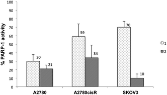
Afterwards, PARP-1 activity was evaluated on cell extracts from A2780, A2780cisR and SKOV3 cells. Thus, incubation of protein cell extracts with the compounds for 24 h at room temperature was followed by PARP-1 activity determination. Fig. 1 shows the residual PARP-1 activity in protein extracts treated with the complexes at a fixed concentration (10 μM).
Attractively, both compounds can induce PARP-1 inhibition to a similar extent in A2780 and A2780cisR cell lines, while a marked difference could be detected in the case of SKOV3 cells, where 2 is able to inhibit PARP-1 until ca. 10% of its residual activity. Instead, 1 is practically ineffective on these cells, in line with the scarce anticancer effects observed above.
Furthermore, PARP-1 activity was evaluated on protein extracts obtained from SKOV3 cells pre-treated with non-cytotoxic doses of each compound for 48 hours. Afterwards, the protein extracts were collected and analyzed for PARP-1 activity. Preliminary results indicate that only the gold complex 2 (20 μM) was able to induce ca. 70% reduction of PARP-1 activity, while ligand 1 was poorly effective.
In order to assess formation of adducts between the gold complex and the zinc finger domain of PARP-1, a peptide model corresponding to the N-terminal ZF domain sequence of PARP-1 was reacted with 2 and the sample was monitored by high-resolution ESI MS as described in the Experimental section. Fig. S3 in the ESI† shows the broadband mass spectrum of the 2–ZF adduct. In agreement with previously reported studies on other Au(III) complexes, when 2 was incubated with the ZF domain in a 3:
1 ratio for 10 min, partial displacement of Zn2+ from the ZF by gold ions leads already to formation of the so-called “gold-finger” adduct.
Afterwards, to evaluate if the different cytotoxic effects of 2 were related to differences in intracellular Au accumulation, ICP-MS analysis of cell extracts out of A2780 and SKOV3 cells, pre-treated with the gold compound for 24 h, demonstrated that the cytotoxicity is somehow proportional to the gold uptake, and the strongest antiproliferative effects correspond to higher values of intracellular gold concentration. In fact, the concentration of Au [pmol Au per 106 cells] measured in A2780 and SKOV3 cells is 1802 ± 209 and 1087 ± 322, respectively. Nevertheless, in spite the reduced accumulation of 2 in SKOV3 cells, the inhibition of PARP-1 activity is more pronounced than in the case of A2780 cells (Fig. 1).
Since TrxR is also a potential target for gold complexes, in vitro inhibition of purified rat TrxR by the two compounds was studied using established protocols as described in the Experimental section. The results are summarized in Table 2 and Fig. S4.† Complex 2 inhibits cytosolic thioredoxin reductases (TrxR1) in the same range as auranofin (IC50 = 14.32 ± 1.62 nM vs. IC50 = 6.88 ± 1.25 nM, respectively). Conversely, ligand 1 is completely ineffective, as expected since it is deprived of the Au(III) centre able to bind the selenol groups (Fig. S1, ESI†). Further studies demonstrated that 2 is also able to inhibit the TrxR closely related, but selenium-free, enzyme glutathione reductase (GR) with IC50 = 0.40 ± 0.06 μM, about 28-fold less efficiently than in the case of TrxR (Table 2).
Complexes | IC50 (nM) | |
---|---|---|
TrxR1 | GR | |
1 | >100 | >10![]() |
2 | 14.32 ± 1.62 | 400 ± 60 |
AF | 6.88 ± 1.25 | >10![]() |
Afterwards, the effect of compounds on TrxR and GR activities was evaluated in cell lysates. For this purpose, SKOV3 cells where the two compounds showed markedly different cytotoxic effects, were pre-treated for 48 h with 20 and 40 μM of 1 and 2, respectively. The obtained results show that 1 does not affect enzymes activities, while 2, causes ca. 50% TrxR inhibition and a slight decrease of GR activity at 40 μM (Fig. 2). In addition, similar experiments were conducted in the A2780 cells, and the obtained results showed no statistically significant inhibition of TrxR at the tested compounds' concentrations (Fig. S5†). Notably, these latter results further corroborate the hypothesis of alternative pharmacological targets for the reported compounds.
Overall, the obtained results on both PARP-1 and TrxR activities, indicate that the cytotoxic gold complex 2 may operate via inhibition of PARP-1, whereas TrxR is only moderately affected. Concerning the observed differences in PARP-1 inhibition by 1 and 2, it may be suggested that the ligand is not so selective for binding to PARP-1 as the gold complex 2, once in the presence of other intracellular components. Nevertheless, in terms of the overall cytotoxic potency, differences in the uptake mechanisms and cellular accumulation between the two compounds should also be taken into account.
The glutathione redox pair (GSH/GSSG) is another fundamental component of the cell redox regulation in cisplatin resistant cells.44 Therefore, our study continued with the analysis of total glutathione content (reduced + oxidized) and of the GSH/GSSG ratio in SKOV3, after treatment with the two compounds for 48 h in comparison to AF. The obtained results are shown in Fig. S6 in the ESI† available. It can be observed that for all tested compounds no statistically significant variation of the total GSH content, as well as of the GSH/GSSG ratio occur, again made exception for 2, which causes a slight increase of GSSG content at 40 μM, in accordance with the compound's above-mentioned inhibition effect of glutathione reductase. This behavior suggests that GSH does not particularly influence the cytotoxic potency of the gold complex, as for cisplatin in the case of certain resistant cancer cells.
Mitochondrial membrane potential (MMP), a consequence of the electrochemical proton gradient maintained for the purpose of ATP synthesis, is an important indicator of functional mitochondria. Previously reported studies showed that gold(III) complexes are able to determine the decrease of MMP depending on the ligands. As an example, gold(III) porphyrin 1a induced apoptosis by mitochondrial death pathways related to reactive oxygen species.45 Similarly, gold(III)-dithiocarbamato derivatives were shown to alter mitochondrial parameters, such as causing a drop of the mitochondrial membrane potential (MMP).46
MMP evaluation was conducted monitoring the fluorescence of tetramethylrhodamine methyl esters (TMRM) according to established protocols (see Experimental for details). Thus, it was possible to determine if the complexes are able to induce a quick drop in mitochondrial membrane potential (Δψm), detectable as a decline in the fluorescence intensity of TMRM.47
Therefore, MMP of SKOV3 cells treated for 18 h with compounds 1 and 2 was measured by cytofluorometric analysis in comparison to auranofin (AF) and CCCP (Fig. 3). Cells were incubated with 25 nM TMRM for 20 min and then analyzed by flow cytometry utilizing an argon laser at 585 nm, as described in the Experimental section.
From the obtained results it has been possible to determine that, at variance with AF and the classical uncoupling agent CCCP (carbonyl cyanide m-chlorophenyl hydrazone), both complexes 1 and 2 do not affect the MMP values with respect to the controls, as it has instead been reported for gold(III) porphyrins and dithiocarbamato complexes.
Finally, since overexpression of PARP in cancer cells has been linked to drug resistance and PARP-1 inhibition has been shown to sensitize tumor cells to chemotherapeutic agents including platinum compounds, we decided to evaluate the cytotoxic effect of cisplatin administered in combination with different concentrations of 2. Initial data were obtained for 72 h co-administration of cisplatin (7.5 μM) and 2 at different concentrations (10–20–30 μM) in SKOV3 cells (see Experimental for details). In Table S1 (ESI†) a comparison of the predicted survival rates (defined as the expected cell viability if the combined activities of the compounds are additive) and the experimentally determined values (the observed viabilities) is reported. Unfortunately, the observed survival rates for the combinations of 2 with cisplatin are similar to those predicted on the basis of an additive effect, ruling out the synergism. Further studies will be necessary to investigate possible synergic effects in different cancer cell types and to validate the possibility of using 2 in combination therapy.
Notably, inhibition of PARP potentiates the activity of DNA-damaging agents, such as alkylators, platinum compounds, topoisomerase inhibitors, and radiation in in vitro and in vivo models. Thus, clinical development to date has focused on PARP inhibitors potential role in combination with DNA-damaging chemotherapy, where efficacy has been limited by enhanced normal tissue toxicity.
Olaparib, a highly potent PARP inhibitor, has recently been approved for ovarian cancer therapy by the FDA and European commission, in patients with platinum-sensitive, recurrent, high-grade serous ovarian cancer with BRCA1 or BRCA2 mutations.48
Within this frame, gold(III) complexes such as 2 may constitute an alternative strategy to PARP-1 inhibition, acting on both the zinc finger DNA binding domain of the protein via gold binding, and on its catalytic domain; therefore, having enhanced efficacy. Further studies are necessary to fully validate this hypothesis and to design compounds with selectivity for PARP-1 with respect to other zinc finger proteins.
Footnotes |
† Electronic supplementary information (ESI) available. See DOI: 10.1039/c6ra11606j |
‡ Valeria Scalcon and Anna Citta: these authors contributed equally. |
This journal is © The Royal Society of Chemistry 2016 |