Selective enhancement of human stem cell proliferation by mussel inspired surface coating

Xiumei Jiangab, Yanfang Lib, Ying Liua, Chunying Chen*a and Menglin Chen*b
aCAS Key Laboratory for Biomedical Effects of Nanomaterials and Nanosafety & CAS Center for Excellence in Nanoscience, National Center for Nanoscience and Technology, Beijing 100190, China. E-mail: chenchy@nanoctr.cn
bInterdisciplinary Nanoscience Center (iNANO), Aarhus University, DK-8000, Aarhus C, Denmark. E-mail: menglin@inano.dk

Received 29th April 2016 , Accepted 10th June 2016

First published on 13th June 2016


Abstract

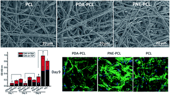
In tissue engineering, promoted cell adhesion and proliferation on a 3D scaffold is desired. Surface functionalization of the scaffolds is often utilized to realize this goal. Polydopamine (PDA) has been extensively studied for promoted cell adhesion and proliferation. Norepinephrine, sharing a similar structure with dopamine, polymerizes slower and forms an even and ultrasmooth surface coating on almost all substrates. However, the effects of polynorepinephrine (PNE) on stem cell adhesion and proliferation have barely been studied. In this study, we compared the biocompatibility and cell adhesion properties of mussel inspired PDA and PNE surface coatings on poly(ε-caprolactone) (PCL) fibers for human mesenchymal stem cells (hMSCs) and human induced pluripotent stem cell derived mesenchymal stem cells (hiPS-MSCs). The surface modification of PDA and PNE on PCL fibers was characterized by environmental scanning electron microscopy (ESME), X-ray photoelectron spectroscopy (XPS) and contact angle measurement. The biocompatibility of the PDA and PNE coatings with hMSCs and hiPS-MSCs was measured using lactase dehydrogenase (LDH) activity, live dead cell staining and cell counting kit 8 (CCK-8) assays. The results show that both PDA and PNE successfully formed a surface coating on the PCL fiber, which dramatically increased the hydrophilicity. The PNE coating showed a much thinner and smoother surface while the PDA coating formed uneven aggregates among the fibers. The biocompatibility analysis and cell proliferation results suggest that the PNE coating is more biocompatible with both hiPS-MSCs and hMSCs than the PDA coating. The PNE coating preferentially promoted cell proliferation for hiPS-MSCs but not for hMSCs, while PDA decreased the cell proliferation of hiPS-MSCs on the PCL fibers. These results suggest that the effect of the PDA and PNE surface coatings on cell proliferation can be cell-dependent. The surface roughness of the PDA coating can negatively affect cell adhesion and proliferation. Different mechanisms of interaction of the PDA and PNE coatings might affect cell adhesion and proliferation, and this needs to be carefully investigated before their application in specific cell type based tissue engineering.


Introduction

Tissue engineering has been developed for around thirty years as a promising strategy to repair damaged tissues or organs. Surface modification of scaffolds or implants with biocompatible adhesive molecules to promote cell adhesion and proliferation could benefit the success of tissue engineering. PDA coating is inspired from the marine organism, the mussel (mytiloida), and has the unique capacity of functionalizing virtually any material surface, including hydrophobic, synthetic polymer,1 noble metal and metal oxide2,3 and carbon material4,5 surfaces, and so on. PDA, a catechol molecule, has been widely reported to increase cell adhesion and proliferation, and reduce inflammation and toxicity both in vitro and in vivo.1,3,6 However, a few studies showed that a PDA coating reduced the proliferation of smooth muscle cells, possibly due to catechol oxidation induced ROS production.7,8 Polymerization of dopamine involves oxidation of catechol to quinine under alkaline condition (pH 8.5), which further reacts with other catechols or quinines to from an adherent polymer film.2 The deposited polydopamine coating is chemically heterogeneous. Despite the advantage of its material-independent functionalizability, the uncontrollable surface roughness after PDA polymerization has been an obstacle for its potential applications. To combat this challenge, a new surface coating derived from the catecholamine, polynorepinephrine (PNE), was introduced.9 Unlike polydopamine coatings, PNE has the ability to activate surface-initiated, ring-opening polymerization in alkaline conditions due to the presence of the alkyl hydroxyl group in norepinephrine, forming nearly perfect smoothness at the nanometer scale on the substrate surface.9,10 The residual quinine is stable under alkaline conditions and reacts with amine and thiol containing biomolecules, enabling facial conjugation of the proteins with the terminal amine.2,10,11 Our previous study suggested that PNE surface functionalized PCL fibers could facilitate PC12 cell differentiation.12

In tissue engineering, the cell source is another critical factor for regenerating or restoring damaged tissues and organs. Mesenchymal stem cells (MSCs) are commonly used for their multi-lineage differentiation capacities. However, their low population and limited proliferation capacity hampered their application in tissue engineering. Induced pluripotent stem (iPS) cells were a ground-breaking discovery by Takahashi and Yamanaka in 2006 that enabled reprogramming of somatic cells back to a pluripotent state with the capacity to differentiate into cell types in three germ layers.13 Based on this technique, MSCs derived from human iPS cells (hiPS-MSCs) have been reported by several groups.14,15 hiPS-MSCs share similar in vitro and in vivo characteristics to MSCs,16,17 and outperform MSCs with a greater cell proliferation capacity to proliferate for 120 population doublings without losing their renewal capacity and MSC characteristics,18,19 which makes them an excellent cell source for tissue engineering.

Designing and fabricating a suitable scaffold to mimic the extracellular matrix (ECM) to facilitate cell adhesion and proliferation is critical for tissue engineering. Among many techniques developed for fabricating fibrous scaffolds, electrospinning attracts the most interest due to its straightforwardness, robustness and versatile capability to generating fibers with diameters ranging from 100 nm to 10 μm.20 Electrospun fibers have a larger surface area for cell interaction and communication than 2D cell culture plates. Poly(ε-caprolactone) (PCL) is a synthetic, biocompatible and biodegradable polymer, and has been approved by the FDA for implants, drug delivery devices and sutures. PCL and PCL composites have been used as scaffolds in tissue engineering for the regeneration of cartilage, bone, nerve and vascular tissue.21 Our recent study used PCL and a hydrogel composed 3D ECM mimicking scaffold to support hiPS-MSC differentiation to fibroblasts.22

Based on a mussel inspired surface coating, PDA has been extensively studied as a functional surface coating in tissue engineering for stem cell adhesion and proliferation.8,23–26 However, PNE, which shares a similar chemical structure with PDA but forms a more even and smooth surface coating, has not been well-explored for stem-cell-based tissue engineering.10,12,27 Therefore, in the present work, we compared the adhesion and proliferation effects of PDA and PNE on hMSCs and hiPS-MSCs on both tissue culture plates (TCPs) and PCL fibers. The surface functionalization with PDA or PNE on the PCL fibers was characterized by ESEM, XPS and contact angle measurement. Cell adhesion and proliferation on the surface functionalized substrate were further analyzed with a cell viability assay and cell morphological characterization. By comparing stem cell proliferation on PDA and PNE surface coatings, this research was intended to enrich the study of mussel chemistry on stem cell therapy for tissue engineering.

Experimental section

Electrospinning of the PCL fibers and surface modification

The polycaprolactone solution was prepared by dissolving PCL (Mw = 70–90 kDa, Sigma, Germany) in 8[thin space (1/6-em)]:[thin space (1/6-em)]2 chloroform/ethanol (v/v) to give a 12% solution (w/v). The PCL solution was then electrospun at 20 kV through a 20 G blunt end needle with a flow rate of 8 ml h−1. The obtained fibers were lyophilized in a freeze drier overnight. The mesh was punched into circular shapes of 12 mm in diameter to fit in 48-well cell culture plates. Before use, the fibers were sterilized with 75% ethanol for 30 min and ultraviolet irradiation for 30 min. The surface was coated with PDA or PNE by simple immersion of the electrospun PCL fibers or TCPs into a dopamine hydrochloride (Sigma, Germany) solution or norepinephrine (Sigma, Germany) solution (2 mg ml−1 in 10 mM Tris–HCL, pH 8.5) at 25 °C for 17 h. After coating, the fibers and TCPs were washed three times with PBS and lyophilized in a freeze drier, then kept at 4 °C until use. From here on, the TCPs coated with PDA or PNE are referred to as PDA–TCP and PNE–TCP, and the PCL fibers coated with PDA or PNE are referred to as PDA–PCL and PNE–PCL.

X-ray photoelectron spectroscopy

The atomic chemical composition of the PCL fibers before and after PDA or PNE coating was analyzed using X-ray photoelectron spectroscopy (XPS, ESCALAB250Xi, Thermo Fisher Scientific, US).

Contact angle

The contact angles of PCL fibers and PDA– and PNE–PCL fibers were measured with the DSA 100 (Germany) using the method indicated by the manufacturer at 25 °C and 60% relative humidity. In brief, 2 μl ddH2O was added on the surface of the fibers and pictures were taken.

Environmental scanning electron microscopy

Environmental scanning electron microscopy (ESEM, Quanta 200 FEG, FEI, US) was applied to observe the surface morphology of the PCL fibers before and after PDA or PNE coating and the cell morphology of the hMSCs on these fibers. The cells were seeded on the fibers for 9 days and then washed 3 times with PBS and fixed with 4% paraformaldehyde for 15 min. After they were washed 3 times with PBS, the PDA and PNE coated PCL fibers and the fibers with cells were dehydrated with 30%, 50%, 70%, 85%, 95% and 100% ethanol, then dried at room temperature. The fibers and cells were observed using ESEM with an acceleration voltage of 4 kV under low vacuum conditions.

Cell culture

Human bone marrow stem cells (hMSCs) were purchased from Lonza (PT-2501) and cultured according to the instructions. hiPS-MSCs, generated from human iPS cells as previously reported,28 were kindly provided by Dr Yonglun Luo from the Department of Biomedicine, Aarhus University. Both cell lines were cultured in H-DMEM (Gibco, UK) plus 10% fetal bovine serum (FBS, Biowhittaker, Walkersville, MD), 10% penicillin and streptomycin (Gibco, Grand Island, NY) in a humidified 37 °C incubator with 5% CO2. Cells were used between 7–8 passages.

LDH assay

A LDH assay was applied to study the cytotoxicity of PDA– and PNE–TCP and the PCL fibers. Briefly, the cells were seeded on the TCPs and PCL fibers in 48-well plates. After 24 h, 200 μl of the cell culture medium was collected into 1.5 ml eppendorf tubes and centrifuged at 2000 rpm for 5 min at 4 °C to remove possible cell contamination. Then 50 μl of the supernatant was transferred from the eppendorf tube to 96-well plates and a 50 μl mixture of the enzyme mix was added to each well and allowed to react for 30 min at RT, protected from light. The absorbance at 490 nm was measured with a Victor X5 microplate reader. Cells cultured on TCPs were set as the normal control (0% cell death), while cells cultured on TCPs treated with 1% Triton X-100 were set as the positive control (100% cell death). The percentage of cytotoxicity was calculated using the equation:
image file: c6ra11173d-t1.tif

Live dead cell staining

Live dead cell staining was applied to study the biocompatibility of PDA– and PNE–TCP for hiPS-MSCs and hMSCs. Briefly, the cells were cultured on PDA– and PNE–TCP for 1, 3 and 5 days. Then the cells were washed with PBS and incubated with calcein-AM (2 μM) and ethidium (4 μM) at 37 °C for 30 min. The cells were then washed with PBS and observed under an inverted fluorescence microscope. The Ex/Em for calcein-AM is 494/517 nm and for ethidium bromide it is 528/617 nm.

Cell viability assay

The cell viability of hiPS-MSCs and hMSCs cultured on the TCP and PCL fibers was measured by a cell counting kit 8 (CCK-8, Dojindo, Kumamoto, Japan) assay. In brief, the cells were seeded and cultured for 1, 3 and 5 days on TCPs or 1, 5 and 9 days on PCL fibers in 48-well plates. The cell culture medium was changed every other day. After that, the PCL fibers were transferred into new 48-well plates to measure the viability of cells on the fibers and the cells that fell on the TCPs. The cell culture medium was discarded and 20% CCK-8/medium (300 μl per well) was added into the 48-well plates followed by incubation at 37 °C for 2 h. 100 μl aliquots from each sample were pipetted into a 96-well plate and the absorbance at 450 nm was measured with a Victor X5 microplate reader.

Cell morphology characterization

The cell morphology of the hiPS-MSCs and hMSCs cultured on PDA– and PNE–PCL fibers were observed under a Zeiss LSM 700 laser confocal microscope (Carl Zeiss Micro-Imaging GmbH, Germany). Briefly, the cells were seeded on the PDA– and PNE–PCL fibers and cultured for 1, 5 and 9 days. The cell culture medium was changed every other day. The fibers were washed 3 times with PBS and fixed with 4% paraformaldehyde for 15 min at room temperature (RT), permeabilized with 0.05% Triton for 5 min and then stained with phalloidin for 30 min. After this, they were washed 3 times with PBS, 10 min for each wash, and the fibers were placed on a slide with a drop of mounting medium containing DAPI. The samples were then observed and pictures were taken under a confocal laser scanning microscope.

Statistical analysis

Data are expressed as mean ± standard deviation (SD). The statistical analysis was performed using Student’s t-test to compare the significance in multiple data groups. Values of p < 0.05 were considered as statistically significant.

Results and discussion

Electrospinning and surface morphology of PDA and PNE coated PCL fibers

Electrospinning technology has been used for the fabrication of tissue-engineered scaffolds because of its ability to mimic the extracellular matrix (ECM) structures.29,30 PCL was chosen as the scaffold material due to its good biocompatibility and mechanical properties. The diameter of the fibers can be controlled by adjusting the electrospinning parameters, including the voltage, speed, needles, etc. The PCL fibers we electrospun are randomly arranged with an average diameter of 1.13 ± 0.5 μm (Fig. 1). The PDA and PNE coatings were prepared by dissolving dopamine or norepinephrine in 10 mM Tris buffer (pH 8.5) to make a 2 mg ml−1 solution. After 17 h of incubation, ESEM was applied to observe the surface morphology of the PCL fibers and the PDA and PNE surface coatings. As is shown in Fig. 1 and S1, the PDA–PCL fibers had uneven surfaces with significant aggregates on the fibers. The average diameter of the PDA–PCL fibers is 1.25 ± 0.58 μm. Compared with the PDA–PCL fibers, the PNE–PCL fibers have much smaller aggregates and smoother surfaces. The average diameter of the PNE–PCL fibers is 1.18 ± 0.45 μm. This is consistent with a previous study in which the PNE coating formed a thin and ultra-smooth surface while the PDA coating formed uncontrolled aggregates.9 The emergence of the uncontrollable roughness during PDA polymerization is proposed to be due to the PDA particles in solution simultaneously attaching and growing directly from the surface.9 Studies at the molecular level found that it is the intermediate, 3,4-dihydroxybenzaldehyde (DHBA), formed during the polymerization of norepinephrine, that resulted in the remarkable difference in surface morphology.
image file: c6ra11173d-f1.tif
Fig. 1 Surface morphology characterization of PCL fibers, PDA–PCL fibers and PNE–PCL fibers.

XPS was applied to quantify the surface elemental species of the PCL fibers and PDA– and PNE–PCL fibers. As is shown in Fig. 2, the uncoated PCL fibers consist of only carbon (C 1s) and oxygen (O 1s), while after PDA or PNE coating, a nitrogen (N 1s) peak was observed. The chemical compositions of the three fibers were quantified and are listed in Table S1 in the ESI. As the coating time was extended from 0.5 h to 17 h, the nitrogen composition increased in both the PDA–PCL and PNE–PCL fibers. Interestingly, under the same incubation time, the PDA–PCL fibers showed a stronger nitrogen signal compared with the PNE–PCL fibers. This is due to the fast polymerization and large aggregates formed during the PDA coating on the PCL fibers.


image file: c6ra11173d-f2.tif
Fig. 2 XPS spectra of PCL fibers before and after PDA and PNE coating. Chemical composition analysis of the PCL fibers after 0 h, 6 h or 17 h coating in (A) PDA and (B) PNE solution. C 1s, O 1s and N 1s peaks are compared in parallel.

The water contact angle of the surface was measured in order to analyze the change of the surface wettability after surface modification with PDA or PNE. As is shown in ESI Fig. S2, the uncoated PCL fibers typically exhibited a water contact angle of 125.5° ± 1.1°, while the contact angles for the surface coated with PDA or PNE significantly decreased and were close to 0°, which means the surface changed from hydrophobic to hydrophilic.

Cell viability of hiPS-MSCs and hMSCs on PDA– and PNE–TCP

Before analyzing the cell proliferation on the PCL fibers, we used TCPs to evaluate the biocompatibility of the PDA and PNE coatings by an LDH assay and live dead cell staining. LDH is a stable cytoplasmic enzyme that is present in all cells. It is rapidly released into the cell culture supernatant upon damage of the plasma membrane. Therefore it is a good biomarker for a cytotoxicity assay. As is shown in Fig. 3, compared with cells cultured on TCPs, the LDH release in hiPS-MSCs cultured on PDA–TCP significantly increased, while for the PNE–TCP group it is as low as the TCP group, suggesting the PNE coating is more biocompatible than the PDA coating for hiPS-MSCs. The live dead cell staining results showed that neither the PDA nor PNE coating induced significant cell death compared with cells on TCPs but the number of live cells and the cell morphology is different on PNE–PCL fibers in comparison to those of PDA–PCL and the uncoated PCL fibers, with more cells on PNE–TCP. This was further confirmed by the proliferation assays of hiPS-MSCs on PDA– and PNE–TCP for 1, 3 and 5 days as determined by the CCK-8 assay. As shown in Fig. 3C, the hiPS-MSC cell viability increased over time and the cell proliferation in PNE–TCP is higher than cells on TCPs at the same incubation time, while PDA–TCP significantly decreased the hiPS-MSC cell viability compared with hiPS-MSCs on TCPs. This is in accordance with the LDH assay results that PDA–TCP increased the LDH release, which is a sign of cytotoxicity. These results suggest that hiPS-MSCs adhere, spread and survive better on PNE–TCP than on uncoated TCPs. PDA–TCP decreased the cell viability of hiPS-MSCs compared with uncoated TCPs, while for hMSCs, as shown in Fig. 4, PDA–TCP increased the LDH release of hMSCs compared to uncoated TCPs. The live dead cell staining suggested that neither PDA nor PNE induced significant cell death. The cell viability assay showed that PNE–TCP did not significantly affect hMSC cell viability, while the PDA coating decreased the hMSC cell viability on day 1 compared with hMSCs on TCPs and showed no significant difference after 3 and 5 days incubation. Taken together, according to our results, the PNE coating is more biocompatible with both hiPS-MSCs and hMSCs than the PDA coating. PNE enhanced the cell adhesion and proliferation of hiPS-MSCs, but had a negligible effect on hMSCs. This is interesting because both dopamine and norepinephrine are catecholamine molecules; the only difference between these two molecules is that PNE has a hydroxyl group on the side chain and formed a smoother surface than PDA when polymerized in pH 8.5 Tris buffer. As has been reported before, PDA polymerization is rather fast and uncontrollable, during which large aggregates are formed in solution and attached to the functionalized surfaces, thus resulting in significant variations in surface roughness.9 Yang et al. and Luo et al. have reported inhibitory effects of a PDA-coated surface on smooth muscle cell (SMC) proliferation. They proposed that the phenolic/quinine groups present on the PDA coating played a key role in modulating the vascular cell behaviour.7,8 Ding et al. further indicated that the quinone group on the PDA coating induces a substantially higher amount of protein adsorption, which plays a key role in promoting epithelial cell attachment and proliferation. Meanwhile, the reactive phenolic hydroxyl group on the PDA coating potently inhibits SMC proliferation.31
image file: c6ra11173d-f3.tif
Fig. 3 hiPS-MSC cell viability on TCPs and PDA– and PNE–TCP. (A) LDH assay of hiPS-MSCs cultured on TCPs and PDA– and PNE–TCP. (B) Live dead cell staining of hiPS-MSCs cultured on TCPs and PDA– and PNE–TCP. (C) Cell viability of hiPS-MSCs cultured on TCPs and PDA– and PNE–TCP for 1, 3 and 5 days. Data are presented as mean ± SD; n = 3; the statistical significance is presented as p < 0.05.

image file: c6ra11173d-f4.tif
Fig. 4 hMSC cell viability on TCPs and PDA– and PNE–TCP. (A) LDH assay of hMSCs cultured on TCPs and PDA– and PNE–TCP. (B) Live dead cell staining of cells cultured on TCPs and PDA– and PNE–TCP. (C) Cell viability of hMSCs cultured on TCPs and PDA– and PNE–TCP for 1, 3 and 5 days. Data are presented as mean ± SD; n = 3; the statistical significance is presented as p < 0.05.

Cell proliferation and cell morphology of hiPS-MSCs and hMSCs on PDA– and PNE–PCL fibers

Electrospun fibers, with a three-dimensional architecture, high porosity, interconnected pore structure, and high surface-to-volume ratio provide great advantages in in vitro cell culture for tissue engineering. PCL, as a FDA approved synthetic polymer, has been widely used due to its good biocompatibility and suitable mechanical properties for tissue engineering. The biocompatibility of uncoated PCL fibers and PDA– and PNE–PCL fibers for hiPS-MSCs and hMSCs was evaluated by LDH and CCK-8 assays. As is shown in Fig. 5A, for hiPS-MSCs the PDA fibers induced a significant increase in LDH release compared to the uncoated PCL fibers, while the PNE–PCL fibers significantly decreased the LDH release. The LDH release for PDA–PCL possibly originated from cells that failed to adhere to the fiber, which was further verified by the cell viability assay. The cell proliferation data (Fig. 5B) suggest that PDA–PCL significantly decreased the cell viability compared with the uncoated fibers, while PNE–PCL increased the cell viability, especially on day 9. This is in accordance with the cell proliferation results on PDA– and PNE–TCP. However, for hMSCs, as shown in Fig. 6, neither PDA nor PNE induced a significant increase in LDH release. The cell viability of hMSCs in PDA– and PNE–PCL fibers is significantly lower than the uncoated PCL fibers, while if we only look at fiber-attached cells (cells in fiber), the cell viability of hMSCs grown on PNE–PCL fibers is similar to those grown on uncoated PCL fibers and much higher than cells grown on PDA–PCL fibers.
image file: c6ra11173d-f5.tif
Fig. 5 Biocompatibility of PCL fibers and PDA– and PNE–PCL fibers with hiPS-MSCs. (A) LDH release of hiPS-MSCs on PCL fibers and PDA– and PNE–PCL fibers. (B) Cell viability of hiPS-MSCs after 1, 5, and 9 days incubation on PCL fibers and PDA– and PNE–PCL fibers. (C) Cell attachment and morphology of hiPS-MSCs after 1, 5, and 9 days incubation on PCL fibers and PDA– and PNE–PCL fibers. The data represent mean ± SD; n = 3; the statistical significance compared with the PCL fibers is presented as p < 0.05.

image file: c6ra11173d-f6.tif
Fig. 6 Biocompatibility of PCL fibers and PDA– and PNE–PCL fibers with hMSCs. (A) LDH release of hMSCs on PCL fibers and PDA– and PNE–PCL fibers. (B) Cell viability of hMSCs after 1, 5, and 9 days incubation on PCL fibers and PDA– and PNE–PCL fibers. (C) Cell adhesion and morphology of hMSCs after 1, 5, and 9 days incubation on PCL fibers and PDA– and PNE–PCL fibers. The data represent mean ± SD; n = 3; the statistical significance compared with the PCL fibers is presented as p < 0.05.

In general, mammalian cells undergo a cell adhesion process of substrate attachment, spreading and cytoskeleton development.32,33 We further investigated the cell morphology of hiPS-MSCs and hMSCs on PDA– and PNE–PCL fibers by staining the cytoskeleton actin filament with FITC-phalloidin. As is shown in Fig. 5C, on day 1, hiPS-MSCs adhered more on PNE–PCL fibers than on PDA–PCL and uncoated PCL fibers. The cytoskeleton of hiPS-MSCs on uncoated and PDA–PCL fibers is not as spread and stretched as cells on PNE–PCL fibers. For the 5 and 9 days cultures, the difference in cell morphology and cell density on the three fibers became more evident; the PNE coating facilitates the hiPS-MSC cell adhesion and proliferation on the PCL fibers. However, as shown in Fig. 6C, the hMSC cell morphology on the PDA– and PNE–PCL fibers did not show much difference to the cells on the uncoated PCL fibers. PDA coating has been reported to selectively reduce the cell proliferation of human umbilical artery smooth muscle cells but increase human umbilical vein endothelial cell proliferation due to the reactive phenolic hydroxyl group on the PDA.8 A study indicated that dopamine polymerized in air is rough due to agglomerate deposition and inhomogeneous stacking of the deposited molecules.34 The decreased cell adhesion and proliferation is possibly due to the surface roughness of the PDA aggregates. In contrast, PNE forms a smooth surface and reacts with thiol and amine containing molecules; therefore the enhanced cell proliferation could be attributed to the immobilization of serum proteins from the cell culture medium on the PNE layer.

Cell morphology of hMSCs on PDA– and PNE–PCL fibers

To investigate interactions between the fibers and the hMSCs, we applied ESEM to observe the cell morphology and localization of the hMSCs on the PCL fibers. As is shown in Fig. 7, most of the cells are located on the surface of the fibers and there are also some cells that migrated into the fibers. The uncoated PCL fibers had more cells adhered to the fiber surface, while PDA–PCL and PNE–PCL had a lower cell number but with more stretched cell morphology. PDA and PNE were reported to promote cell proliferation and differentiation by immobilizing the thiol and amine containing molecules or growth factors,35 but in our study this is true for hiPS-MSCs but not for hMSCs. The underlying mechanisms need further investigation.
image file: c6ra11173d-f7.tif
Fig. 7 ESEM images of hMSCs on PCL fibers with and without PDA and PNE coatings. hMSCs on PCL fibers (A–C), PDA–PCL fibers (D–F) and PNE–PCL fibers (H, I, G).

Conclusions

Enhanced cell adhesion and proliferation are desired in tissue engineering. Surface modification with biofunctional molecules is a good way to make this possible. Mussel inspired surface coating was introduced into tissue engineering as a functional surface coating to promote cell adhesion and proliferation. Dopamine has been extensively studied for cell proliferation on different substrates. However, norepinephrine, which shares a similar molecular structure with dopamine but forms superior homogenous coatings, has not been as well studied as dopamine for cell adhesion and proliferation. Therefore, we herein performed a study comparing the biocompatibility and cell adhesion properties of PDA and PNE using two kinds of stem cells, human mesenchymal stem cells and human pluripotent stem cell derived mesenchymal stem cells. Our results suggest that both PDA and PNE successfully formed a surface coating on PCL fibers and dramatically increased the hydrophilicity as characterized by ESEM, XPS and contact angle measurements. The PNE coating showed a much thinner and smoother surface in comparison with the PDA coating. The biocompatibility analysis and cell proliferation results showed that the PNE coating is more biocompatible with both hiPS-MSCs and hMSCs than the PDA coating. The PNE coating preferentially promoted hiPS-MSC cell proliferation, while PDA decreased the cell proliferation of hiPS-MSCs on the PCL fibers. These results suggest that the effect of PDA and PNE surface coatings on cell proliferation can be cell-dependent. The surface roughness of the PDA coating can negatively affect cell adhesion and proliferation. A careful investigation should be taken before using PDA or PNE on scaffold surface coatings for stem cell therapy and tissue engineering. Further study is needed to illustrate the different mechanisms of PDA and PNE coatings on different cells.

Acknowledgements

The authors gratefully acknowledge the Danish Council for Strategic Research for the funding to the ElectroMed Project at the iNANO Center, and the Aarhus University Research Foundation and the Carlsberg Foundation for their financial support. This work was also financially supported by MOST 973 program (2012CB934000) and the NSFC Distinguished Young Scholars (11425520).

Notes and references

  1. S. H. Ku and C. B. Park, Biomaterials, 2010, 31, 9431–9437 CrossRef CAS PubMed.
  2. H. Lee, S. M. Dellatore, W. M. Miller and P. B. Messersmith, Science, 2007, 318, 426–430 CrossRef CAS PubMed.
  3. S. Hong, K. Y. Kim, H. J. Wook, S. Y. Park, K. D. Lee, D. Y. Lee and H. Lee, Nanomedicine, 2011, 6, 793–801 CrossRef CAS PubMed.
  4. B. H. Kim, D. H. Lee, J. Y. Kim, D. O. Shin, H. Y. Jeong, S. Hong, J. M. Yun, C. M. Koo, H. Lee and S. O. Kim, Adv. Mater., 2011, 23, 5618–5622 CrossRef CAS PubMed.
  5. X. Liu, R. H. Hurt and A. B. Kane, Carbon, 2010, 48, 1961–1969 CrossRef CAS PubMed.
  6. M. E. Lynge, R. Ogaki, A. O. Laursen, J. Lovmand, D. S. Sutherland and B. Stadler, ACS Appl. Mater. Interfaces, 2011, 3, 2142–2147 Search PubMed.
  7. R. Luo, L. Tang, S. Zhong, Z. Yang, J. Wang, Y. Weng, Q. Tu, C. Jiang and N. Huang, ACS Appl. Mater. Interfaces, 2013, 5, 1704–1714 Search PubMed.
  8. Z. Yang, Q. Tu, Y. Zhu, R. Luo, X. Li, Y. Xie, M. F. Maitz, J. Wang and N. Huang, Adv. Healthcare Mater., 2012, 1, 548–559 CrossRef CAS PubMed.
  9. S. Hong, J. Kim, Y. S. Na, J. Park, S. Kim, K. Singha, G. I. Im, D. K. Han, W. J. Kim and H. Lee, Angew. Chem., Int. Ed., 2013, 52, 9187–9191 CrossRef CAS PubMed.
  10. S. M. Kang, J. Rho, I. S. Choi, P. B. Messersmith and H. Lee, J. Am. Chem. Soc., 2009, 131, 13224–13225 CrossRef CAS PubMed.
  11. J. H. Waite, Nat. Mater., 2008, 7, 8–9 CrossRef CAS PubMed.
  12. M. B. Taskin, R. Xu, H. Zhao, X. Wang, M. Dong, F. Besenbacher and M. Chen, Phys. Chem. Chem. Phys., 2015, 17, 9446–9453 RSC.
  13. K. Takahashi and S. Yamanaka, Cell, 2006, 126, 663–676 CrossRef CAS PubMed.
  14. T. Teramura, Y. Onodera, T. Mihara, Y. Hosoi, C. Hamanishi and K. Fukuda, Cell. Reprogram., 2010, 12, 249–261 CrossRef CAS PubMed.
  15. L. G. Villa-Diaz, S. E. Brown, Y. Liu, A. M. Ross, J. Lahann, J. M. Parent and P. H. Krebsbach, Stem Cells, 2012, 30, 1174–1181 CrossRef CAS PubMed.
  16. W. Gruenloh, A. Kambal, C. Sondergaard, J. McGee, C. Nacey, S. Kalomoiris, K. Pepper, S. Olson, F. Fierro and J. A. Nolta, Tissue Eng., Part A, 2011, 17, 1517–1525 CrossRef CAS PubMed.
  17. Y. Jung, G. Bauer and J. A. Nolta, Stem Cells, 2012, 30, 42–47 CrossRef CAS PubMed.
  18. Q. Lian, Y. Zhang, J. Zhang, H. K. Zhang, X. Wu, Y. Zhang, F. F. Lam, S. Kang, J. C. Xia, W. H. Lai, K. W. Au, Y. Y. Chow, C. W. Siu, C. N. Lee and H. F. Tse, Circulation, 2010, 121, 1113–1123 CrossRef PubMed.
  19. R. Kang, Y. Luo, L. Zou, L. Xie, H. Lysdahl, X. Jiang, C. Chen, L. Bolund, M. Chen and F. Besenbacher, RSC Adv., 2014, 4, 5734–5739 RSC.
  20. S. Agarwal, J. H. Wendorff and A. Greiner, Adv. Mater., 2009, 21, 3343–3351 CrossRef CAS PubMed.
  21. B. D. Ulery, L. S. Nair and C. T. Laurencin, J. Polym. Sci., Part B: Polym. Phys., 2011, 49, 832–864 CrossRef CAS PubMed.
  22. R. Xu, M. B. Taskin, M. Rubert, D. Seliktar, F. Besenbacher and M. Chen, Sci. Rep., 2015, 5, 8480,  DOI:10.1038/srep08480.
  23. M. Xu, Y. Zhang, D. Zhai, J. Chang and C. Wu, Biomater. Sci., 2013, 1, 933–941 RSC.
  24. S. Zhong, R. Luo, X. Wang, L. Tang, J. Wu, J. Wang, R. Huang, H. Sun and N. Huang, Colloids Surf., B, 2014, 116, 553–560 CrossRef CAS PubMed.
  25. S. R. Nielsen, F. Besenbacher and M. Chen, Phys. Chem. Chem. Phys., 2013, 15, 17029–17037 RSC.
  26. N. G. Rim, S. J. Kim, Y. M. Shin, I. Jun, D. W. Lim, J. H. Park and H. Shin, Colloids Surf., B, 2012, 91, 189–197 CrossRef CAS PubMed.
  27. M. Park, M. Shin, E. Kim, S. Lee, K. I. Park, H. Lee and J.-H. Jang, J. Nanomater., 2014, 6, 8288–8294 Search PubMed.
  28. L. Zou, Y. Luo, M. Chen, G. Wang, M. Ding, C. C. Petersen, R. Kang, F. Dagnaes-Hansen, Y. Zeng and N. Lv, Sci. Rep., 2013, 3, 2243 CrossRef PubMed.
  29. Q. P. Pham, U. Sharma and A. G. Mikos, Tissue Eng., 2006, 12, 1197–1211 CrossRef CAS PubMed.
  30. U. Boudriot, R. Dersch, A. Greiner and J. H. Wendorff, Artif. Organs, 2006, 30, 785–792 CrossRef CAS PubMed.
  31. Y. Ding, Z. Yang, C. W. C. Bi, M. Yang, J. Zhang, S. L. Xu, X. Lu, N. Huang, P. Huang and Y. Leng, J. Mater. Chem. B, 2014, 2, 3819–3829 RSC.
  32. U. Hersel, C. Dahmen and H. Kessler, Biomaterials, 2003, 24, 4385–4415 CrossRef CAS PubMed.
  33. Y. Qiao, Z. Zhai, L. Chen and H. Liu, Chin. Sci. Bull., 2015, 60, 1193–1202 CAS.
  34. H. W. Kim, B. D. McCloskey, T. H. Choi, C. Lee, M.-J. Kim, B. D. Freeman and H. B. Park, ACS Appl. Mater. Interfaces, 2012, 5, 233–238 Search PubMed.
  35. C.-Y. Chien and W.-B. Tsai, ACS Appl. Mater. Interfaces, 2013, 5, 6975–6983 Search PubMed.

Footnote

Electronic supplementary information (ESI) available. See DOI: 10.1039/c6ra11173d

This journal is © The Royal Society of Chemistry 2016
Click here to see how this site uses Cookies. View our privacy policy here.