DOI:
10.1039/C6RA11140H
(Paper)
RSC Adv., 2016,
6, 59389-59394
Detailed theoretical investigation on ESIPT process of pigment yellow 101
Received
29th April 2016
, Accepted 16th June 2016
First published on 17th June 2016
Abstract
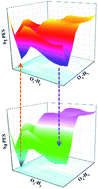
Based on density functional theory (DFT) and time-dependent density functional theory (TDDFT) methods, the detailed excited state intramolecular proton transfer (ESIPT) mechanism of 2,2′-dihydroxy-1,1′-naphthalazine (P.Y. 101) has been investigated theoretically. Unlike previous theoretical investigation of P.Y. 101, our calculated results not only reproduce the absorption and fluorescence spectra reported in the previous experiment, but also were completed with considering solvent effect. It further demonstrates that the TDDFT theory we adopted is very reasonable and effective. The calculations of main bond lengths and bond angles involving in the hydrogen bondings (O1–H2⋯N3 and O4–H5⋯N6) as well as the infrared vibrational spectra and as well as the calculated hydrogen bonding energies demonstrated the intramolecular hydrogen bond was strengthened in the S1 state. In addition, qualitative and quantitative intramolecular charge transfer based on the frontier molecular orbitals provided the possibility of the ESIPT reaction. The potential energy surfaces of ground state and the first excited state have been constructed to illustrate the ESIPT mechanism. Based on our calculations, the equilibrium ESIPT process exists in the S1 state. And after the radiative transition, reversed GSIPT can also occur in the S0 state.
1. Introduction
It is well known that the excited-stated inter- or intra-molecular proton transfer (ESIPT) reaction, incorporating transfer of a hydroxyl (amino) proton to the imine nitrogen (carbonyl oxygen) through a pre-existing nitrogen bonding, plays important roles in various chemical and biological processes.1–3 In general, the ESIPT process is ultrafast, occurring in less than 100 fs, to result in the absence of emission from the reactant (the enol isomer), which in turn contributes to a large Stocks shift ranging from 6000 to 10
000 cm−1.4,5 Both the ground- and excited-state proton transfers have become more and more significant from the mechanistic point of view,6–8 which consequently find their way in wide variety of technological applications, including the development of proton transfer lasers, photo-stabilizers, white-light-emitting diodes, fluorescence sensors, molecular switches, and so forth.9–16 Furthermore, some of these molecules are highly sensitive to the change in the microenvironments,17–20 based on which Sytnik et al. reported how the ESIPT chromophores can be used in the study of protein conformations and binding site polarity.17,18 In fact, the general processes could be concluded as follow: upon the photoexcitation, the studied molecule can be projected on the excited-stated potential energy surface, which makes the position of the proton unstable. The gap of energy between the local excited state and the relaxed excited state provides driving force for proton transfer, and the slope of corresponding potential surface connecting these two points determines the relative kinetics. Therefore, the resulting proton-transfer tautomer possess significant changes in structures and electronic configurations, which leads to the broaden applications mentioned above.
2,2′-Dihydroxy-1,1′-naphthalazine (P.Y. 101), as an organic fluorescent yellow pigment, has been known and produced since 1899 industrially.21,22 In the solid state, P.Y. 101 is special among the available organic pigments with its luminous fluorescence, which is different from other organic pigments.22 Particularly, since its bright yellow color and high photostability, it is usually used as one kind of commercial colorant.23 Even though P.Y. 101 has been known more than a century, its fluorescence properties and its excited-stated dynamic processes are not particularly clear with just a few previous work reported about this molecular.24–28 Lorenz et al. investigated the ultrafast photoinduced dynamics of P.Y. 101 through comparing P.Y. 101 with a closely related molecule 1,1′-naphthalazine experimentally and theoretically.25 Through transient absorption and femtosecond time-resolved spectroscopy, the ESIPT exists in the S1 state in dichloromethane (DCM) solvent. Based on their theoretical calculations (without considering solvent effect), however, the absorption and fluorescence spectra do not match their experimental results very much, which was interpreted that it is typical for TDDFT calculations.25,26 Very recently, mainly based on steady-state absorption and emission spectra as well as time-resolved fluorescence spectrum, Lee et al. assigned the two spectra to enol and keto isomers of P.Y. 101 in the S1 state, which demonstrates that the intramolecular proton transfer process occurs in the first excited state,27 while in their work, all the theoretical calculations were finished in gas phase. In 2015, Hibbs and co-workers investigated the charge density distribution of P.Y. 101 system using high-resolution X-ray diffraction and multipole refinement methods.28 Whereas the Scheme 1 of ref. 28 shows the double proton transfer forms in both S0 and S1 states, which were considered to be nonexistent based on previous theoretical work with ignoring solvent effect.24–27 As far as we know, all the previous theoretical work about P.Y. 101 was done without considering solvation effect, which may provide imprecise spectra compared with experiment. So we doubt whether the big gap of absorption and emission between experimental and theoretical results is not just due to the TDDFT method.25 Besides, the double proton transfer structures drawn in Scheme 1 of ref. 28 triggers our thought that whether P.Y. 101 can occur double proton transfer process when taking the solvation effect into consideration theoretically. In addition, it is well known that only spectroscopic techniques, such as steady-state absorption spectroscopy or the time resolved fluorescence spectroscopy, can just provide indirect information about photochemical properties and reflect few excited-stated dynamic process.29–35 Since we major in investigating the PT or ESPT process in this work, other processes mentioned in previous are not within the scope of our study. Therefore, in the present work, density functional theory (DFT) and time-dependent density functional theory (TDDFT) have been selected to comprehend the detailed mechanism and clarify the fundamental aspects concerning the different electronic states and structures occurring in the PT and ESPT reactions. The relevant configurations of S0 state and S1 state were optimized (shown in Fig. 1), further vertical excitation energies, infrared (IR) vibration spectra, hydrogen bond energies, the frontier molecular orbitals (MOs), Mulliken's charge distribution analysis as well as Hirshfeld charge distribution analysis, and homologous potential energy curves were calculated and analyzed to provide the clear and definite excited-stated process.
 |
| Scheme 1 The ESIPT scheme among enol–enol, enol–keto and keto–keto. | |
 |
| Fig. 1 Views of optimized structures for P.Y. 101 system based on B3LYP/TZVP theoretical level (enol–enol: normal P.Y. 101 form; enol–keto: single proton transfer P.Y. 101 form; keto–keto: double proton transfer P.Y. 101 form). | |
2. Computational details
In this present work, all the theoretical calculations presented were accomplished using the DFT and TDDFT methods with Becke's three-parameter hybrid exchange function with the Lee–Yang–Parr gradient-corrected correlation functional (B3LYP)36–41 as well as the triple-ζ valence quality with one set of polarisation functions (TZVP)42 basis set by Gaussian 09 programs.43 Unlike all the previous theoretical work of P.Y. 101, solvent effects (DCM) were included in all calculations based on the Polarizable Continuum Model (PCM) using the integral equation formalism variant (IEF-PCM)44–46 in our work. All the ground-stated geometries of all the relative structures were optimized without constraint based on DFT methodology, in addition, vibrational frequencies were analysed at the optimized forms to confirm that all these configurations corresponds to the local minima on the S0 potential energy surface (PES). Vertical excitation energies calculations were finished from the S0 state optimized configuration using TDDFT with six low-lying absorbing transitions. Moreover, from the ground equilibrium structures, the S1 state was optimised with analyzing vibrational frequencies with TDDFT method to ensure the local minima on S1 PES. Herein, it is worth mentioning that the TDDFT method has become a very useful tool to investigate the hydrogen bonding in the excited states of the hydrogen-bond system theoretically,47–58 even though this kind of calculations are very time-consuming. The PESs of the S0 and S1 states were also carried out for the proton transfer process in DCM. All the stationary points along the reaction coordinate were scanned by constraining optimizations and frequency analyses (no imaginary frequency) to obtain the thermodynamic corrections in the corresponding electronic state.
Fine quadrature grids of size 4 were employed. The self-consistent field (SCF) convergence thresholds of the energy for both the ground state and excited state optimization were set at 10−8 (default settings are 10−6). Harmonic vibrational frequencies in the ground and excited state were determined by diagonalization of the Hessian. The excited-stated Hessian was obtained by numerical differentiation of the analytical gradients using central differences and default displacements of 0.02 Bohr. The infrared intensities were determined from the gradients of the dipole moment.41
3. Results and discussion
3.1 Structures analysis
The ground state and the first excited stated structures of the P.Y. 101 chromophore (enol–enol, enol–keto and keto–keto forms) were obtained based on the B3LYP function with TZVP basis set level of theory, with a subsequent vibrational frequency analysis to insure the validity of the stationary points in DCM solvent. For these three stable configurations, the most dominating structure parameters involved in the intramolecular hydrogen bondings have been listed in Table 1 after comparing other parts with no obvious big changes among them. It should be noticed that the calculated bond lengths of O1–H2, H2⋯N3, O4–H5 and H5⋯N6 of the enol–enol form are 0.999, 1.690, 0.999 and 1.690 Å, respectively. After the photo-excitation to the S1 state, these bond lengths change to be 1.008, 1.653, 1.008 and 1.653 Å, respectively. Obviously, hydrogen bondings (H2⋯N3 and H5⋯N6) are shortened, which demonstrates that the intramolecular hydrogen bondings (i.e., O1–H2⋯N3 and O4–H5⋯N6) are strengthened in the S1 state. In addition, the bond angle θ (O–H–N) changes from 146.3° in the S0 state to 148.1° in the S1 state. That is to say, the hydrogen bonding angle of S1 state is closer to 180 degrees than that of the S0 state, which further indicates that hydrogen bondings are strengthened in S1 state.
Table 1 The primary bond lengths (Å) and bond angles (°) of enol–enol, enol–keto and keto–keto forms in S0 and S1 states based on the DFT/TDDFT methods in dichloromethane solvent
Electronic state |
Enol–enol |
Enol–keto |
Keto–keto |
S0 |
S1 |
S0 |
S1 |
S0 |
S1 |
O1–H2 |
0.999 |
1.008 |
1.732 |
1.685 |
1.614 |
1.576 |
H2–N3 |
1.690 |
1.653 |
1.039 |
1.049 |
1.054 |
1.066 |
O4–H5 |
0.999 |
1.008 |
0.989 |
0.995 |
1.614 |
1.576 |
H5–N6 |
1.690 |
1.653 |
1.728 |
1.697 |
1.054 |
1.066 |
θ(O1–H2–N3) |
146.3° |
148.1° |
134.1° |
138.6° |
138.1° |
141.8° |
θ(O4–H5–N6) |
146.3° |
148.1° |
145.0° |
146.4° |
138.1° |
141.8° |
Furthermore, investigating the vibrational frequencies of O–H stretching vibration involved in the hydrogen bondings can provide a clear-cut signature of hydrogen bonding dynamics.47–58 Therefore, we used vibrational frequencies to detect whether the intramolecular hydrogen bondings O1–H2⋯N3 and O4–H5⋯N6 are strengthened or not. It should be noted that the calculated both O–H synchronous stretching vibrational frequency in the S0 state is located at 3073.2 cm−1. However, it changed to be 2887.5 cm−1 in the S1 state. A strong red-shift of 185.7 cm−1 for the O–H stretching band was induced by the intramolecular hydrogen bonds O1–H2⋯N3 and O4–H5⋯N6. So it indicates that the intramolecular hydrogen bonds should be strengthened in the S1 state, which provides the possibility of ESPT.
3.2 Mechanism analysis of ESIPT
In order to reveal the proton transfer process of P.Y. 101 in the S1 state, we optimized all the ground-stated and excited-stated geometrical structures with fixed O1–H2 and O4–H5 lengths at the DFT/TDDFT B3LYP/TZVP level with the IEF-PCM solvation model of DCM, respectively. It is well known that potential energy barriers can be able to tell how easy it will be to occur ESIPT process, therefore, the method of constructing potential energy surfaces was adopted in our work. The constructed PESs of the S0 and S1 states as functions of both O1–H2 and O4–H5 lengths (from 0.89 to 1.79 Å) have been shown in Fig. 2. In fact, previous work had indicated the TDDFT/B3LYP method can be reliable to provide qualitative energetic pathways for intra- and inter-molecular proton transfer process,60,61 even though it may not be expected to be sufficiently accurate to yield the correct ordering of the closely spaced excited state. In our present work, it can be found clearly that both the S0 and S1 state PESs are symmetrical along with the diagonal. In our calculations, the enol–enol is the most stable form and the enol–keto is the secondary stable form in both S0 and S1 states. For the S0 state, the coordinates of main stable structures are A (1.69, 0.99), B (0.99, 1.69) and C (0.99, 0.99). In order to reveal the potential energies more intuitively, the relationships of barriers among these stable structures have been displayed in Table 2. It can not be sure whether the ground state intramolecular proton transfer (GSIPT) process can happen due to the potential barrier 5.98 kcal mol−1. Whereas the reverse GSIPT process can occur from A or B point to C point, since the potential energy barrier is just 2.18 kcal mol−1.
 |
| Fig. 2 The constructed potential energy surfaces of both S0 and S1 states of P.Y. 101 as functions of both O1–H2 and O4–H5 bond lengths. (a) S0 PES; (b) S1 PES. | |
Table 2 The calculated potential energy barriers (kcal mol−1) among these three stable points on both S0 and S1 state PESs
S0 |
S1 |
A or B → C |
2.18 |
a or b → c |
2.78 |
C → A or B |
5.98 |
c → a or b |
3.26 |
Similarly, three main stable configurations can be found in the S1 PES, i.e. a (1.69, 0.99), b (0.99, 1.69) and c (0.99, 0.99). Due to the symmetrical structure of PES, the energy of a point equals to that of b point. Apparently, the low 3.26 kcal mol−1 potential barrier separates c point from a or b point, which demonstrates that the excited-stated single proton transfer process can occur in the S1 state. In addition, in view of the potential barrier 2.78 kcal mol−1 from a or b to c point, the enol–keto form can also occur reverse ESIPT process returning to enol–enol structure. Furthermore, the keto–keto configuration is also a stable point with coordination (1.69, 1.69), however, it can be found that there are high potential energy barriers no matter from enol–keto to keto–keto or from enol–enol to keto–keto (seen in Scheme 1). It further demonstrates that the 540 nm fluorescence from previous work should be assigned to the enol–keto rather than keto–keto form reasonably. Therefore, herein, we summarize the ESIPT process of P.Y. 101 molecule as follow: following the photo-excitation process, the P.Y. 101 enol–enol chromophore is excited to the S1 state. Due to the low potential energy barriers between a, b and c points, it transfers the single proton forming enol–keto structure along with one of the hydrogen bondings (O1–H2⋯N3 or O4–H5⋯N6) rather than double protons forming keto–keto structure. Due to the low potential energy barriers in the S1 state, the equilibrium ESIPT process exist between enol–enol and enol–keto structures. In turn, through the process of the radiative transition, enol–keto returns to the ground state with the fluorescence of about 551 nm. Then, because of the lower barrier of 2.18 kcal mol−1 between C point and A or B point, enol–keto configuration can occur reversed GSIPT process back to the enol–enol structure.
3.3 Electronic spectra and frontier molecular orbitals (MOs)
Based on the TDDFT/B3LYP/TZVP calculated level, the corresponding absorption and fluorescence spectra of P.Y. 101 structures were calculated (seen in Fig. 3) in DCM solvent. In addition, Table 3 lists the electronic excitation energies and corresponding oscillator strengths (f) of first three low-lying single excited states for P.Y. 101 enol–enol form. It can be found clearly that an intense S0 → S1 transition for enol–enol is predicted at around 438.2 nm with large oscillator strength of 0.5027 based on our calculated result, which is a good reproduction of experimental result (411 nm).25–27 It is well-known that charge distribution over the studied molecule can be changed upon the process of photoexcitation, which effectively has an effect on excited-stated dynamics.47–58 Particularly, the frontier molecular orbitals (MOs) can provide information about properties of excited-stated structures on qualitative discussion of charge distribution. It can be assigned as a dominant ππ* type transition from the highest occupied molecular orbital (HOMO) to the lowest unoccupied molecular orbital (LUMO) with the percentage of 99.09% (shown in Fig. 4). It should be noticed that the electron density of N3 and N6 atoms increases, while that of O1 and O4 atoms decreases upon the transition from HOMO to LUMO. Since the extent of change is not very large, this change tendency is not very clear in Fig. 4. Therefore, quantitative electron density changes of primary atoms were also calculated in our present work. Firstly, based on analysis of Mulliken's charge distribution, we found that the contribution of both N3 and N6 atoms increases from 10.474% to 14.344%, while that of both O1 and O4 atoms drops from 7.728% to 4.292%. In order to avoid the inaccuracy of Mulliken's charge, the Hirshfeld charges were also calculated, which demonstrates that contribution of both N3 and N6 atoms augments from 12.086% to 13.472%, at the same time, that of both O1 and O4 atoms decreases from 7.654% to 5.132%.59 That is to say, the same charge tendency of these two charge distribution indicates that increased electron density of N3 and N6 atoms could enhance intramolecular hydrogen bondings, which should promote the ESIPT process. Therefore, based on charge distribution analysis, we once again explain the reason why ESIPT occurs and replenish part 3.2.
 |
| Fig. 3 Our calculated absorption and fluorescence spectra of correlative P.Y. 101 chromophore in dichloromethane solvent based on B3LYP/TZVP theoretical level. | |
Table 3 The calculated first three low-lying transition, absorption energies λ (nm), homologous oscillator strengths (f), corresponding composition and percentage (%) for P.Y. 101 enol–enol form
|
Transition |
λ (nm) |
f |
Composition |
CI (%) |
P.Y. 101 |
S0 → S1 |
438.2 |
0.5027 |
H → L |
99.09% |
S0 → S2 |
365.9 |
0.0000 |
H → L+1 |
94.49% |
S0 → S3 |
335.4 |
0.1533 |
H-2 → L |
94.06% |
 |
| Fig. 4 Views of HOMO-2, HOMO and LUMO orbitals for P.Y. 101 enol–enol structures. | |
Furthermore, our calculation shows that the transition of S0 → S2 is at about 365.9 nm, while the oscillator strength of 0.0000 (dark state) demonstrates that no absorption band can be obtained from S0 to S2 state, which is in good agreement with previous experiment.25–27 In order to show more obvious, we compare our calculated absorption and fluorescence spectra with previous experimental results (listed in Table 4). In previous experimental work, it was found two absorption bands (340 and 411 nm),25–27 for the second absorption band (340 nm), our theoretical 335.4 nm from S0 to S3 again reappears previous work. And affiliation from HOMO-2 to LUMO with the oscillator strength of 0.1533 was obtained. In addition, when it comes to the emission band, our calculated fluorescence of P.Y. 101 enol–enol is 540 nm, which is consistent with previous 512 nm.25 Moreover, the experimental 550 nm assigned to P.Y. 101 enol–keto form is also reproduction in our calculated 551 nm. Whereas the P.Y. 101 keto–keto structure in the S1 state was also calculated in our work, which has an around 561 nm fluorescence that is also close to experimental 550 nm.25–27 Herein, since our present work is based on TDDFT method, it is worth mentioning that the gap between experimental and theoretical result in ref. 25 is not just due to the problem of TDDFT method. The dominating difference between our work and previous theoretical work is that solvent effect is considered in all our work. That is to say, we deem that previous discrepancy may be due to the error of TDDFT method, but also the neglect of considering solvent effect.
Table 4 The calculated absorption and fluorescence spectra, compared with previous experimental results
|
Absorption (nm) |
Fluorescence (nm) |
cal./exp.a |
cal./exp.a |
The experimental data are taken from ref. 25–27. |
Enol–enol |
438/412 |
540/512 |
Enol–keto |
— |
551/550 |
Keto–keto |
— |
561/— |
4. Conclusions
In conclusion, this study investigated the ESIPT mechanism of P.Y. 101 chemosensor based on the DFT/TDDFT method. The absorption and fluorescence spectra were well represented by the vertical transition energies calculated on the basis of optimized geometries of the S0 and S1 states. Our theoretical study demonstrates that two intramolecular hydrogen bondings (O1–H2⋯N3 or O4–H5⋯N6) of P.Y. 101 enol–enol form are strengthening in the first excited state by comparing staple bond lengths and IR spectra, which can facilitate the proton transfer process effectively. Based on the frontier MOs analysis, intramolecular charge transfer phenomenon can be found. The Mulliken's charge distribution and Hirshfeld charge analysis, as the reasonable evidences, confirm the occurrence of the ESPT process. Through the comparations of potential barriers among these stable structures, the excited-stated single proton transfer process can be concluded rather than ESDPT process. In addition, due to the low potential energy barriers in the S1 state, the equilibrium ESIPT process exist between enol–enol and enol–keto structures. After the radiative transition to S0 state, reversed GSIPT occurs returning to the normal P.Y. 101 enol–enol structure.
Acknowledgements
This work was financially supported by the National Natural Science Foundation of China (Grant No. 11304135 and 11544015), the Program for Liaoning Excellent Talents in University, China (LJQ2014001), and the Shenyang Natural Science Foundation of China (F15-199-1-04). Liaoning Provincial Department of Education Project (Grant No. L2015200).
Notes and references
- C. C. Hsieh, C. M. Chang and P. T. Chou, Acc. Chem. Res., 2010, 43, 1364 CrossRef CAS PubMed.
- A. P. Demchenko, K. C. Tang and P. T. Chou, Chem. Soc. Rev., 2013, 42, 1379 RSC.
- K. C. Tang, C. L. Chen, H. H. Chuang, J. L. Chen, Y. J. Chen, Y. C. Lin, J. Y. Shen, W. P. Hu and P. T. Chou, J. Phys. Chem. Lett., 2011, 2, 3063 CrossRef CAS.
- A. Weller, Prog. React. Kinet., 1961, 1, 188 Search PubMed.
- A. Weller, Z. Elektrochem., 1956, 60, 1144 CAS.
- L. G. Arnaut and S. J. Formosinho, J. Photochem. Photobiol., A, 1993, 75, 1 CrossRef CAS.
- L. G. Arnaut and S. J. Formosinho, J. Photochem. Photobiol., A, 1993, 75, 21 CrossRef.
- S. Y. Park, Y. S. Lee and D. J. Jang, Phys. Chem. Chem. Phys., 2011, 13, 3730 RSC.
- R. F. Service, Science, 2005, 310, 1762 CrossRef CAS PubMed.
- D. A. Parthenopoulos, D. P. McMorrow and M. Kasha, J. Phys. Chem., 1991, 95, 2668 CrossRef CAS.
- A. Sytnik and M. Kasha, Proc. Natl. Acad. Sci. U. S. A., 1994, 91, 8627 CrossRef CAS.
- M. T. Sun, Y. Ding, G. L. Cui and Y. J. Liu, J. Phys. Chem. A, 2007, 111, 2946 CrossRef CAS PubMed.
- M. T. Sun, J. Chen and H. Xu, J. Chem. Phys., 2008, 128, 064106 CrossRef PubMed.
- K. C. Tang, M. J. Chang, T. Y. Lin, H. A. Pan, T. C. Fang, K. Y. Chen, W. Y. Hung, Y. H. Hsu and P. T. Chou, J. Am. Chem. Soc., 2011, 133, 17738 CrossRef CAS PubMed.
- P. T. Chou, M. L. Martinez and S. L. Studer, Appl. Spectrosc., 1991, 45, 918 CrossRef CAS.
- P. T. Chou, S. L. Studer and M. L. Martinez, Appl. Spectrosc., 1991, 45, 513 CrossRef CAS.
- A. Sytnik, D. Gormin and M. Kasha, Proc. Natl. Acad. Sci. U. S. A., 1994, 91, 11968 CrossRef CAS.
- A. Sytnik and I. Litvinyuk, Proc. Natl. Acad. Sci. U. S. A., 1996, 93, 12959 CrossRef CAS.
- S. Mandal, V. G. Rao, C. Ghatak, R. Pramanik, S. Sarkar and N. Sarkar, J. Phys. Chem. B, 2011, 115, 12108 CrossRef CAS PubMed.
- P. Song and F. C. Ma, Int. Rev. Phys. Chem., 2013, 32, 589 Search PubMed.
- W. Herbst and K. Hunger, Industrial Organic Pigments: Production, Properties, Applications, Wiley, 2006 Search PubMed.
- W. Herbst and K. Hunger, Industrial Organic Pigments, Wiley-VCH, Weinheim, Germany, 3rd edn, 2004 Search PubMed.
- J. Plötner, A. Dreuw, L. Lorenz, J. Wachtveitl, J. E. Djanhan, B. Brünind, T. Metz, M. Bolte and M. U. Schmidt, Angew. Chem., Int. Ed., 2005, 44, 7783 CrossRef PubMed.
- J. Plötner and A. Dreuw, Phys. Chem. Chem. Phys., 2006, 8, 1197 RSC.
- L. Lorenz, J. Plötner, V. V. Matylitsky, A. Dreuw and J. Wachtveitl, J. Phys. Chem. A, 2007, 111, 10891 CrossRef CAS PubMed.
- J. Plötner and A. Dreuw, Chem. Phys., 2008, 347, 472 CrossRef.
- S. N. Lee, J. Park, M. Lim and T. Joo, Phys. Chem. Chem. Phys., 2014, 16, 9394 RSC.
- J. J. Du, L. Varadi, J. Tan, Y. Zhao, P. W. Groundwater, J. A. Platts and D. E. Hibbs, Phys. Chem. Chem. Phys., 2015, 17, 4677 RSC.
- J. F. Zhao, J. S. Chen, Y. L. Cui, J. Wang, L. X. Xia, Y. M. Dai, P. Song and F. C. Ma, Phys. Chem. Chem. Phys., 2015, 17, 1142 RSC.
- Y. Li, T. Pullerits, M. Zhao and M. T. Sun, J. Phys. Chem. C, 2011, 115, 21865 CrossRef CAS.
- J. F. Zhao, H. B. Yao, J. Y. Liu and M. R. Hoffmann, J. Phys. Chem. A, 2015, 119, 681 CrossRef CAS PubMed.
- J. F. Zhao, J. S. Chen, J. Y. Liu and M. R. Hoffmann, Phys. Chem. Chem. Phys., 2015, 17, 11990 RSC.
- Y. F. Liu, D. P. Yang, D. H. Shi and J. F. Sun, J. Comput. Chem., 2011, 32, 3475 CrossRef CAS PubMed.
- J. F. Zhao, P. Song and F. C. Ma, Communications in Computational Chemistry, 2014, 2, 117 Search PubMed.
- G. Y. Li and T. S. Chu, Phys. Chem. Chem. Phys., 2011, 13, 20766 RSC.
- C. T. Lee, W. T. Yang and R. G. Parr, Phys. Rev. B: Condens. Matter Mater. Phys., 1988, 37, 785 CrossRef CAS.
- B. Miehlich, A. Savin, H. Stoll and H. Preuss, Chem. Phys. Lett., 1989, 157, 200 CrossRef CAS.
- W. Kolth, A. D. Becke and R. G. Parr, J. Phys. Chem., 1996, 100, 12974 CrossRef.
- S. H. Vosko, L. Wilk and M. Nusair, Can. J. Phys., 1980, 58, 1200 CrossRef CAS.
- O. Treutler and R. Ahlrichs, J. Chem. Phys., 1995, 102, 346 CrossRef CAS.
- F. Furche and R. Ahlrichs, J. Chem. Phys., 2002, 117, 7433 CrossRef CAS.
- D. Feller, J. Comput. Chem., 1996, 17, 1571 CrossRef CAS.
- M. J. Frisch, G. W. Trucks, H. B. Schlegel, G. E. Scuseria, M. A. Robb, J. R. Cheeseman, G. Scalmani, V. Barone, B. Mennucci, G. A. Petersson, H. Nakatsuji, M. Caricato, X. Li, H. P. Hratchian, A. F. Izmaylov, J. Bloino, G. Zheng, J. L. Sonnenberg, M. Hada, M. Ehara, K. Toyota, R. Fukuda, J. Hasegawa, M. Ishida, T. Nakajima, Y. Honda, O. Kitao, H. Nakai, T. Vreven, J. A. Montgomery Jr, J. E. Peralta, F. Ogliaro, M. Bearpark, J. J. Heyd, E. Brothers, K. N. Kudin, V. N. Staroverov, T. Keith, R. Kobayashi, J. Normand, K. Raghavachari, A. Rendell, J. C. Burant, S. S. Iyengar, J. Tomasi, M. Cossi, N. Rega, J. M. Millam, M. Klene, J. E. Knox, J. B. Cross, V. Bakken, C. Adamo, J. Jaramillo, R. Gomperts, R. E. Stratmann, O. Yazyev, A. J. Austin, R. Cammi, C. Pomelli, J. W. Ochterski, R. L. Martin, K. Morokuma, V. G. Zakrzewski, G. A. Voth, P. Salvador, J. J. Dannenberg, S. Dapprich, A. D. Daniels, O. Farkas, J. B. Foresman, J. V. Ortiz, J. Cioslowski and D. J. Fox, Gaussian 09, revision C.01, Gaussian, Inc., Wallingford, CT, 2009 Search PubMed.
- B. Mennucci, E. Cances and J. Tomasi, J. Phys. Chem. B, 1997, 101, 10506 CrossRef CAS.
- E. Cances, B. Mennucci and J. Tomasi, J. Chem. Phys., 1997, 107, 3032 CrossRef CAS.
- R. Cammi and J. Tomasi, J. Comput. Chem., 1995, 16, 1449 CrossRef CAS.
- Y. Liu, Y. Yang, K. Jiang, D. Shi and J. Sun, Bull. Chem. Soc. Jpn., 2011, 84, 191 CrossRef CAS.
- Y. Liu, Y. Yang, K. Jiang, D. Shi and J. Sun, Phys. Chem. Chem. Phys., 2011, 13, 15299 RSC.
- J. F. Zhao and P. Li, RSC Adv., 2015, 5, 73619 RSC.
- M. T. Sun, L. W. Liu, Y. Ding and H. X. Xu, J. Chem. Phys., 2007, 127, 084706 CrossRef PubMed.
- Y. G. Yang, Y. F. Liu, D. P. Yang, H. Li, K. Jiang and J. F. Sun, Phys. Chem. Chem. Phys., 2015, 17, 32132 RSC.
- Y. Wang, H. Yin, Y. Shi, M. Jin and D. Ding, New J. Chem., 2014, 38, 4458 RSC.
- L. Cong, H. Yin, Y. Shi, M. Jin and D. Ding, RSC Adv., 2015, 5, 1205 RSC.
- H. Yin, H. Li, G. Xia, G. Ruan, Y. Shi, H. Wang, M. Jin and D. Ding, Sci. Rep., 2016, 6, 19774 CrossRef CAS PubMed.
- Y. Liu, J. Ding, R. Liu, D. Shi and J. Sun, J. Photochem. Photobiol., A, 2009, 201, 203 CrossRef CAS.
- H. Li, Y. Shi, H. Yin, Y. Wang, L. Cong, M. Jin and D. Ding, Spectrochim. Acta, Part A, 2015, 141, 211 CrossRef CAS PubMed.
- Y. Liu, J. Ding, D. Shi and J. Sun, J. Phys. Chem. A, 2008, 112, 6244 CrossRef CAS PubMed.
- Y. H. Liu, S. C. Lan, C. Zhu and S. H. Lin, J. Phys. Chem. A, 2015, 119, 6269 CrossRef CAS PubMed.
- T. Lu and F. Chen, J. Comput. Chem., 2012, 33, 580 CrossRef CAS PubMed.
- L. Serrano-Andres and M. Merchan, J. Photochem. Photobiol., C, 2009, 10, 21 CrossRef CAS.
- Y. Saga, Y. Shibata and H. Tamiaki, J. Photochem. Photobiol., C, 2010, 11, 15 CrossRef CAS.
|
This journal is © The Royal Society of Chemistry 2016 |
Click here to see how this site uses Cookies. View our privacy policy here.