DOI:
10.1039/C6RA10994B
(Paper)
RSC Adv., 2016,
6, 72586-72595
Novel nanostructural contrast for magnetic resonance imaging of endothelial inflammation: targeting SPIONs to vascular endothelium†
Received
28th April 2016
, Accepted 8th July 2016
First published on 19th July 2016
Abstract
This study aimed to develop superparamagnetic iron oxide nanoparticles (SPIONs) targeted to the areas of vascular endothelium changed in the initial inflammation process, a first step of numerous cardiovascular diseases. Iron oxide nanoparticles coated with a cationic derivative of chitosan (CCh) and having attached monoclonal antibodies (anti VCAM-1 and anti P-selectin) were successfully prepared. Owing to electrostatic stabilization, they form a stable colloidal dispersion in aqueous media. The superparamagnetic properties of the resulting SPION-CCh-anti-VCAM-1 maghemite nanoparticles were proved by magnetometric and Mössbauer measurements. In vitro studies confirmed the specific interaction of anti-VCAM-1 antibodies bound to the surface of SPIONs with endothelial cells of aorta of db/db mice, known to display endothelial inflammation associated with diabetes. The nanoparticles obtained were also visualized using MRI in the aortic arch of ApoE/LDLR−/− mice displaying endothelial inflammation associated with atherosclerosis.
Introduction
Cardiovascular diseases, including atherosclerosis, myocardial infarction and stroke, cause death of millions of people every year.1 All of these diseases begin with inflammation of the endothelium – the tissue lining the blood vessels.2 Early detection of this pathological condition would allow effective therapy to be implemented, increasing the chances for patients' recovery. It would also be helpful in development of more effective prevention procedures for cardiovascular diseases. However, to make it happen, a new methodology for diagnosing endothelial inflammation has to be developed. Although it is still not possible to diagnose endothelial changes at the very early stage of inflammation, there are studies suggesting that magnetic resonance imaging (MRI) might be useful for that purpose.3 MRI allows non-invasive imaging of internal organs and distinguishes healthy from diseased structures at the molecular and cellular level.2 Unfortunately, it usually also requires application of a contrast agent. For this purpose, complexes of gadolinium are currently used. They are, however, toxic, and their administration is associated with a number of side effects, including life-threatening episodes in patients suffering from kidney failure.4 Superparamagnetic iron oxide particles have been used in diagnostics since the nineties, but their performance is still not satisfactory. Currently, the main area of research on these systems is focused on the possible improvement of contrast agents with magnetic properties, for example by decreasing their size to the nanoscale region.5 We have recently developed superparamagnetic iron oxide nanoparticles (SPIONs) coated with ionic derivatives of chitosan and shown that they are promising as MRI contrast agents because of their high values of saturation magnetization and transverse relaxivity.6,7 Compared with gadolinium contrast agents, SPIONs have better magnetic properties and provide higher sensitivity for MRI, which translates to a lower dose required to obtain satisfactory images.8 Application of suitable coatings9,10 to iron oxide nanoparticles may also increase their biocompatibility and decrease possible unwanted interactions with endothelial cells11 and with the other organs of the patient's body.6,8 Various polymeric coating materials, such as dextran and its derivatives, starch, poly(ethylene glycol) (PEG), arabinogalactan, glycosaminoglycan and organic siloxane, have recently been proposed.12,13 It was demonstrated that both the nature of the coating and the hydrodynamic size of the coated particle affect the efficacy of the superparamagnetic particles in MRI, and also their stability, biodistribution, opsonization tendency and metabolism, as well as their clearance from the vascular system.14,15
Based on the results of our experiments and the literature data, one can assume that the side effects related to the application of SPIONs as contrast agent are rather rare and they do not represent a long-term threat to the functioning of the body of the patient examined. However, to increase the effectiveness and minimize the possible side effects, selective and targeted contrast agents are being sought.16 To design such a system for detection of the early stages of arteriosclerosis, one should take into consideration that inflammation of endothelium occurs through several stages. These involve impairment of vasoprotective NO production and increased expression of cell adhesion molecules, which facilitate monocyte recruitment, their differentiation into macrophages, and further events leading to vascular inflammation and thrombosis.2,17,18 Increased expression of VCAM-1 (vascular cell adhesion molecule-1) and P-selectin plays an important role in endothelial inflammation – these molecules take part in the recruitment of monocytes and other leukocytes which have a pathogenetic role in the development of vascular inflammation. Their expression at the surface of vascular endothelial cells occurs in areas endangered by atherosclerosis before the accumulation of macrophages in arterial intima.19–21 Therefore, they may be suitable “targets” for site-specific endothelial diagnosis and therapy.22
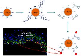
The aim of the current study was to develop a novel site-specific contrast agent for MRI, effective in diagnosis of early endothelial inflammation, the first step in the majority of cardiovascular diseases. Iron oxide particles with attached anti-VCAM-1 and anti-P-selectin antibodies have previously been used as probes for MRI and their combination has proved to be a more efficient targeting approach than either antibody alone.23–25 These systems have been used for non-invasive in vivo imaging of activated endothelial cells at the site of inflammation in the course of the development of atherosclerosis. However, these commercially available tosylated particles are of micrometer size (MPIOs). The nanoparticles, SPIONs, seem to be better suited for the purpose, as they are more easily available to the specific cells. They can also penetrate the endothelial tissue, while not posing the risk of plugging the smaller blood vessels. Unfortunately they also have a stronger tendency to aggregate than MPIOs and are characterized by slower blood clearance; they are also less stable in aqueous media. These properties may, however, be improved by applying appropriate coating and surface modifications. We proposed to use an ionic derivative of chitosan as a coating in order to provide sufficiently stable SPIONs and to introduce anti-VCAM-1 and anti-P-selectin antibodies to achieve the targeted localization of the contrast agent to detect endothelial inflammation.
Experimental section
Materials
Low molecular weight chitosan was obtained from Sigma-Aldrich. A cationic derivative of chitosan, N-[(2-hydroxy-3-trimethylammonium)propyl] chitosan chloride (CCh), was synthesized and characterized according to procedures described earlier;26 the degree of modification with trimethylammonium groups was estimated as 62.2%. Iron(III) chloride hexahydrate (Sigma-Aldrich, puriss. p.a., ≥99.0%), iron(II) chloride tetrahydrate (Sigma-Aldrich, puriss. p.a., ≥99.0%), ammonia solution (25%, POCH S.A., puriss. p.a., ≥99.0%), p-toluenesulfonyl chloride (Sigma-Aldrich, p.a., ≥99.0%), sodium chloride (Sigma-Aldrich), pyridine (Firma Chempur, Sigma-Aldrich, p.a.), hydrochloric acid (36–38%, Sigma-Aldrich, p.a.), boric acid (Sigma-Aldrich, ≥99.5%), ammonium sulfate (Sigma-Aldrich, for molecular biology, ≥99.0%), Tween 20® (Fluka), tri-sodium citrate (POCH, p.a.), Texas Red® goat anti-rat IgG (H + L) antibodies (Life Technologies, 2 mg mL−1), rat anti-mouse CD106/VCAM-1-UNLB antibodies (Southern Biotech), purified rat anti-CD62P antibodies (clone RB40.34, Biosciences) and Hoechst 33258 (Sigma; for nuclei counterstaining) were used as received. Millipore-quality water was used during the experiments.
Methods
Malvern Nano ZS apparatus (Malvern Instrument Ltd, Worcestershire, UK) was used for both dynamic light scattering (DLS) and zeta potential measurements. Zeta potential was determined using laser Doppler velocimetry (LDV). The time-dependent autocorrelation function of the photocurrent was acquired every 10 s, with 15 acquisitions for each run. The sample was illuminated using a 633 nm laser, and the intensity of light scattered at an angle of 173° was measured by an avalanche photodiode. The z-averaged hydrodynamic mean diameter (dz) and dispersity index (DI) of the samples were calculated using the software provided by Malvern. Fluorescence spectra were measured using a Hitachi F-2700 fluorescence spectrophotometer with excitation wavelength 593 nm (based on absorption maximum, see Fig. 2B) and scanning speed 1500 nm min−1, using a quartz cuvette (1 cm optical path) at room temperature. Samples for Fourier transform infrared (FTIR) analysis were isolated from dispersion by lyophilization. The FTIR spectra were recorded in the range 400 to 4000 cm−1 using a Nicolet IR200FT-IR spectrometer with an ATR accessory. Elemental analysis was performed with a CHNS Vario Micro Cube Elemental Analyzer. SEM observations were carried out using a cold field emission scanning electron microscope (FESEM) HITACHI S-4700 equipped with a NORAN Vantage energy dispersion spectrometer. Samples for the measurements were prepared by placing a drop of solution on a silicon plate. The solvent was allowed to evaporate at room temperature and subsequently a thin film of gold was deposited on the sample by sputtering.
Synthesis of SPIONs
The synthesis of SPIONs was carried out in aqueous solution following the co-precipitation method described earlier.6 Briefly, the iron salts in molar ratio Fe(III)
:
Fe(II) = 2
:
1, (0.1622 g FeCl3·6H2O and 0.0596 g FeCl2·4H2O) were dissolved in 50 mL of an aqueous solution of CCh (3 g L−1 in 0.1 M NaCl). The solution was deoxygenated by purging with argon and sonicated (Sonic-6, Polsonic, 480 W, 1 s; 1 pulse every 5 s) for 10 minutes in an ultrasound bath at 20 °C. In the next step, 5 mL of 2.44 M NH3(aq) was added dropwise and the sonication of the solution was continued for 30 minutes. Magnetic chromatography was used to purify the surface-modified nanoparticles (SPION-CCh) and then the suspension obtained was filtered using cellulose syringe filters (0.22 μm).
Modification of the SPION surface with active tosyl groups
A 5 mL aqueous solution of SPION-CCh was deoxygenated by purging with argon for 10 minutes in an ultrasound bath at 20 °C, and 0.33 g of p-toluenesulfonyl chloride in 10 mL of pyridine was added dropwise. Before sealing the system, the mixture was deoxygenated by purging with argon for 2 minutes in an ice bath. The reaction was carried out in an ice bath for 2 hours and continued for 2 hours at room temperature. To purify the SPION-CCh-Ts, magnetic filtration was used. The particles entrapped in the column were washed with 0.1 M HCl (12 mL) and water (24 mL) to remove traces of pyridine.
Modification of SPIONs with anti-VCAM-1 and P-selectin antibodies
In order to bind the antibodies to tosylated SPIONs, 80 μL of primary antibody solution (0.5 mg mL−1) was mixed with 0.3 mL of 0.1 M borate buffer (pH 9.5) in a small vial. Ammonium sulfate (135 mg) was added and thoroughly mixed. Next, 0.5 mL of SPION-CCh-Ts (pH 6; sonicated for 5 minutes) was added to the mixture. The sample was incubated at 37 °C for 24 h while being vigorously shaken. The sample was then centrifuged (10
000 rpm, 15 minutes) and the supernatant was removed. In order to purify the nanoparticles, 2 mL of borate buffer was added to the precipitate, and the sample was vortexed and again centrifuged. The procedure was repeated three times. The purified nanoparticles were re-suspended in 1 mL of aqueous 5% glucose solution.
In order to confirm the permanent attachment of primary antibodies (anti-VCAM-1 or anti-P-selectin) to the surface of the SPIONs, immunostaining with IgG-TR (Texas Red) antibodies was performed. The Texas Red chromophore (sulforhodamine 101 acid chloride) is a popular red fluorescent dye used for staining and sorting cells in fluorescence microscopy applications and in immunohistochemistry. It exhibits strong absorption at 589 nm and fluorescence at 615 nm. To 1 mL of modified SPIONs, 20 μL of IgG-TR was added. The sample was protected against light using metal foil. It was then vortexed and incubated at 37 °C for 1 hour, under vigorous mixing. Next the sample was centrifuged (10
000 rpm) for 15 minutes and the supernatant was removed. In order to purify the nanoparticles 2 mL of borate buffer was added to the precipitate, and the sample was vortexed and again centrifuged. The procedure was repeated until no significant traces of the free IgG-TR antibodies were detected (by spectrofluorimetry). The purified nanoparticles were re-suspended in borate buffer and the fluorescence emission spectrum of the sample was measured.
Determination of the content of Fe in the samples of SPIONs
To determine the content of iron in the samples of SPIONs, a classical colorimetric method based on absorbance measurements of the complex of Fe(II) with phenanthroline was used. The SPION dispersion (1 mL) was dissolved in 1 mL of hot 1 M HCl. Then, an excess of vitamin C (with respect to the anticipated iron content) was introduced in order to reduce Fe(III) to Fe(II). Finally, 3 mL of a 0.2% solution of phenanthroline in 0.08 M HCl was added to the solution obtained. The sample was left in the dark for 10 min, and its absorbance at 512 nm (characteristic for the formed complex) was measured. Based on the calibration curve, the concentration of iron in the sample was determined.
Magnetic studies
The magnetic properties of dried nanoparticle sample were determined with the Vibrating Sample Magnetometer option of a Quantum Design Physical Property Measurement System (PPMS) equipped with a superconducting 9 tesla magnet. DC magnetic susceptibility and hysteresis loops were measured. 57Fe Mössbauer measurements were carried out using a constant acceleration spectrometer in transmission mode. A 10 mCi 57Co/Rh source was used.
Biological studies
Immunostaining of endothelial VCAM-1. In order to confirm the specific interaction of the SPION-CCh-anti-VCAM-1 and SPION-CCh-anti-P-selectin nanoparticles with endothelium in a state of early inflammation, 10 μm-thick cross-section slides of the aorta of 24-week-old diabetic db/db mice with endothelial dysfunction were incubated for 1 hour with SPION-CCh-anti-VCAM-1 and SPION-CCh-anti-P-selectin samples containing 0.12 mg of antibodies per 1 mg of Fe. Aorta cross-section slides taken from age-matched db + mice were acetone-fixed (10 min) and used as a control. The material was pre-incubated for 30 minutes in blocking solution containing 5% normal goat serum and 2% filtered dry milk. The samples were then immunostained with IgG-TR antibodies (incubation for 30 minutes; dilution 1
:
600) and counterstained with Hoechst 33258 for 10 minutes. Images of the samples were taken using a Carl Zeiss Axio Observer.D1 inverted fluorescent microscope (Axiocam) – image acquisition: Hrm; exposure time: 1 ms (DAPI), 30 ms (FITC), 200 ms (Cy3).
MRI measurements in vitro. Measurements were performed using a 9.4 T Bruker BioSpec 94/20 MR imaging system (Bruker, Germany). A home-built solenoid coil (5 turns, internal diameter 3 mm, length 7 mm) adopted to the Bruker system was used. 3D maps were collected with the use of 3D MGE (multi-gradient echo) sequence with parameters as follows: FOV, 7.9 × 2.7 × 2.7 mm3; image matrix, 256 × 86 × 86; spatial resolution, 31 × 31 × 31 μm3; echo time TE, 3.5 ms; repetition time (TR), 0.5 s; flip angle (FA), 35°.Tissue sections examined consisted of the brachiocephalic artery including the branching point of the right common subclavian artery and the right common carotid artery as well as a slice of the adjacent aorta. The measurements were done for ApoE/LDLR−/− mice at 32 weeks of age. Excised sections were placed in polypropylene capillaries (outer diameter 3 mm; from Microvette 100 K3E, Sarstedt, Germany) in 4% formalin solution; then the capillary was carefully rinsed out with the solution. After sealing one end of the capillary the sample was placed vertically in a larger container and subjected to sonication for 5 minutes. Then the sample tube was closed and mounted in the RF coil. The coil was mounted on a standard animal bed and positioned inside the magnet.
Raw data were processed using the susceptibility gradient mapping (SGM)27 method implemented in-house using Interactive Data Language scripts (IDL v. 6.4, ITT Visual Information Solutions, USA). Echo shift was estimated after sparse fast Fourier transform (SFFT) by means of first lobe sinus function fitting by the simplex method (IDL). Echo shift maps were calculated in all three directions, resulting in the full susceptibility gradient vector for all voxels in a 3D volume. Then magnitude maps were calculated and a threshold was applied (equal to 1). Calculations were performed independently for the first three echoes obtained from the MGE sequence. The resulting maps were combined with the corresponding anatomical maps (Fig. 10).
All animal procedures used in the present study conformed to EU directive 2010/63/EU for animal experiments and all experimental procedures were approved by the First Local Ethical Committee on Animal Testing at the Jagiellonian University in Krakow (955/2012).
Results and discussion
Activation of the SPION surface using tosyl chloride
In order to modify the SPIONs coated with cationic derivative of chitosan, obtained as described above, with monoclonal antibodies, their surfaces were first activated by tosylation using p-toluenesulfonyl chloride (tosyl chloride). The presence of tosyl groups on the SPIONs' surface would allow further covalent binding of proteins or antibodies via primary amino or sulfhydryl groups.26 Tosylate esters are commonly used in organic synthesis as protecting group for amines and for the binding of biomolecules to polysaccharides.28 The mechanism consists of an activation step, where active tosylate esters are formed by reacting hydroxyl groups of the polymer with p-toluenesulfonyl chloride, and a further coupling reaction with primary amine. The activation step is preferentially performed in anhydrous conditions owing to possible hydrolysis of tosyl chloride. In our case CCh-stabilized SPIONs could not be separated from the aqueous solution, so we decided to perform the reaction in a 1
:
2 mixture of water and pyridine, which played the role of both organic solvent and catalyst. The removal of oxygen from the reaction mixture was also important, as the effective oxidation of alcohols in tosyl chloride/pyridine systems is described in the literature.29 The reaction was performed in an ice bath and the product was purified by magnetic filtration. The success of the tosylation reaction was confirmed by FTIR analysis of modified SPIONs and comparing the results with those obtained for unmodified SPIONs (Fig. 1). The occurrence of tosylation was confirmed by the appearance of a band at 1332 cm−1 characteristic for –SO2 asymmetric stretching vibrations and one at 804 cm−1, which could be ascribed to the –S–O–C– stretching of the tosyl group. The intensity of a broad band with a maximum at 1628 cm−1 significantly increased, most probably due to the presence of the aromatic ring of tosyl groups being active in the same region. Owing to the lower pH of the suspension, in the spectrum of tosylated SPIONs two bands at 3141 cm−1 and 3046 cm−1, characteristic for primary amines, were no longer visible, while a broad band at 3342 cm−1 and a smaller one at 1562 cm−1 appeared, which could be ascribed to the asymmetric stretching and asymmetric deformation vibrations of –NH3+. Finally there were visible bands at around 1024 cm−1 corresponding to the pyranose unit of chitosan.
 |
| Fig. 1 FTIR spectra of SPION-CCh (A) and SPION-CCh-Ts (B). | |
The successful tosylation was also confirmed by the presence of sulfur in the SPION-CCh-Ts sample by elemental analysis. Based on elemental analysis and taking into consideration both the degree of deacetylation of the chitosan used (0.79) and the degree of its modification with the trimethylammonium groups (0.622) it was calculated that there were around 1.9 tosyl groups per glucose unit of CCh, on average. Such a high number of active tosyl groups on the surface of the nanoparticles should allow effective attachment of antibodies.
DLS and LDV analysis
Both size (dz) and surface charge (ζ) are important parameters determining the stability of particles and their fate after administration. In one of our previous works6 we demonstrated that the core size of SPION-CCh nanoparticles was slightly above 10 nm, but that in aqueous suspensions they formed larger, charged aggregates. It was also shown that upon aggregation these nanoparticles do not lose their magnetic properties. Based on dynamic light scattering (DLS) measurements of SPION-CCh-Ts we found that the tosylation procedure had a positive effect on both size and surface charge of the aggregates. The average size of the nanoparticles was decreased, most probably due to the additional filtration step, from 144 nm to 91 nm, and the distribution profile became narrower (see ESI, Fig. S1†). The zeta potential of the tosylated SPIONs in aqueous suspension was increased to +44 mV and was in the range characteristic for stable, electrostatically stabilized colloids. We believe that this increase resulted from the protonation of free amino groups of chitosan chains on the SPIONs' surfaces induced by decrease in pH. Despite colloidal stability of the aqueous suspension of SPION-CCh-Ts, the system was not stable in the buffer solution and some precipitation was observed. That can be explained by considering that a shielding effect of the counter-ions on the electrostatic repulsion between particles resulted from the presence of salt at high concentration. Thus all the measurements were done in aqueous media, without buffer. The results obtained are presented in Table 1.
Table 1 The physicochemical characteristics of the aqueous suspension of cationic chitosan-stabilized SPIONs before (SPION-CCh) and after (SPION-CCh-Ts) tosylation
|
pH |
ζ [mV] |
dz [nm] |
DI |
cFe [mg mL−1] |
SPION-CCh |
10 |
32.55 ± 4.17 |
143.83 |
0.23 |
0.58 |
SPION-CCh-Ts |
6 |
43.82 ± 8.22 |
91.24 |
0.20 |
0.29 |
We have also tested the effect of the concentration of tosyl chloride used in the reaction mixture on the parameters discussed. No significant changes in size or zeta potential were observed in the range of 0.002–0.17 mg mL−1 of the tosyl chloride concentration.
To characterize the yield of the SPIONs obtained after tosylation, the concentration of iron in 1 mL of the suspension (cFe) before and after the process was determined, as described in the Experimental section. It was done by colorimetric analysis of the sample after reduction of Fe3+ to Fe2+ and formation of the orange complex between Fe2+ and 1,10-phenanthroline. About 50% of the nanoparticles were lost during the tosylation step.
Binding anti-VCAM-1 and anti-P-selectin monoclonal antibodies to the surface of SPIONs
The sulfonyl ester is an electrophilic active group that can couple covalently with nucleophiles e.g. with primary amines. The reaction occurs under basic conditions in aqueous buffers, with pH in the range 8.5–10.30 This reaction was also applied to commercially available Dynabeads® marketed as suitable to covalently couple antibodies with optimal antibody orientation. These beads were applied by McAteer et al.31 to covalently attach anti-VCAM-1 and anti-P-selectin antibodies without losing their activity. We have adopted the protocol described by McAteer et al. to attach anti-VCAM-1 and anti-P-selectin antibodies to the tosylated surface of SPIONs. SPIONs were incubated in buffered (borate buffer, pH 9.5) solution of appropriate monoclonal antibody (anti-VCAM-1 or anti-P-selectin) in the presence of ammonium sulfate, then centrifuged, washed with borate buffer and finally re-suspended in 5% aqueous solution of glucose. The presence of borate buffer provided optimal conditions for binding reaction and facilitated separation of the nanoparticles from unreacted antibodies. To prepare the final suspension of SPIONs we used glucose owing to its relatively high viscosity, which prevents aggregation, and because of its standard use for injections as intravenous sugar solution.
The success of the binding reaction was confirmed using immunostaining. The secondary IgG antibodies labelled with Texas Red (IgG-TR) chromophore were used to label nanoparticles. After the monoclonal antibodies were attached to the nanoparticles' surface, they were incubated with IgG-TR for 1 hour and washed until there was no visible TR fluorescence in the supernatant (Fig. 2). Finally they were re-suspended and their fluorescence was measured. The presence of TR fluorescence in the spectrum of the SPION sample confirmed successful binding.
 |
| Fig. 2 (A) Fluorescence spectra of the supernatants obtained in the purification process after binding anti-VCAM-1 antibodies to the activated surface of SPIONs: first filtrate (black dashed line), second filtrate (black dotted line), third filtrate (grey solid line), SPION-CCh-anti-VCAM-1 sample (red solid line). (B) Absorption spectrum of the SPION-CCh-anti-VCAM-1 sample. | |
This optimized procedure allowed us to attach both types of antibodies – anti-VCAM-1 and anti-P-selectin – to the surface of SPIONs (Fig. 3A and B, respectively).
 |
| Fig. 3 (A) Fluorescence spectra of SPION-CCh-Ts (dotted line) and SPION-CCh-anti-VCAM-1/IgG-TR (solid line); (B) fluorescence spectra of tosylated SPION-CCh (dotted line) and SPION-CCh-anti-P-selectin/IgG-TR (solid line). All solutions were prepared in glucose. | |
To quantify the amount of monoclonal antibodies attached to nanoparticles, we determined the concentration of the TR chromophore in 1 mL of the sample by fluorescence measurements and compared it with the amount of iron in 1 mL of the same suspension. A calibration curve was obtained by fluorescence measurements of a series of standard solutions of IgG-TR. The concentration of iron was obtained by the phenanthroline method, described earlier for tosylated samples. The average amount of antibodies attached to SPIONs' surface was calculated as 0.016 mg per 1 mg of iron. Based on the proportion of the tosylated SPIONs to anti-VCAM-1 antibodies used for the attachment reaction we estimated that only around 6% of the antibodies were successfully attached. Decreasing the amount of antibodies led to a decreased degree of modification (based on immunostaining measurements), while increasing the amount of antibodies did not allow significantly better results to be obtained; therefore we decided to keep the starting proportions. This result may be due to the low efficiency of the binding reaction in this heterogeneous system or because of the tendency of the SPIONs to aggregate, thus limiting the surface available for reacting.
Scanning electron microscopy (SEM)
The morphology of the nanoparticles obtained was also studied using scanning electron microscopy (SEM). Fig. 4 presents the resulting SEM micrographs of tosylated SPIONs and SPIONs with antibody attached. It was observed that both types of particles form round structures that have tendency for aggregation. The images of the aggregates clearly revealed the presence of many closely stacked particles with diameter significantly lower than 50 nm.
 |
| Fig. 4 Scanning electron microscopy images of SPION-CCh-Ts (A) and SPION-CCh-anti-VCAM-1 (B). | |
The discrepancy between the particle sizes obtained by DLS and SEM resulted from the differences in these experimental techniques. Usually the mean sizes (diameters) of the particles observed on SEM micrographs are lower than those estimated from DLS measurements and this is noticeable for both types of particles developed. There are different reasons for this fact. The DLS method measures the mean hydrodynamic diameter, that is, the size of the particle together with the layer of ions and solvent molecules surrounding it. The influence of the aqueous medium is even more significant for the particles coated with chitosan, as this polymer has a tendency to swell in water. Additionally, owing to the intensity of the scattered light which increases with increasing size of objects, the z-averaged hydrodynamic mean diameter obtained by cumulant analysis is overestimated. Thus the results obtained from these techniques are expected to differ significantly, but they provide us with complementary information. While SEM informs us about the size and shape of the core, DLS gives an insight into the change of the particle's diameter when it is transferred to the aqueous medium. The average hydrodynamic diameter of the antibody obtained from DLS measurements was 8.6 nm (data not shown). In view of these results we consider that, although the sizes of the antibody and single nanoparticle were comparable, in reality we obtained larger stable aggregates of around 100 nm that were modified with antibodies, such that the size ratio of the nanoparticle to antibody is more or less 1
:
10. The hydrodynamic diameter of the SPION-CCh-anti-VCAM-1 system, obtained by DLS measurements, was in the region of 200 nm, probably owing to the combined effect of size increase and further aggregation caused by the presence of the antibodies on the surface of the nanoparticles. The observed small size of the core should ensure the superparamagnetic behavior of the sample, while hydrodynamic diameters in the range of 100–200 nm in aqueous media seem suitable for biomedical applications.
Magnetic measurements – magnetic susceptibility
Measurements of the magnetic susceptibility versus temperature have been carried out for dried SPION-CCh-anti-VCAM-1 nanoparticle samples at the applied DC magnetic field of 50 Oe in the zero-field-cooled (ZFC) and field-cooled (FC) modes. The data are presented in Fig. 5.
 |
| Fig. 5 The temperature dependence of the magnetic susceptibility of dried SPION-CCh-anti-VCAM-1 samples measured at 50 Oe field in zero-field-cooled (ZFC) and field-cooled (FC) modes. | |
The ZFC and FC curves coincide at temperatures above 200 K, indicating superparamagnetic behavior in this temperature range. Below 200 K they diverge, revealing the static character of the magnetic moments of nanoparticles in the 50 Oe applied field. For selected temperatures, magnetization loops were measured in the field range −89 kOe to 89 kOe. The results are presented in Fig. S2 (ESI†) and 6. The overall character of the magnetic field dependence at all the temperatures is similar to that observed for soft ferro- or ferrimagnets. The saturation magnetization amounts to 26 emu g−1 at 3 K, which is one-third of that for bulk maghemite, and decreases only slightly with increasing temperature, to reach 21 emu g−1 at 300 K. For other details of the magnetization loops a closer inspection of the plots close to the origin (Fig. 6) is needed.
 |
| Fig. 6 The region of magnetization loops in the −1 kOe to 1 kOe range for dried SPION-CCh-anti-VCAM-1 sample at selected temperatures. In the inset the −80 Oe to 80 Oe range is shown. | |
The small field range depicted in Fig. 6 shows hysteresis loops characterized by the remanent magnetization decreasing from 7 emu g−1 at 3 K through 0.5 emu g−1 at 100 K to zero at 200 K and 300 K. The coercive field decreases from about 300 Oe at 3 K through 12 Oe at 100 K to zero at 200 K and 300 K. The vanishing width of the hysteresis loop at 200 K and 300 K as well as the observed significant attraction of the sample to a small permanent magnet indicate that it is superparamagnetic at these temperatures. The magnetic properties of the material are similar to those reported by us for dual modal contrast agents.32
Magnetic measurements–Mössbauer measurements
The Mössbauer spectrum obtained at 300 K for a dried nanoparticle sample is presented in Fig. 7.
 |
| Fig. 7 Mössbauer spectrum obtained at 300 K for dried SPION-CCh-anti-VCAM-1 sample. | |
The spectrum consists of a broad structure spanning from −8 mm s−1 to 8 mm s−1 with a pronounced double peak feature in the center. The overall splitting of the spectrum is close to that of a “magnetic” sextet observed for the microcrystalline maghemite but its shape is typical for the “relaxational case” observed for superparamagnets. It is characterized by a pronounced broadening of the inner sides of the lines, strongest for the outer lines, and weakest for the inner lines. The relaxational spectrum obtained for the sample studied shows that the nanoparticles are superparamagnetic at the temperature of measurement (300 K) and the characteristic time of their magnetic fluctuations is in the time window of Mössbauer transition, i.e. in the megahertz range. This result, together with the magnetic data, proves the superparamagnetic character of the SPION-CCh-anti-VCAM-1 maghemite nanoparticles synthesized and studied in this work.
Biological studies
Measurement of the physicochemical characteristics of SPIONs stabilized with a cationic coating and attached to appropriate antibodies was followed by biological studies. The experiments were designed to determine the ability of SPIONs with attached antibodies to bind in vitro to dysfunctional endothelium that is known to be associated with overexpression of VCAM-1 and selectin P. Aortic rings obtained from the diabetic db/db mice with endothelial dysfunction, and from db + mice for comparison, were used, embedded and frozen in OCT (optimal cutting temperature compound). Material from both db/db and control mice was immunostained first using free monoclonal antibodies in order to confirm their specificity for the biological material studied. The samples were incubated with both types of antibodies and then washed and immunostained with secondary IgG antibodies labeled with Texas Red (red fluorescence with maximum at 615 nm). The results were visualized using fluorescence microscopy. The nuclei were additionally visualized with Hoechst 33258 dye (blue fluorescence with maximum at 461 nm); green autofluorescence of elastin was also clearly visible. The images obtained showed high specificity of anti-VCAM-1 antibodies towards the aortic endothelium of the mice with diabetes-induced endothelial dysfunction (clearly visible red fluorescence at the surface of endothelium in Fig. 8 and relatively lower specificity of anti-P-selectin antibodies – see Fig. S3 in ESI†). Therefore we chose to use SPIONs decorated with anti-VCAM-1 antibodies for further studies.
 |
| Fig. 8 Cross sections of VCAM1-immunostained aortic rings taken from diabetic db/db mice with endothelial dysfunction: (A) control sample and (B) sample incubated with rat anti-mouse VCAM-1 antibodies, followed by secondary goat anti-rat Cy3-conjugated antibodies (orange fluorescence). Immunostained endothelial layer is marked with white arrows. | |
In the next step we verified the activity of the anti-VCAM-1 antibodies attached to the surface of SPION-CCh towards endothelial cells in the state of early inflammation, thus confirming their targeting abilities. The experiment was performed similarly to the previously described ones, with the exception that the material was incubated with SPION-CCh-anti-VCAM-1 suspension. The ability of SPION-CCh-anti-VCAM-1 to attach to the dysfunctional endothelium was also compared with the activity of the unbound anti-VCAM-1 antibodies. The results obtained confirmed that synthesized nanoparticles bound specifically to the surface of endothelial cells in a state of endothelial inflammation, as seen in Fig. 9.
 |
| Fig. 9 Cross sections of aortic rings taken from diabetic db/db mice with endothelial dysfunction: (A) immunostained with rat-anti-mouse VCAM-1 antibodies; (B) incubated with SPION-CCh-anti-VCAM-1 particles. SPION-VCAM1 nanoparticle binding sites are marked with white arrows. | |
To verify further the possibility of application of the obtained SPION-CCh-anti-VCAM-1 as MRI contrast agent, the immunostaining experiments were followed by MRI. Samples of aortic arch with part of the descending aorta and brachiocephalic artery (BCA) aortic ring obtained from ApoE/LDLR−/− mice with endothelial dysfunction were incubated in a suspension of SPION-CCh-anti-VCAM-1 nanoparticles and, after thorough multiple rinsing, examined using MRI. Susceptibility gradient maps (SGM) were obtained, analyzed and compared with the results obtained for the control vessel. The results are presented in Fig. 10. Representative slices taken from the 3D data sets were collected using MGE sequence for the specimen incubated in SPION-CCh-anti-VCAM-1 suspension (10e–h) and control sample (10a–d). The top row (slices 10a and 10e) shows the anatomical images. The next rows show the images of the same slice resulting from the SGM method applied to the first, the second, and the third echo of the gradient echo train combined with the corresponding anatomical image. An increased T2* contrast is seen in the anatomical image of the incubated sample (10e) due to increasing echo time. The threshold above which the change caused by the local field disturbance was considered significant was set arbitrarily in the SGM images. Numerous regions of the local field disturbance (above the threshold, marked red) with a trend towards intensification with increasing echo number are seen for the incubated artery (slices 10f–h). In contrast, only isolated small regions occurred in the control section (10b–d). This confirms the presence of the superparamagnetic nanoparticles in the incubated sample and the fact that they can be visualized using MRI.
 |
| Fig. 10 Anatomical images (first row) and susceptibility gradient maps (rows 2–4) obtained using 3D MRI for the samples of aortic arch of ApoE/LDLR−/− mouse with endothelial dysfunction: (a–d) (control) and (e–h) (incubated in SPION-CCh-anti-VCAM-1 suspension). | |
The presence of the nanoparticles in the sample after multiple rinsing and sonication steps suggests their strong binding to the surface of the aorta. This may confirm the presence of specific interactions between the antibodies on the surface of SPIONs and the tissue. However, one can notice that the agglomerates of nanoparticles are present also in the outer part of the aorta, which may mean that the antibodies used are not sufficiently selective towards inflamed endothelium and may also interact with perivascular tissue. Further studies, including in vivo analysis, are thus necessary.
Conclusions
Novel, well defined, superparamagnetic iron oxide nanoparticles with monoclonal antibodies anti-VCAM-1 and anti-P-selectin attached to their surfaces were obtained. Owing to the high value of zeta potential the nanoparticles form stable dispersions in aqueous media. Magnetic studies confirmed that the SPION-CCh-anti-VCAM-1 particles obtained exhibit superparamagnetic behavior above 200 K. Their saturation magnetization was found to be 26 emu g−1 at 3 K, decreasing only slightly with increasing temperature to reach 21 emu g−1 at 300 K. Their saturation magnetization was found to be 26 emu g−1 at 3 K, and decreased only slightly to reach 21 emu g−1 at 300 K. The data obtained show that attachment of the antibodies to SPION-CCh particles did not have a significant influence on their magnetic properties. Mössbauer spectroscopy further confirmed the superparamagnetic properties of the nanoparticles and allowed us to identify the core iron oxide structure as maghemite. In vitro immunostaining experiments carried out using samples of aorta taken from diabetic db/db mice demonstrated that SPION-CCh-anti-VCAM-1 attaches to dysfunctional endothelium featuring inflammatory endothelial phenotype. MRI measurements confirmed that SPION-CCh-anti-VCAM-1 can be visualized using MRI. These results indicate that SPION-CCh-anti-VCAM-1 can be considered as a potential novel, highly sensitive, site-specific MRI contrast agent allowing detection of endothelial inflammation which represents a hallmark of numerous diseases associated with endothelial dysfunction, for example atherosclerosis, diabetes and cancer metastasis.
Acknowledgements
This work was supported by the European Union from the resources of the European Regional Development Fund under the Innovative Economy Programme (grant coordinated by JCET-UJ, no. POIG.01.01.02-00-69/09). The research was carried out with the equipment purchased thanks to the financial support of the European Regional Development Fund in the framework of the Polish Innovation Economy Operational Programme (contract no. POIG.02.01.00-12-023/08). Partial support from the National Science Centre, Poland, Project no. 2012/07/B/ST8/03109 is acknowledged.
References
- E. S. Nicholls, A. Peruga and H. E. Restrepo, World Health Stat. Q., 1993, 46, 134 CAS.
- M. A. McAteer and R. P. Choudhury, Vasc. Pharmacol., 2013, 58, 31 CrossRef CAS PubMed.
- C. Naim, M. Douziech, E. Therasse, P. Robillard, M. F. Giroux, F. Arsenault, G. Cloutier and G. Soulez, Can. Assoc. Radiol., J., 2014, 65, 275 CrossRef PubMed.
- A. K. Gupta and M. Gupta, Biomaterials, 2005, 26, 3995 CrossRef CAS PubMed.
- L. Ternent, D. A. Mayoh, M. R. Lees and G. L. Davies, J. Mater. Chem. B, 2016, 4, 3065 RSC.
- A. Szpak, G. Kania, T. Skórka, W. Tokarz, S. Zapotoczny and M. Nowakowska, J. Nanopart. Res., 2013, 15, 1372 CrossRef PubMed.
- J. Lewandowska-Łańcucka, M. Staszewska, M. Szuwarzyński, M. Kępczyński, M. Romek, W. Tokarz, A. Szpak, G. Kania and M. Nowakowska, J. Alloys Compd., 2014, 586, 45 CrossRef.
- G. Kania, U. Kwolek, K. Nakai, S. Yusa, J. Bednar, T. Wójcik, S. Chłopicki, T. Skórka, M. Szuwarzyński, K. Szczubiałka, M. Kepczynski and M. Nowakowska, J. Mater. Chem. B, 2015, 3, 5523 RSC.
- J. Thevenot, H. Oliveira, O. Sandre and S. Lecommandoux, Chem. Soc. Rev., 2013, 42, 7099 RSC.
- J. V. Jokerst, T. Lobovkina, R. N. Zare and S. S. Gambhir, Nanomedicine, 2011, 6, 715 CrossRef CAS PubMed.
- J. Matuszak, J. Zaloga, R. P. Friedrich, S. Lyer, J. Nowak, S. Odenbach, C. Alexiou and I. Cicha, J. Magn. Magn. Mater., 2015, 380, 20 CrossRef CAS.
- L. H. Reddy, J. L. Arias, J. Nicolas and P. Couvreur, Chem. Rev., 2012, 112, 5818 CrossRef CAS PubMed.
- C. Corot, P. Robert, J. M. Idee and M. Port, Adv. Drug Delivery Rev., 2006, 58, 1471 CrossRef CAS PubMed.
- K. Yan, P. H. Li, H. E. Zhu, Y. J. Zhou, J. D. Ding, J. Shen, Z. Li, Z. S. Xu and P. K. Chu, RSC Adv., 2013, 3, 10598 RSC.
- L. H. Reddy, J. L. Arias, J. Nicolas and P. Couvreur, Chem. Rev., 2012, 112, 5818 CrossRef CAS PubMed.
- M. Fréchou, V. Beray-Berthat, J. S. Raynaud, S. Mériaux, F. Gombert, E. Lancelot, M. Plotkine, C. Marchand-Leroux, S. Ballet, P. Robert, G. Louin and I. Margaill, Contrast Media Mol. Imaging, 2013, 8, 157 CrossRef PubMed.
- R. Ross, N. Engl. J. Med., 1999, 340, 115 CrossRef CAS PubMed.
- R. P. Choudhury, V. Fuster and Z. A. Fayad, Nat. Rev. Drug Discovery, 2004, 3, 913 CrossRef CAS PubMed.
- N. Hanyu M' Kume, T. Ikeda, M. Minami, T. Kita and M. Komeda, Atherosclerosis, 2001, 158(2), 313 CrossRef.
- M. I. Cybulsky and M. A. Gimbrone Jr, Science, 1991, 251, 788 CAS.
- A. Sakai, N. Kume, E. Nishi, K. Tanoue, M. Miyasaka and T. Kita, Arterioscler., Thromb., Vasc. Biol., 1997, 17, 310 CrossRef CAS.
- M. A. McAteer, J. E. Schneider, Z. A. Ali, N. Warrick, C. A. Bursill, V. Von zur Muhlen, D. R. Greaves, S. Neubauer, K. M. Channon and R. P. Choudhury, Arterioscler., Thromb., Vasc. Biol., 2008, 28, 77 CrossRef CAS PubMed.
- L. C. Hoyte, K. J. Brooks, S. Nagel, A. Akhtar, R. Chen, S. Mardiguian, M. A. McAteer, D. C. Anthony, R. P. Choudhury, A. M. Buchan and N. R. Sibson, J. Cereb. Blood Flow Metab., 2010, 30, 1178 CrossRef CAS PubMed.
- M. A. McAteer, K. Mankia, N. Ruparelia, A. Jefferson, H. B. Nugent, L. A. Stork, K. M. Channon, J. E. Schneider and R. P. Choudhury, Arterioscler., Thromb., Vasc. Biol., 2012, 32(6), 1427 CrossRef CAS PubMed.
- S. Serres, S. Mardiguian, S. J. Campbell, M. A. McAteer, A. Akhtar, A. Krapitchev, R. P. Choudhury, D. C. Anthony and N. R. Sibson, FASEB J., 2011, 25, 4415 CrossRef CAS PubMed.
- P. A. Bertin, J. M. Gibbs, C. Kwang-Fu Shen, C. S. Thaxton, W. A. Russin, C. H. A. Mirkin and S. T. Nguyen, J. Am. Chem. Soc., 2006, 128(13), 4168 CrossRef CAS PubMed.
- H. Dahnke, W. Liu, D. Herzka, J. A. Frank and T. Schaeffter, Magn. Reson. Med., 2008, 60, 595 CrossRef PubMed.
- K. Nilsson and K. Mosbach, Eur. J. Biochem., 1980, 112, 397 CrossRef CAS PubMed.
- S. Moulay and O. Goucem, Synth. Commun., 2010, 40, 1595 CrossRef CAS.
- G. T. Hermanson, Bioconjugate Techniques, Elsevier, Oxford, UK, 3rd edn, 2013, vol. 3, p. 235 Search PubMed.
- M. A. McAteer, N. R. Sibson, C. von zur Mühlen, J. E. Schneider, A. S. Lowe, N. Warrick, K. M. Channon, D. C. Anthony and R. P. Choudhury, Nat. Med., 2007, 13, 1253 CrossRef CAS PubMed.
- A. Szpak, S. Fiejdasz, W. Prendota, T. Strączek, C. Kapusta, J. Szmyd, M. Nowakowska and S. Zapotoczny, J. Nanopart. Res., 2014, 16(2678), 1 CAS.
Footnote |
† Electronic supplementary information (ESI) available. See DOI: 10.1039/c6ra10994b |
|
This journal is © The Royal Society of Chemistry 2016 |
Click here to see how this site uses Cookies. View our privacy policy here.