The effect of surfactants and their concentration on the liquid exfoliation of graphene

Shuai Wang*ab, Min Yib and Zhigang Shena
aBeijing Key Laboratory for Powder Technology Research and Development, Beihang University, Beijing 100191, China. E-mail: wang@mfm.tu-darmstadt.de
bInstitute of Materials Science, Technische Universität Darmstadt, Jovanka-Bontschits-Strasse 2, 64287 Darmstadt, Germany. E-mail: yimin@buaa.edu.cn; Fax: +49-6151-16-6023; Tel: +49-6151-16-75681

Received 27th April 2016 , Accepted 6th June 2016

First published on 8th June 2016


Abstract

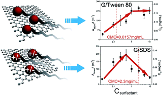
We investigated the effect of surfactants and their concentration on the final graphene concentration via the liquid-phase exfoliation method. Six typical surfactants including ionic and non-ionic ones are explored and the optimized concentration for each surfactant is determined. For ionic surfactants, the graphene concentration increases with surfactant addition and then decreases after reaching its maximum value. In contrast, for non-ionic surfactants, graphene concentration firstly increases with surfactant concentration and then saturates. The different mechanisms of ionic and non-ionic surfactants in stabilizing graphene dispersions are explained by the theory for colloidal stability. Surfactant molecules can adhere to exfoliated graphene sheets and provide an available repulsive force for their stabilization. The as-prepared graphene sheets are verified to be highly exfoliated through transmission electron microscopy and atomic force microscopy studies. The defect level is investigated by Raman spectra and X-ray photoelectron spectroscopy.


1 Introduction

Graphene with its excellent mechanical, optical, electrical and thermal properties has gained great interest since it was first discovered in 2004.1–3 Synthesis methods are vitally important for application. Comparing with chemical vapor deposition4 and other routes, direct exfoliation of graphite in the liquid phase opens a new vista for industrially synthesizing graphene with simplicity and efficiency since Hernandez et al. first accomplished it in 2008.5 Extensive efforts in this approach have been made to improve the yield and the quality of graphene.6–11 Readers are referred to previous reviews, for instance Yi and Shen12 and Nicolosi et al.13 Organic solvent based exfoliation,14,15 polymer based exfoliation16 and surfactant based exfoliation17 are three different approaches in the liquid-phase exfoliation method. Despite some defects brought by the surfactant, the last one was proven to be an ideal way of preparing graphene dispersion with high graphene concentration (CG), and more importantly, with excellent stability. Ionic surfactants were first introduced to assist the exfoliation process. For instance, Vadukumpully et al.18 used a cation surfactant cetyltrimethylammonium bromide (CTAB), Hernandez et al.5 used sodium dodecyl benzene sulfonate (SDBS), sodium cholate (SDOC) and another ten kinds of surfactants19 to exfoliate graphite flakes. Nuvoli et al. designed a series of works to get extremely high CG. Graphene concentration as high as 5.33 mg mL−1 is obtained in a commercial ionic liquid 1-hexyl-3-methyl-imidazolium hexafluorophosphate, 2.21 mg mL−1 in N-methylpyrrolidone solution, 9.45 mg mL−1 in polymerizing media, and 8.00 mg mL−1 in organosilanes.20–23 Du et al. introduced some organic salt to assist exfoliation and enhanced the exfoliation efficiency.24 For a non-ionic surfactant, Guardia et al. first explored the differences between ionic and non-ionic surfactants in assisting exfoliation and verified the ascendancy of the non-ionic surfactant, and then extended the method to synthesize inorganic graphene analogues.25,26 In addition, Niu et al.27 obtained graphene dispersion with enhanced CG with the assistant of inorganic salts. Wang et al.28 introduced ethanol into the surfactant/water solution to reduce exfoliation energy in the surfactant/water medium and enhanced CG up to 3 times. Samoilov et al.29 adopted an effective fluorinated surfactant for graphene production, which is environmentally friendly.

All the previous works show the advantage and the potential of surfactant-assisted liquid-phase exfoliation method. The continuous research in this field is thus necessary and meaningful. To make improvements in this approach, at least two significant problems should be considered. Firstly, what are the main factors that can influence the degree of exfoliation. Secondly, which parameters can represent the effectiveness of a method. For the first question, based on the predecessors’ works, some particular factors, for instance surfactant type, sonication time (tsonic), centrifugation (CF) speed and initial graphite concentration (CGi) were discussed as a function of CG. For surfactant type, Smith et al.19 proposed that ionic and non-ionic surfactants have different mechanisms for stabilizing graphene dispersions. For tsonic, 5 hours of sonication may have a decent marginal benefit over longer or shorter sonication times.25 For CF, the increase of CF will negatively affect CG and graphene sheet quantity.30 For CGi, CG equals a factor (0.01 for SDBS) multiplied by the square root of CGi.17 However, to the best of our knowledge, the influence of surfactant concentration (Csur) on CG have not been deeply explored. For the second question, Coleman et al. first used the dispersion absorption as a main index for exfoliation according to the Lambert–Beer Law, and used transmission electron microscopy (TEM) and other characterization tools to examine the quality of the dispersion.5 Whether graphene dispersion with high CG has the same quality as the one with relatively low concentration is not solidly confirmed.

In this paper, six kinds of surfactants are used to give possible answers to these questions (see Table 1). The relationship between Csur and CG is discussed, and the underlying mechanism of the result is explained. The optimum concentrations of all six surfactants for exfoliation are found. Two models are introduced to explain the differences between ionic and non-ionic surfactants in the sedimentation process. In order to get a full understanding of the factors that influence CG, many controlled experiments are carried out. Characterization methods are performed to examine the quality (sheet size, number of the layers and structural defects) of the product. The results provide valuable data and references for graphene exfoliation in a water/surfactant dispersion.

Table 1 List of surfactants and their acronyms used throughout the text
Acronym Surfactant name
SDOC Sodium deoxycholate
SDBS Sodium dodecylbenzenesulfonate
SDS Sodium dodecyl sulfate
HTAB Hexadecyltrimethylammonium bromide
Tween 80
Triton X-100


2 Experimental

The natural graphite flakes are purchased from Alfa Aesar Co., Ltd. (−325 mesh, 99.8%). SDOC and other surfactants are purchased from Sinopharm Chemical Reagent Co., Ltd. The purified water is purchased from Beijing Kebai’ao Biotech. Co., Ltd. All the materials employed in the experiments are used as received.

SDOC is taken as an example to demonstrate the exfoliation process. Graphite dispersion is firstly prepared by adding 1 g of graphite powder to 200 mL of a SDOC/water mixture in a 300 mL capped round-bottomed flask (CGi = 5 mg mL−1). The solvent is prepared by adding different quantities of SDOC in purified water, by which we can tune the SDOC concentration, i.e. 0.025, 0.05, 0.1, 0.25, 0.5, 1 and 2.5 mg mL−1. It is worth noting that SDOC becomes hard to dissolve in water as the concentration of SDOC rises, hence ultrasonication for about 1 minute is needed to accelerate the dissolution procedure. The SDOC solution mixed with graphite is transferred into six reagent bottles (30 mL). All the graphite solutions are then ultrasonicated in a 100 W ultrasonic bath (KX-1730T Shenzhen Kexi Chemical Co., Ltd) for 8 hours. In order to remove the possible massive graphite sediment, the bottles are placed still and kept overnight. Subsequently, the supernatant is carefully transferred into a 10 mL test tube for CF. The centrifuged supernatant is extracted for the measurement of absorption by a UV-visible light spectrophotometer (TV-1900 Beijing Purkinje General Instrument Co., Ltd.; 660 nm wave length), through which the concentration of graphene dispersed in the solution can be measured. According to the Lambert–Beer Law, A = α660nmCGi, and CG can be obtained from absorption with the coefficient of α660nm = 1458 mL mg−1 m−1 (see results and Discussion section).

3 Results and discussion

In order to measure CG, one convenient method is ultraviolet (UV) absorption analysis based on the Beer–Lambert Law.5 However, one should notice that, this method lacks reliability when comparing with the gravimetric method. To combine the advantage of UV analysis and gravimetric method, a calibration procedure was carried out.

Firstly, the exact value for CG was obtained through gravimetric analysis.20,31 The mass of the membrane was first weighted, denoted as m1. The graphene dispersion was then prepared through liquid-phase exfoliation. The absorption of the dispersion, A, was measured thereafter. The dispersion (volume V) was filtrated through the pre-weighted membrane by a vacuum filtration method. The membrane together with graphene was dried in vacuum and weighted. The total mass of the membrane and the filtrated graphene is denoted as m2. The graphene concentration is finally determined by the formula image file: c6ra10933k-t1.tif.

After calculating CG, the coefficient α in the Beer–Lambert Law for surfactant/water/graphene dispersion is calculated by linearly fitting the curve of absorbance per unit length, A/l, versus CG. Fig. 1 shows the relationship between A/l and CG. Parameter α660nm can be confirmed by the slope of the fitted line (dashed line). The dispersion with high Csur (=5 mg mL−1) has a relatively low value of α660nm (578.5 mg mL−1 m−1), while dispersion with low Csur (Csur = 0.1 mg mL−1) has a relatively high value of α660nm (1458.3 mg mL−1 m−1). This result should be attributed to the fact that for the dispersion with high Csur, the surfactant cannot be totally washed away during the filtration process and the value of α660nm is smaller than the actual value. Hence, α660nm of low Csur dispersion, i.e., 1458.3 mg mL−1 m−1, is recommended as the reference value for both cases with high Csur and low Csur.


image file: c6ra10933k-f1.tif
Fig. 1 Absorption per unit length as a function of CG. One dot shape represents one kind of surfactant. A different color represents different surfactant concentration (black for Csur = 0.1 mg mL−1; red dots for Csur = 5 mg mL−1). Inset: ultraviolet-visible spectrum of graphene dispersion from 200 nm to 900 nm (CGi = 5 mg mL−1, tsonic = 8 h, CF: 1500 rpm for 30 min).

Five vital factors, tsonic, CGi, CF rate, surfactant type and Csur, have influence on CG. In order to get a full view of CG’s dependence on Csur, tsonic, the other factors, i.e., CGi and CF rate are identical in the following experiment. To get the optimum value of these three factors for exfoliation, different experimental conditions have been attempted. Firstly, an exfoliation procedure with different CGi has been made and the results are shown in Fig. 2. From the 3-D bars in Fig. 2a and b, one can find that, both CGi and Csur affect CG. For low CGi, i.e. 1 mg mL−1, CG reaches its peak at a low Csur (SDOC: 0.1 mg mL−1, Tween 80: 0.5 mg mL−1), while for high CGi, i.e., 10 mg mL−1, CG reaches its peak at a relatively high Csur (SDOC: 1 mg mL−1, Tween 80: 1 mg mL−1). The reason is that, for larger numbers of graphite flakes in the dispersion, more surfactant molecules are needed to adhere on the flakes. The best Csur for exfoliation is, obviously, related to CGi. Hence, it is not advisable to only emphasize the best Csur for exfoliation while not mention CGi. The influence of other factors, such as tsonic and CF rate, on CG are shown in S3 (ESI), and the optimum values for exfoliation are obtained and applied in the following experiments.


image file: c6ra10933k-f2.tif
Fig. 2 3-D bar of graphene dispersion concentration as a function of Csur and CGi with the assistance of (a) SDOC (b) Tween 80 (tsonic = 8 h, CF: 1500 rpm (×320g) for 30 min).

An experiment was then designed to find out the impact of the surfactant with different concentrations on CG. The results are shown in Fig. 3. The experimental conditions are set as follows: tsonic = 8 h, CF rate = 1500 rpm, and CF time = 30 min (S3, ESI). The critical micelle concentrations (CMCs) of each surfactant are depicted as a vertical red dash line. CMC was originally thought as the minimum Csur required for successful dispersion of graphite. Then negative experimental results were presented, from which no relations between CMC and Csur could be detected.32 The comparison between the optimum Csur and the CMC of the surfactant in Fig. 3, again confirms the results by Lotya et al.32


image file: c6ra10933k-f3.tif
Fig. 3 (a)–(f) Optical absorbance per unit length (A600 nm L−1) and CG as a function of Csur (tsonic = 8 h, CF: 1500 rpm for 30 min). CMC of each surfactant was shown by the vertical red dashed line. (g) Sedimentation curves for six surfactants after CF. Inset: graphene dispersion after CF using Triton X-100 as the surfactant. Csur from left to right: 0.025 mg mL−1, 0.05 mg mL−1, 0.1 mg mL−1, 0.25 mg mL−1, 0.5 mg mL−1, 1 mg mL−1 and 5 mg mL−1.

Each ionic surfactant has a best Csur for exfoliation, i.e., 0.1 mg mL−1 for SDOC, 0.05 mg mL−1 for SDBS, 0.5 mg mL−1 for SDS and 0.5 mg mL−1 for HTAB. An interesting phenomenon shown in Fig. 3 is that, as Csur increases, the CG curves show different tendencies between ionic and non-ionic surfactants. For ionic surfactants, i.e., SDOC, HTAB, SDS and SDBS, CG reaches its climax and decreases. For non-ionic surfactants, i.e., Tween 80 and Triton X-100, CG reaches its peak value and then is maintained at a high level when Csur is further increased. Excellent stability of the dispersion was confirmed in Fig. 3g. After standing for 700 h, the concentration of the dispersion is lowered by only 5%, on average. This result shows at least one privilege, i.e., better stability of the surfactant-based method over other liquid-phase ones.

The different shapes of the curves in Fig. 3a–g may be explained by the fact that the ionic surfactant and non-ionic surfactant have different mechanisms for stabilizing the colloid according to the theory of Coleman’s group.19 We then expound these two different mechanisms by two different models.

For the ionic surfactant, graphite flakes are first exfoliated by sonication-induced cavitation and shear force, and the exfoliated graphene sheets are then adhered by charged surfactant molecules. When the two charged graphene sheets approach each other, Derjaguin–Landau–Verwey–Overbeek (DLVO) theory33 can be used to explain the anti-aggregation mechanism. The potential energy per unit area between two infinitely extended solids with a gap of x can be calculated by the formula:34

 
image file: c6ra10933k-t2.tif(1)
in which, Ψ0 represents the surface potential, C0 represents the bulk concentration of the salt, kb represents Boltzmann constant, T represents the local temperature, λD = κ−1 represents the decay length,
 
image file: c6ra10933k-t3.tif(2)

AH = π2CABρAρB represents the Hamaker constant. The first part of the formula stands for the double layer electrostatic repulsion force, while the last part of the formula stands for the van der Waals attraction force (v.d.w. force). As the salt concentration ρ increases, AH, as well as the v.d.w force increases, while the repulsion force is almost unchanged. From the formula, the potential barrier w(x) becomes smaller for the same distance x. The graphene sheets are more inclined to aggregate and the dispersion become less stable. Therefore, the trend of the curve for the ionic surfactant in Fig. 3 can be explained. When Csur is comparatively small, the exfoliated graphene sheets are in great demand for surfactant molecules to adhere, and all the surfactants are adhere to the exfoliated graphene sheets. The addition of the surfactant can help with the exfoliation process and thus enhance the stabilization of the dispersion and CG. However, at relatively high Csur, the excess of the surfactant molecules will make w(x) smaller, and the exfoliated graphene sheets are more likely to aggregate. After CF, the aggregated flakes are precipitated. As the result, CG decreases with the excessive ionic surfactant.

For non-ionic surfactants, the graphene sheets adhered by the surfactant molecules are merely moved, and the double layer electrostatic force becomes much lower. As the hydrophobic tails (hydrocarbon chain) of the surfactant molecules from two coated graphene sheets begin to interact, the steric repulsion force plays a more important role. Steric force was used to explain the lack of adhesion or aggregation of uncharged lipids, and it was proven to be dominating over the electrostatic force and v.d.w. force in a short distance.35 De Gennes established a model to calculate the interaction force and corresponding energy between two polymer-coated sheets, and the force per unit area was given by formula:36

 
image file: c6ra10933k-t4.tif(3)
in which hc represents the chain length of the adhered molecule which adhered to the sheet and D represents the average distance between two junction points which connect the sheet and the adhered molecule. The first term in the bracket represents the osmotic force, and the second term represents the elastic force. At strong compressions, the osmotic term should dominate completely. One can find that the interaction force is in inverse proportion to the third power of D. As Csur increases, more surfactant molecules were attached onto the exfoliated graphene sheets, and filled the surface, which lead to a decrease of D. A stronger repulsion force F can avoid graphene sheets from aggregation. When the surface of the graphene sheets are fully occupied by the surfactant molecules, the addition of the surfactant has little effect on CG. On the other hand, the exfoliated graphene sheets cannot stack together because of the surfactant, therefore CG does not decease with the increase of Csur as shown in Fig. 3b, d and f. Sedimentation curves show great stability of the as-made graphene dispersion. Over 80% of the graphene remained in the dispersion after 20 days of standing.

The exfoliation degree of graphene sheets are examined by TEM and AFM. Fig. 4 shows typical TEM images of the exfoliated graphene flakes. The edge of the graphene in Fig. 4a is a protruding few-layers of graphene. Fig. 4b shows the wrapped edge of Fig. 4a, from which the few-layers of graphene sheets can be easily seen. The HRTEM picture in Fig. 4c shows that the graphene sheet in Fig. 4b has a sheet number less than five. Fig. 5a shows some graphene sheets with the thickness of 1 nm (surfactant: Triton X-100) by AFM. As shown in Fig. 5b, a large number of graphene sheets can be observed, with the average area of 46.83 μm2 and thickness of 1 to 3 nm (surfactant: SDOC). In Fig. 5a and b, the white dots on the graphene flakes could be the agglomerated surfactant molecules. One may notice that the shapes of the white dots and the graphene sheets in two figures are different. The shape of the sheets is cotton-shaped in Fig. 5a, while the shape is uniformly block-shaped in Fig. 5b. The reason for this phenomenon is still not clear yet. The XRD spectrum in Fig. 6b supports the AFM results. Compared with pristine graphite filtered film, the exfoliated graphene sheets show a very weak peak appearing at 2θ – 26.6° corresponding to the (002) planes, which is symbolic for graphite powder. Hence, we can draw a conclusion that, after exfoliation, the distance between sp2 hybrid constructed carbon layers was not changed, but the number of this layer to the layer gap was decreased. Furthermore, no (004) peak can be found, which indicates that a long-range order greater than four layers is eliminated by the exfoliation procedure.


image file: c6ra10933k-f4.tif
Fig. 4 (a) and (b) TEM images of one graphene flake. The cyan arrow indicates a graphene sheet protruding from a curly sheet. (c) A HRTEM image of the edge area in (b). Inset: intensity profile recorded in the region of the marked red line showing the edge fringe separation of 0.37 nm. (d) An image of a GNR.

image file: c6ra10933k-f5.tif
Fig. 5 (a) and (b) AFM images of graphene sheets exfoliated with the assistance of (a) Triton X-100 and (b) SDOC. (c) and (d) Histogram of size and thickness of the graphene sheets, respectively.

image file: c6ra10933k-f6.tif
Fig. 6 (a) Comparison of Raman spectra between graphite powder and filtered graphene films. (b) Upper: XRD spectra of the graphene filtered film (CSDOC = 5 mg mL−1, tsonic = 8 h, CF: 1500 rpm, 30 min) and pristine graphite filtered film. Lower: the comparison of XRD spectra of the graphene filtered film with a different surfactant (Triton X-100) concentration. Inset a photograph of a filtered graphene film.

What should be noticed in the XRD characterization is that, in Fig. 3, a graphite/water dispersion with a Triton X-100 concentration (CTriton) of 1 mg mL−1 has a relatively poor performance compared with CTriton = 5 mg mL−1. But in Fig. 6b, XRD spectra show that the (002) peak of the curve of CTriton = 1 mg mL−1 is relatively higher than that of 5 mg mL−1. Moreover, (004) peak can be found in the curve with CTriton = 1 mg mL−1, which indicates that the graphene sheet with CTriton = 1 mg mL−1 is thicker than that with CTriton = 5 mg mL−1. All these clues indicate that although CG is higher for CTriton = 1 mg mL−1 than 5 mg mL−1, the latter one is more effective in the exfoliation process. Hence CG could not be the only index used for evaluating and instructing the exfoliation method.

The defect level of graphene sheets was evidenced by Raman spectra and X-ray photoelectron spectroscopy (XPS). From Fig. 6a, the graphene flakes may mainly suffer from edge defects rather than basal-plane disorder defects. Because in the filtered film the D band is relatively weak and the G band is not broadened. In previous works, a higher D band is often considered as the symbol of disorder defects in the basal plane and a largely broadened G band is commonly found in GO or chemically reduced graphene.37,38 The XPS survey of graphene sheets shows a richer C atom content compared to the O atom (97.09% of C atoms to 2.91% of O atoms; CTriton = 5 mg mL−1), which indicates that surfactants with a high concentration did not oxidize the graphite flakes during the exfoliation process. The fitting peaks of C1s indicate that the non-sp2 C atoms match up with the Triton X-100 structure, so C atoms in graphene are mainly sp2 hybridized (S4, ESI).

4 Conclusion

In conclusion, we have demonstrated the influence of Csur on graphene concentration. The best concentrations of the six surfactants for exfoliation are measured. The results show no relations between the best Csur and the critical micelle concentration of the surfactant. For ionic surfactants, CG reaches its maximum value with the increase of Csur, and then falls down. For non-ionic surfactants, CG reaches its maximum value and maintains the high level. The different trends of CsurCG curves between ionic and non-ionic surfactants can be explained by two different colloidal models. The as-prepared graphene sheets were proven to be large sized and few-layered. These results will give us guidance for future graphene preparation in a water/surfactant medium.

Acknowledgements

This work was supported by Beijing Natural Science Foundation (Grant No. 2132025), the Special Funds for Co-construction Project of Beijing Municipal Commission of Education, the Fundamental Research Funds for the Central Universities in China.

References

  1. K. S. Novoselov, A. K. Geim, S. Morozov, D. Jiang, Y. Zhang, S. Dubonos, I. Grigorieva and A. Firsov, Science, 2004, 306, 666–669 CrossRef CAS PubMed.
  2. A. Kuzmenko, E. Van Heumen, F. Carbone and D. Van Der Marel, Phys. Rev. Lett., 2008, 100, 117401 CrossRef CAS PubMed.
  3. A. A. Balandin, S. Ghosh, W. Bao, I. Calizo, D. Teweldebrhan, F. Miao and C. N. Lau, Nano Lett., 2008, 8, 902–907 CrossRef CAS PubMed.
  4. A. N. Obraztsov, Nat. Nanotechnol., 2009, 4, 212–213 CrossRef CAS PubMed.
  5. Y. Hernandez, V. Nicolosi, M. Lotya, F. M. Blighe, Z. Sun, S. De, I. McGovern, B. Holland, M. Byrne and Y. K. Gun’Ko, et al., Nat. Nanotechnol., 2008, 3, 563–568 CrossRef CAS PubMed.
  6. Z. Shen, J. Li, M. Yi, X. Zhang and S. Ma, Nanotechnology, 2011, 22, 365306 CrossRef PubMed.
  7. M. Yi and Z. Shen, Carbon, 2014, 78, 622–626 CrossRef CAS.
  8. M. Yi, Z. Shen, S. Liang, L. Liu, X. Zhang and S. Ma, Chem. Commun., 2013, 49, 11059–11061 RSC.
  9. J. N. Coleman, M. Lotya, A. O’Neill, S. D. Bergin, P. J. King, U. Khan, K. Young, A. Gaucher, S. De and R. J. Smith, et al., Science, 2011, 331, 568–571 CrossRef CAS PubMed.
  10. K. R. Paton, E. Varrla, C. Backes, R. J. Smith, U. Khan, A. O’Neill, C. Boland, M. Lotya, O. M. Istrate and P. King, et al., Nat. Mater., 2014, 13, 624–630 CrossRef CAS PubMed.
  11. C. Yeon, S. J. Yun, K.-S. Lee and J. W. Lim, Carbon, 2015, 83, 136–143 CrossRef CAS.
  12. M. Yi and Z. Shen, J. Mater. Chem. A, 2015, 3, 11700–11715 CAS.
  13. V. Nicolosi, M. Chhowalla, M. G. Kanatzidis, M. S. Strano and J. N. Coleman, Science, 2013, 340, 1226419 CrossRef.
  14. P. K. Srivastava, P. Yadav and S. Ghosh, RSC Adv., 2015, 5, 64395–64403 RSC.
  15. R. Zhang, B. Zhang and S. Sun, RSC Adv., 2015, 5, 44783–44791 RSC.
  16. T. Kim, H. Lee, J. Kim and K. S. Suh, ACS Nano, 2010, 4, 1612–1618 CrossRef CAS PubMed.
  17. M. Lotya, Y. Hernandez, P. J. King, R. J. Smith, V. Nicolosi, L. S. Karlsson, F. M. Blighe, S. De, Z. Wang and I. McGovern, et al., J. Am. Chem. Soc., 2009, 131, 3611–3620 CrossRef CAS PubMed.
  18. S. Vadukumpully, J. Paul and S. Valiyaveettil, Carbon, 2009, 47, 3288–3294 CrossRef CAS.
  19. R. J. Smith, M. Lotya and J. N. Coleman, New J. Phys., 2010, 12, 125008 CrossRef.
  20. S. BittoloáBon, et al., J. Mater. Chem., 2011, 21, 3428–3431 RSC.
  21. V. Alzari, D. Nuvoli, S. Scognamillo, M. Piccinini, E. Gioffredi, G. Malucelli, S. Marceddu, M. Sechi, V. Sanna and A. Mariani, J. Mater. Chem., 2011, 21, 8727–8733 RSC.
  22. V. Alzari, D. Nuvoli, R. Sanna, S. Scognamillo, M. Piccinini, J. M. Kenny, G. Malucelli and A. Mariani, J. Mater. Chem., 2011, 21, 16544–16549 RSC.
  23. D. Nuvoli, V. Alzari, R. Sanna, S. Scognamillo, M. Piccinini, L. Peponi, J. M. Kenny and A. Mariani, Nanoscale Res. Lett., 2012, 7, 1–7 CrossRef PubMed.
  24. W. Du, J. Lu, P. Sun, Y. Zhu and X. Jiang, Chem. Phys. Lett., 2013, 568, 198–201 CrossRef.
  25. L. Guardia, M. Fernández-Merino, J. Paredes, P. Solis-Fernandez, S. Villar-Rodil, A. Martinez-Alonso and J. Tascón, Carbon, 2011, 49, 1653–1662 CrossRef CAS.
  26. L. Guardia, J. I. Paredes, R. Rozada, S. Villar-Rodil, A. Martínez-Alonso and J. M. Tascón, RSC Adv., 2014, 4, 14115–14127 RSC.
  27. L. Niu, M. Li, X. Tao, Z. Xie, X. Zhou, A. P. Raju, R. J. Young and Z. Zheng, Nanoscale, 2013, 5, 7202–7208 RSC.
  28. S. Wang, M. Yi, Z. Shen, X. Zhang and S. Ma, RSC Adv., 2014, 4, 25374–25378 RSC.
  29. V. Samoilov, E. Danilov, A. Nikolaeva, G. Yerpuleva, N. Trofimova, S. Abramchuk and K. Ponkratov, Carbon, 2015, 84, 38–46 CrossRef CAS.
  30. A. A. Green and M. C. Hersam, Nano Lett., 2009, 9, 4031–4036 CrossRef CAS PubMed.
  31. Y. Arao and M. Kubouchi, Carbon, 2015, 95, 802–808 CrossRef CAS.
  32. M. Lotya, P. J. King, U. Khan, S. De and J. N. Coleman, ACS Nano, 2010, 4, 3155–3162 CrossRef CAS PubMed.
  33. E. J. W. Verwey, J. T. G. Overbeek and J. T. G. Overbeek, Theory of the stability of lyophobic colloids, Courier Corporation, 1999 Search PubMed.
  34. H.-J. Butt, K. Graf and M. Kappl, Physics and chemistry of interfaces, John Wiley & Sons, 2006 Search PubMed.
  35. J. N. Israelachvili, Intermolecular and surface forces: revised third edition, Academic press, 2011 Search PubMed.
  36. P. De Gennes, Adv. Colloid Interface Sci., 1987, 27, 189–209 CrossRef CAS.
  37. M. Yi, Z. Shen, X. Zhang and S. Ma, J. Phys. D: Appl. Phys., 2012, 46, 025301 CrossRef.
  38. A. Ferrari, J. Meyer, V. Scardaci, C. Casiraghi, M. Lazzeri, F. Mauri, S. Piscanec, D. Jiang, K. Novoselov and S. Roth, et al., Phys. Rev. Lett., 2006, 97, 187401 CrossRef CAS PubMed.

Footnote

Electronic supplementary information (ESI) available. See DOI: 10.1039/c6ra10933k

This journal is © The Royal Society of Chemistry 2016