DOI:
10.1039/C6RA10477K
(Paper)
RSC Adv., 2016,
6, 57977-57985
A new organic NLO material isonicotinamidium picrate (ISPA): crystal structure, structural modeling and its physico-chemical properties†
Received
22nd April 2016
, Accepted 3rd June 2016
First published on 6th June 2016
Abstract
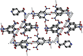
A novel organic nonlinear optical (NLO) crystal, isonicotinamidium picrate (ISPA) was synthesized and grown by the slow evaporation method. Formation of the new crystalline compound was confirmed by a single crystal X-ray diffraction study. The title compound crystallizes in the monoclinic crystal system with the space group of P21/n. DFT was carried out to calculate the bond lengths and bond angles. Structural parameters obtained from single crystal XRD and DFT studies are in good agreement with each other. The frontier energy gaps calculated theoretically are useful to understand the charge transfer taking place in the molecule. Polarizability of the ISPA compound is found using Penn gap analysis and the Clausius–Mossotti equation. The UV-vis-NIR spectral study showed that the grown crystal is transparent in the entire visible region with the lower cut-off wavelength of 469 nm. The four independent tensor coefficients of dielectric permittivity were found to be ε11 = 1.42, ε22 = 4.62, ε33 = 6.23 and ε13 = 5.56 from the dielectric measurements. The titular compound ISPA is thermally stable up to 229 °C assessed by thermogravimetric and differential thermal (TG-DTA) analyses. The specific heat capacity of ISPA is found to be 3.49 J g−1 K−1. The laser induced surface damage threshold of the grown crystal was measured to be 0.22537 GW cm−2 for 1064 nm Nd:YAG laser radiation. Nonlinear refractive index (n2), absorption coefficient (β) and third order nonlinear susceptibility (χ(3)) were determined using the Z-scan technique. The optical limiting study revealed that the transmitted output power increases linearly with the input power up to the irradiance of around 3.25 mW mm−2.
1. Introduction
The developments in ultrafast signal processing are attributed mainly to materials possessing a nonlinear optical susceptibility of a higher order originating from higher order nonlinearity.1 In this context, organic nonlinear optical (NLO) materials have gained much attention in the past due to their relatively high nonlinearity and fast response. Notable benefits of organic systems over inorganic counterparts include their high electronic susceptibility through high molecular hyperpolarizability and facile modification through standard synthetic methods.2,3 It is also feasible to fine tune the chemical structure for desirable properties of organic NLO materials, such as optical switching, power limiting for sensor protection and optical computing.4 The amide linkage [–NHC(O)–] is known to be strong enough to form protein architectures and is utilized to create various molecular devices for a spectrum of purposes in organic chemistry. There has been much focus on amide-type compounds and their metal ion complexes for their properties and inherent applications including molecular recognition, ion electrodes and photochemistry.5 Due to the perturbation under external electric field the conjugated π-electron moiety contributes a footpath for the redistribution of electronic charge across the entire length of conjugation. The second order nonlinearity of a compound arises from the ground-state charge asymmetry of the molecule provided by the donor and acceptor groups. Picric acid forms crystalline picrates of various organic molecules through hydrogen bonding and π–π interactions.6 Picric acid acts as an acidic ligand to form salts through specific electrostatic or hydrogen bond interactions apart from being an acceptor to form various π-stacking complexes with other aliphatic and aromatic molecules.7 Therefore, research is actively progress in the field of nonlinear optical mechanism; the new organic material bis isonicotinamide perchlorate has been reported recently.8 Herein, we report the synthesis, growth, structural, structure–property relationship, optical transmittance, frontier molecular orbitals, thermal, specific heat capacity, dielectric tensor, laser damage threshold, third order nonlinear optical and optical limiting behavior of isonicotinamidium picrate single crystal.
2. Experimental
2.1. Material synthesis and crystal growth
The ISPA compound was synthesized by adding analytical grade isonicotinamide (Himedia) to the picric acid solution in a 1
:
1 equimolar ratio using acetonitrile and water in a (2
:
1) ratio as solvent. The entire experimental procedure has been given as the (ESI).† The crystals grown at different temperatures are shown in Fig. 1(a)–(c) respectively. The reaction scheme is shown in Fig. S1 (ESI).† The solubility of the grown crystal is shown in Fig. S2 (ESI).†
 |
| Fig. 1 (a) As grown bushy ISPA crystal with 2 : 1 acetonitrile + water solvent at room temperature. (b) As grown needle shaped ISPA crystal with 2 : 1 acetonitrile + water solvent at 45 °C. (c) As grown bulk ISPA crystal with 2 : 1 acetonitrile + water solvent at 40 °C. | |
3. Computational details
The molecular structure of ISPA in the ground state was optimized by DFT method using B3LYP, hybrid function consisting three-parameters of Becke's non-local exchange function with the correlation function of Lee, Yang and Parr.9 DFT calculations were carried out using Gaussian 03, B3LYP/6-31G(d) basis set.10
4. Results and discussion
4.1. Single crystal X-ray diffraction studies
X-ray diffraction intensity data were collected at room temperature (293 K) on a Bruker AXS SMART APEXII single crystal X-ray diffractometer equipped with graphite monochromatic MoKα (λ = 0.71073 Å) radiation and CCD detector. A crystal of dimensions 0.30 × 0.25 × 0.15 mm3 was mounted on a glass fiber using cyanoacrylate adhesive. The unit cell parameters were determined from 36 frames measured (0.5° phi-scan) from three different crystallographic zones using the method of difference vectors. The intensity data were collected with an average four-fold redundancy per reflection and optimum resolution (0.75 Å). The intensity data collection, frames integration, Lorentz and polarization corrections and decay correction were carried out using SAINT-NT (version 7.06a) software. An empirical absorption correction (multi-scan) was performed using the SADABS program.11 The crystal structure was solved by direct methods using SHELXS-97 and refined by full-matrix least-squares using SHELXL-97.12 Molecular geometry was calculated using PARST.13 All non-hydrogen atoms were refined using anisotropic thermal parameters. The hydrogen atoms were included in the structure factor calculation at idealized positions using a riding model, but not refined. Images were created with the ORTEP-PLATON program.14,15
The hydrogen atoms were placed in calculated positions with C–H = 0.93 Å and N–H = 0.86 Å refined in the riding model with fixed isotropic displacement parameters: Uiso(H) = 1.5Ueq(C) for methyl groups and Uiso(H) = 1.2Ueq(C) for C aromatic.
The title compound crystallizes in the monoclinic P21/n space group with four molecules in the unit cell with cell parameters, a = 14.290(5) Å, b = 7.430(5) Å, c = 14.697(5) Å, β = 116.638(5)°. The morphology of the grown crystal is shown in Fig. S3 (ESI).† The three dimensional molecular structure of this compound was determined by X-ray crystallography using SHELXS97 and later refined by SHELXL97 to a final R-value 6.22%. The crystal parameters, selected bond lengths and bond angles (experimental and theoretical), selected torsion angles and hydrogen bonds are given in Tables S1–S4 respectively (ESI).†
The ORTEP of the ISPA molecule is shown in Fig. 2(a). The pyridine ring (C1–C2–C3–C4–C5–N1) and phenyl ring (C7–C8–C9–C10–C11–C12) almost have planar conformation with maximum deviation of atom N1 = −0.006 Å and C10 = 0.018 Å, respectively. In this structure, the phenyl ring of nitro group of atoms O4 and O5 lie in a plane which is evidenced by torsion angle values of [O4–N4–C10–C9] = 179.3° and [O5–N4–C10–C11] = 176.7°. The oxygen atom O8 attached with the phenyl ring lies in a plane which is evidenced by torsion angle value of [O8–C7–C12–C11] = 179.5°. The nitro groups O2–O3–N5–C12 and O6–O7–N3–C8 be deviate from the phenyl ring by 35.10° and 23.39° respectively. The acetamide group O1–N2–C3–C6 deviates from the pyridine ring by 14.42°.
 |
| Fig. 2 (a) ORTEP diagram of the ISPA molecule, showing the atom labelling. Displacement ellipsoids are drawn at the 30% probability level. (b) Packing diagram of the ISPA compound running along ‘bc’ plane (c) packing diagram of the ISPA compound viewed down ‘b’ axis. (d) C–H⋯O types of crystal packing diagram of the ISPA compound. | |
In the title crystal, the packing is stabilized by intermolecular N–H⋯O and C–H⋯O types of hydrogen bonds. The N1–H1A⋯O7 and N1–H1A⋯O8 interactions constitute a pair of bifurcated donor bonds generating an R12 (6) ring motif formation and N2–H2A⋯O4 and N2–H2A⋯O5 interactions constitute a pair of bifurcated donor bonds generating an R12 (4) ring motif formation as shown in Fig. 2(b). These two bifurcated molecules were connected with alternative molecules forming a chain running along bc plane. In this molecule, N2–H2B⋯O3 interaction was forming 101 plane and its view down b axis is shown in Fig. 2(c). In the crystal, C5–H5 interaction forming a bifurcated donor bonds which can interact with two different molecules of O5 and O7 atoms and C1–H1⋯O6 interact molecules perpendicular to each other. These two molecules in contact, generates sheets, which is approximately perpendicular to each other. Within these sheets, groups of four molecules form R44 (24) ring motif formation which is shown in Fig. 2(d). In this molecule, the interaction of C11–H11⋯O1 is shown in a view down ‘b’ axis in Fig. 3(a). In the crystal, the molecules are stacked in layers held together by π–π interaction, with a distance of Cg1 and Cg2 to be 3.794(3) Å, between the centroids of adjacent pyridine ring and phenyl ring (symmetry code of Cg1 is x, y, z; Cg2 is x, 1 + y, z) as shown in Fig. 3(b). Cambridge crystallographic data centre (CCDC No. 1058823) contains the supplementary crystallographic data for the ISPA compound.
 |
| Fig. 3 (a) C–H–O type of packing diagram of the ISPA compound view down ‘b’ axis. (b) π–π stacking interactions of the ISPA compound. | |
Table S2 (ESI)† represents the crystallographic information of selected bond angles and bond lengths of experimental and theoretical values. It is observed that the optimized structural parameters, such as bond angles and lengths are slightly higher than the experimental values. This is because, in real case, the intermolecular columbic interaction with the neighboring molecules occurs, whereas, in the gas phase the interaction is absent.16 The optimized molecular geometry of ISPA is shown in Fig. S4 (ESI).†
The polarizability and plasma energy of ISPA have been calculated by taking into account its molecular weight (351.24 g mol−1) and its total number of valence electrons (Z = 130). The density (ρ) of the title compound is 1.673 g cm−3 and the dielectric constant (ε∞) was found to be 10. The calculations were performed in the same manner as reported in the literature17 and are given as (ESI)† and the parameters which were calculated using the relations are given in Table S5 (ESI).†
4.2. Structure–property relationships
Good functional properties are closely related to its crystal structure. Picrate anion with an electron withdrawing group and isonicotinamidium cation with an acceptor group are delocalized by resonance over benzene and the nitro group (NO2). This charge delocalization results in the enhancement of the nonlinear optical response in the titular material. Furthermore it is noteworthy that the picrate anions were strongly bonded to each other through intermolecular hydrogen bonds with short distances ranging from 1.91 Å to 2.51 Å. The isonicotinamidium cations based on C–H–O hydrogen bonds are stabilized in the title compound with distances ranging from 2.34 Å to 2.59 Å. As a result of these hydrogen bonds, high molecular hyperpolarizability is induced in the ISPA molecule due to charge transfer enhancement to have high nonlinear optical absorption.
4.3. Optical transmittance
The UV-vis-NIR transmission spectrum of ISPA crystal with thickness 0.7 mm was recorded in the wavelength range from 190–900 nm using Labindia 3032 UV-vis-NIR spectrophotometer. From the recorded spectrum, it is observed that ISPA crystal has a high transmittance of nearly 70% up to 900 nm with lower cut-off wavelength at 469 nm and is shown in Fig. 4(a). Crystalline defects affect the optical properties18 such as light absorption, scattering and refractive index. For practical devices, crystals should be free from scattering and absorbing defects are required.19 It has been already reported that an increase in defect density leads to the increase in the possibility of the occurrence of scattering centres,20 which is further responsible for the optical loss into the crystals.21 The reasonable optical transmission in ISPA crystal can be attributed to lesser defects and the absence of solvent inclusions. The absorption coefficient (α) and the optical parameters, such as refractive index (n), reflectance (R) and extinction coefficient (K) can be determined from the transmission (T) spectrum based on the following relations, |
 | (1) |
where ‘T’ is the transmittance and ‘t’ is the thickness of the crystal.
 |
| Fig. 4 (a) Optical transmittance of ISPA crystal (b) Tauc's plot of ISPA crystal. | |
The absorption coefficient ‘α’ is related to the extinction coefficient ‘K’ by,
|
 | (2) |
The reflectance (R) in terms of the absorption coefficient and refractive index (n) can be derived from the relations,
|
 | (3) |
|
 | (4) |
The variation of refractive index and extinction coefficient (complex index of refraction) as a function of wavelength is shown in Fig. S5 (ESI).† The calculated value of refractive index of ISPA crystal is 1.4 at 532 nm. It is observed that the refractive index and extinction coefficient decrease with increasing wavelength and the large values of refractive index at lower wavelength suggests that ISPA crystal absorbs higher energy region. It is also noted that the interaction between photons and electrons leads to the variation in refractive index and extinction coefficient.
The electric susceptibility χc can be calculated according to the relation from the optical constants22
|
εr = ε0 + 4πχc = n2 − K2
| (5) |
|
 | (6) |
where
ε0 is the dielectric constant in the absence of any contribution from free carriers. The value of electric susceptibility
χc is 2.44 at 532 nm.
The optical band gap was estimated from the transmission spectrum and the optical absorption coefficient (α) near the absorption edge was calculated using
where
Eg is the optical band gap of the crystal and
A is a constant. The variation of (
αhν)
2 with
hν in the fundamental absorption region was plotted
23 and is shown as an inset in
Fig. 4(b). The band gap of the crystal estimated by extrapolation of the linear part of the graph is 2.59 eV.
4.4. Frontier molecular orbitals (FMOs)
FMOs are the highest occupied molecular orbital (HOMO) and the lowest unoccupied molecular orbital (LUMO). These FMOs play a major role in the study of electrical, optical and chemical properties of the compounds.24 This also predicts the most reactive position in the π-electron system and various reactions involved in the conjugated systems.25 The HOMO shows various prominent donor orbitals and the LUMO shows that of prominent acceptor orbitals. The HOMO–LUMO energy gap shows the charge transfer interaction taking place within the molecule. The electronic absorption is due to the transition from the ground state to the first excited state i.e., an electron is excited from the highest occupied molecular orbital (HOMO) to the lowest unoccupied molecular orbital (LUMO). The orbital energy level analysis for the titular material showed that HOMO value is −7.78 eV while LUMO value is −2.71 eV, and the ΔE value calculated at the DFT level is 2.53 eV. The theoretical energy band gap value obtained from FMO agrees well with experimental value. The obtained frontier molecular orbitals are shown in Fig. 5.
 |
| Fig. 5 Frontier molecular orbitals of ISPA. | |
4.5. Dielectric studies
The relative dielectric permittivity of ISPA crystal was determined as a function of frequency ranging from 50 Hz to 5 MHz at 40 °C using a HIOKI 3532-50 LCR HITESTER instrument. As the dielectric permittivity of the crystal is a second rank tensor, the monoclinic system has four independent components of permittivity ε11, ε22, ε33 and ε13 corresponding to X, Y and Z directions.26 The dielectric properties of a crystal may be characterized by the magnitude and direction of four principal dielectric permittivities.27 The crystal was cut along a, b, c-axis and the fourth plane was cut along 45° between ‘a’ and ‘c’ directions with the thickness of 2.50 mm and a schematic is shown in Fig. S6(a) (ESI).† The prepared samples on either side were coated with silver to make them as parallel plate capacitors. The fourth component, ε13 can be obtained from the following relation: |
ε′33 = ε11 sin2 θ + 2ε13 sin θ cos θ + ε33 cos2 θ
| (8) |
The variation of dielectric permittivity with frequency along the four crystallographic planes is shown in Fig. S6(b) (ESI).† The calculated relative permittivity values are in the ranges 55.31–1.42, 95.09–4.62, 123.48–6.23, 64.05–9.39. It is observed that at lower frequency region the relative permittivity is high and then it decreases with increase in frequency. The dielectric permittivity ε13 is found to be 5.56. The positive value of the ISPA crystal indicates that the polarization occurs in the positive direction of the c-axis and it also confirms that the stored electric energy per unit volume is a positive number.26
4.6. Thermal analysis
The thermal stability of ISPA was studied by thermogravimetric (TG) and differential thermal analyses (DTA). The ISPA sample weighing 3.2 mg was analysed using a STA 409 PL thermal analyzer in the range 30–600 °C under nitrogen atmosphere is depicted in Fig. S7 (ESI).† From the TG curve, it is evident that the ISPA material is stable up to 229 °C and moisture free. The TG curve shows single stage weight loss pattern. An endotherm in DTA thermogram at 229 °C, confirms the absorption of energy for breaking of bonds during decomposition. The decomposition process was carried upto 600 °C with the removal of material into gaseous products (mixture of CO, CO2, NO and hydrocarbons). The residual mass which remains after all the decomposition process is 30.61%. Thus, the ISPA crystal could be exploited for any applications below 229 °C. It has to be noted that ISPA crystal possesses high thermal stability compared to its counterparts such as (bis)isonicotinamide perchlorate,8 triethylaminium picrate28 and hydroxyethylammonium picrate.29 The data comparison is given in Table 1.
Table 1 Comparison of thermal stability, nonlinear absorption coefficient, third order susceptibility and optical limiting threshold with KDP, isonicotinamide, picrate and some of the organic materials
Name of the crystal |
TG-DTA |
β cm W−1 |
χ(3) esu |
Optical lmt threshold |
Present work. |
ISPAa |
229 °C |
0.43 × 10⁻⁴ |
4.48 × 10⁻⁶ |
3.25 mW mm−2 |
(Bis)isonicotinamide perchlorate8 |
88 °C |
— |
— |
— |
Triethylaminium picrate28 |
224.5 °C |
— |
— |
— |
Hydroxyethylammonium picrate29 |
204 °C |
−14.0374 |
3.246 × 10⁻³ |
— |
2-Amino-4,6 dimethoxypyrimidine p-toluenesulfonic acid monohydrate42 |
93 °C |
0.31 × 10⁻² |
3.143 × 10⁻⁴ |
— |
Guanidinium trichloroacetate43 |
— |
4.88 × 10⁻⁶ |
6.96 × 10⁻⁸ |
— |
KDP44 |
213 °C |
0.71 × 10⁻¹⁹ |
4.02 × 10⁻¹⁴ |
— |
2,6-Diaminopyridinium tosylate45 |
180 °C |
0.063 |
4.256 × 10⁻³ |
— |
Cyclo[8]pyrrole46 |
— |
— |
— |
5 J cm−2 |
Ammonium acid phthalate47 |
— |
— |
6.2 × 10⁻⁹ |
0.89 MW mm−2 |
Triphenylamine based dendrimers48 |
— |
— |
— |
5.85 μJ |
4.7. Specific heat capacity
There are certain parameters which influence the laser damage threshold of crystals; specific heat capacity is one among them. According to the definition of specific heat, the energy change in crystal is related to the variation of temperature and can be expressed aswhere CV is the specific heat and ΔT is the variation of temperature due to thermal absorption by the crystal. From the equation, it can be seen that the temperature gradient is inversely proportional to specific heat, which will induce crystal cracking due to thermal expansion anisotropy. Thus, a large specific heat is desirable for applications at higher temperature.30 The heat capacity of the ISPA sample weighing 7 mg was analysed using a NETZSCH analyzer. From Fig. S8 (ESI),† it is observed that the specific heat capacity of ISPA crystal is linear with temperature. The specific heat capacity increases from 3.49 to 6.24 J g−1 K−1 in the measured temperature range from 65–275 °C.
4.8. Laser induced optical damage study
Optical damage is easily induced with pulsed lasers. The damage in laser rods and components were the motivation behind the early studies of laser damage in materials.31 One of the decisive criteria for an NLO crystal material to accomplish as a device is its resistability to laser damage, since nonlinear processes involve high optical intensities. Hence a crystal's usefulness not only depends on the NLO property, but also greatly on its ability to sustain high intensity laser light.32 The surface damage study of the NLO crystals is extremely important since it limits their performance in NLO applications.33 LDT values depend on several factors such as laser pulse width, wavelength, spatial profile pulse, repetition rate, quality of the surface, type and density of defects in the sample.34 The thermal breakdown due to absorption by impurities, self-focusing, avalanche ionization and multi-photon absorption are the mechanisms for the laser induced damage in crystals.35 In wide band-gap materials, the breakdown caused by ‘free electrons’ are initially produced by multi-photon ionization, which further gets heated by the ultra-short laser pulse leads to avalanche processes. The multi-photon excitation becomes more dominant as the laser pulse duration decreases and it has important consequences for the damage threshold.36 Three methods are reported in literature to measure LDT values: (i) one-shot per site (referred as “1 on 1” method): here one shot is made per site and the next shot after increasing the intensity is made at a fresh site. This process is repeated till the damage occurs; (ii) N shots per site (referred as “N or S on 1” method): here a single site is targeted by a fixed number of N shots of same intensity. If in case no damage occurs, the procedure mentioned above is repeated at another site with increase of intensity. This is continued till the damage occurs; (iii) multiple shots per site with ramped increase of intensity of every shot until the damage occurs to the spot. This is referred as “R on 1” method.37 In the present investigation 1 on 1 method is carried out to find the LDT value of ISPA crystal. The pulse width of 13 ns with a repetition rate of 10 Hz and a fundamental wavelength of 1064 nm was used to measure the laser damage threshold of ISPA crystal. The laser beam of diameter 1 mm was focused on the crystal. The sample was placed at the focus of a plano-convex lens of focal length 30 cm. The pulse energy of each shot was measured using the combination of phototube and oscilloscope. The surface damage threshold of the crystal was calculated using the expression, power density (Pd) = E/τπr2 where E represents the input energy (mJ), τ, the pulse width (ns) and r, the radius of the spot (mm). The laser damage threshold of ISPA crystal is found to be 0.22537 GW cm−2.
4.9. Third order nonlinear optical study
The Z-scan method is an effective tool for the accurate measurement of intensity-dependent nonlinear refractive index (NLR), nonlinear absorption coefficient (NLA) and third-order nonlinear optical susceptibility (χ(3)). The nonlinear absorptive properties and the nonlinear refractive index could be assessed by open aperture and closed-aperture Z-scan experiments, respectively. In general, nonlinear optical absorption can be categorized into two kinds, such as (i) saturable absorption (SA), where the transmittance of the sample increases with increasing optical intensity and (ii) reverse saturable absorption (RSA) where the transmittance decreases while increasing the optical intensity. The latter includes two photon absorption and the former, multi-photon absorption.38 The open and closed aperture Z-scan curves of ISPA crystal was performed using a continuous wave Nd:YAG laser of wavelength 532 nm with power 42 mW. The experimental result indicates that the sample exhibits saturation absorption with a positive absorption coefficient (β). The maximum transmittance at the focus (Z = 0) reveals the saturation of absorption at high intensity. For materials with saturation of absorption, there is a maximum transmittance in focus at peak and for materials with reverse saturable absorption there is a minimum transmittance in the focus at valley. A peak in the open-aperture curve, as seen in Fig. 6(a) confirms the occurrence of multi photon absorption in the ISPA crystal. The peak followed by a valley normalized transmittance in the closed-aperture Z-scan curve as shown in Fig. 6(b) indicates the signature of negative nonlinearity due to the self-defocusing effect.39 This can be understood from the above discussion that an NLO characteristic is the result of two photon absorption. The presence of hydrogen bonding interaction in the ISPA material (N–H⋯O and C–H⋯O with short distances ranging from 1.91–2.51 (Å) and 2.34–2.59 (Å) respectively) is ascribed to be the major cause for influencing the electron density distribution and two photon absorption. Hence the nonlinear absorption of ISPA material is higher when compared with the well known KDP and some of the other organic materials. The experimental results compared with the standard KDP and some of the other organic materials are presented in Table 1. And therefore ISPA could be a potential candidate for third order nonlinear optical applications. The formulae used for calculating third order nonlinear parameters are given as (ESI).†
 |
| Fig. 6 (a) Open aperture curve of ISPA (b) closed aperture curve of ISPA. | |
4.10. Optical limiting study
Optical limiters are devices designed to have high transmittance for low level inputs while blocking the transmittance for high intensity laser beams. Since the development of the first lasers in the late 1960's passive optical limiters have been built and tested to protect optical sensors against laser-induced damage.40 The optical limiting behavior of the ISPA crystal was tested by varying the input laser power systematically and the corresponding output power values were measured by the photo detector. The optical limiting behavior of ISPA crystal is shown in Fig. 7. This study shows that the transmitted output power increases linearly with the input power up to the irradiance of 3.25 mW mm−2. With the further increment of input power the transmitted power reaches a plateau and is saturated at a point defined as the limiting amplitude, i.e., the maximum output power showing an obvious limiting property.41 The experimental results of ISPA is comparable with that of the pre-existing organic materials and the values are presented in Table 1. Hence from the observations, the grown ISPA crystal can be used as a suitable candidate for optical limiting applications.
 |
| Fig. 7 Optical limiting behavior of ISPA. | |
5. Conclusion
In summary, good quality crystals of isonicotinamidium picrate (ISPA) was synthesized and grown by the slow evaporation method. The cell parameters and the crystallographic planes were confirmed by single crystal X-ray diffraction study. The HOMO–LUMO energy gaps explains the charge transfer occurring in the molecule. Polarizability of the ISPA compound was found and concluded to be the same in both Penn gap analysis and Clausius–Mossotti equation. UV-vis-NIR spectral study showed that the grown crystal is transparent in the entire visible region ranging from 469 nm to 900 nm. The four independent dielectric tensor coefficients confirmed the anisotropic behavior of the grown crystal. The thermal behavior was studied from TG and DTA analysis. The laser induced surface damage threshold of grown crystal was found to be 0.22537 GW cm−2 for 1064 nm Nd:YAG laser radiation. Nonlinear refractive index (n2), absorption coefficient (β) and third order nonlinear susceptibility (χ(3)) were measured by Z-scan technique. Optical limiting behavior confirms that the transmitted output power increases linearly with the input power up to the irradiance of around 3.25 mW mm−2.
Acknowledgements
One of the authors (R. M. J) is grateful to the DST - SERB, India for the financial support under the DST-SERB Major Research Project (SR/S2/LOP-0031/2012). He also wishes to thank SAIF, IIT Madras for characterization facilities.
References
- J. L. Freeman, Q. Zhao, Y. Zhang, J. Wang, C. M. Lawsonb and G. M. Gray, Dalton Trans., 2013, 42, 14281–14297 RSC.
- B. Gu, Y. H. Wang, X. C. Peng, J. P. Ding, J. L. He and H. T. Wang, Appl. Phys. Lett., 2004, 85, 3687–3689 CrossRef CAS.
- D. S. Chemla and J. Zyss, Nonlinear Optical Properties of Organic Molecules and Crystals, Academic Press, New York, 1987 Search PubMed.
- P. Nagapandiselvi, C. Baby and R. Gopalakrishnan, RSC Adv., 2014, 4, 22350 RSC.
- M. NizamMohideen, S. Thenmozhi, A. SubbiahPandi, N. Panneer Selvam and P. T. Perumal, Acta Crystallogr., Sect. A: Found. Crystallogr., 2009, 65, o740–o741 CAS.
- V. Matulis, A. Lyakhov, P. Gaponik, S. Voitekhovich and A. Ivashkevich, J. Mol. Struct., 2003, 649, 309–314 CrossRef CAS.
- S. El-Medani, T. Youssef and R. Ramadan, J. Mol. Struct., 2003, 644, 77–87 CrossRef CAS.
- K. Syed Suresh Babu, G. Peramaiyan, M. NizamMohideen and R. Mohan, J. Therm. Anal. Calorim., 2015, 120, 1337–1345 CrossRef CAS.
- C. Lee, W. Yang and R. G. Parr, Phys. Rev. B: Condens. Matter Mater. Phys., 1988, 37, 785–789 CrossRef CAS.
- M. J. Frisch, G. W. Trucks, H. B. Schlegel, G. E. Scuseria, M. A. Robb, J. R. Cheeseman, J. A. Montgomery jr, T. Vreven, K. N. Kudin, J. C. Burant, J. M. Millam, S. S. Iyengar, J. Tomasi, V. Barone, B. Mennucci, M. Cossi, G. Scalmani, N. Rega, G. A. Petersson, H. Nakatsuji, M. Hada, M. Ehara, K. Toyota, R. Fukuda, J. Hasegawa, M. Ishida, T. Nakajima, Y. Honda, O. Kitao, H. Nakai, M. Klene, X. Li, J. E. Knox, H. P. Hratchian, J. B. Cross, C. Adamo, J. Jaramillo, R. Gomperts, R. E. Stratmann, O. Yazyev, A. J. Austin, R. Cammi, C. Pomelli, J. W. Ochterski, P. Y. Ayala, K. Morokuma, G. A. Voth, P. Salvador, J. J. Dannenberg, V. G. Zakrzewski, S. Dapprich, A. D. Daniels, M. C. Strain, O. Farkas, D. K. Malick, A. D. Rabuck, K. Raghavachari, J. B. Foresman, J. V. Ortiz, Q. Cui, A. G. Baboul, S. Clifford, J. Cioslowski, B. B. Stefano, G. Liu, A. Liashenko, P. Piskorz, I. Komaromi, R. L. Martin, D. J. Fox, T. Keith, M. A. Al-Laham, C. Y. Peng, A. Nanayakkara, M. Challacombe, P. M. W. Gill, B. Johnson, W. Chen, M. W. Wong, C. Gonzalez and J. A. Pople, Gaussian 03, Revision A.1, Gaussian Inc, Pittsburgh PA, 2003 Search PubMed.
- Bruker, APEX2, SAINT and SADABS, Bruker AXS Inc, Madison, Wisconsin, USA, 2009 Search PubMed.
- G. M. Sheldrick, Acta Crystallogr., Sect. A: Found. Crystallogr., 2008, 64, 112–122 CrossRef CAS PubMed.
- M. P. Nardelli, J. Appl. Crystallogr., 1995, 28, 659 CrossRef CAS.
- L. J. Farrugia, J. Appl. Crystallogr., 2012, 45, 849–854 CrossRef CAS.
- A. L. Spek, Acta Crystallogr., Sect. A: Found. Crystallogr., 2009, 65, 148–155 CrossRef CAS PubMed.
- H. Tanak, A. Alaman Agar and O. Buyukgungor, J. Mol. Struct., 2013, 1048, 41–50 CrossRef CAS.
- P. Koteeswari, S. Suresh and P. Mani, Am. J. Condens. Matter. Phys., 2012, 2, 116–119 CrossRef.
- M. Senthil Pandian, N. Balamurugan, V. Ganesh, P. V. Raja Shekar, K. Kishan Rao and P. Ramasamy, Mater. Lett., 2008, 62, 3830–3832 CrossRef.
- M. Senthil Pandian, P. Ramasamy and B. Kumar, Mater. Res. Bull., 2012, 47, 1587–1597 CrossRef CAS.
- M. Senthil Pandian and P. Ramasamy, Mater. Res. Bull., 2012, 47, 826–835 CrossRef CAS.
- M. Senthil Pandian and P. Ramasamy, Mater. Chem. Phys., 2012, 132, 1019–1028 CrossRef CAS.
- A. Ashour, N. El-Kadry and S. A. Mahmoud, Thin Solid Films, 1995, 269, 117–120 CrossRef CAS.
- J. Tauc, R. Grigorovici and A. Vancu, Phys. Status Solidi B, 1966, 15, 627–637 CrossRef CAS.
- I. Fleming, Frontier Orbitals, Organic Chemical Reactions, Wiley, London, 1976 Search PubMed.
- C. H. Choi and M. Kertez, Conformational information from vibrational spectra of styrene, trans-stilbene, and cis-stilbene, J. Phys. Chem. A, 1997, 101, 3823–3831 CrossRef CAS.
- F. Yu, S. Zhang, X. Zhao, D. Yuan, C.-M. Wang and T. R. Shrout, Cryst. Growth Des., 2010, 418, 71–1877 Search PubMed.
- B. M. Sornamurthy, G. Peramaiyan, P. Pandi, S. Das, G. Bhagavannarayana, V. Manivannan and R. Mohan Kumar, J. Cryst. Growth, 2014, 397, 1–5 CrossRef CAS.
- G. Peramaiyan, P. Pandi and R. Mohan Kumar, J. Therm. Anal. Calorim., 2015, 119, 291–299 CrossRef CAS.
- N. Sudharsana, B. Keerthana, R. Nagalakshmi, V. Krishnakumar and L. Guru Prasad, Mater. Chem. Phys., 2012, 134, 736–746 CrossRef CAS.
- C. M. Kachhava, Solid State Physics, McGraw-Hill, New Delhi, 1990 Search PubMed.
- Damage in Laser Glass, ed. A. J. Glass and A. H. Guenther, ASTM Special Technical Publication 469 ASTM, Philadelphia, 1969 Search PubMed.
- G. C. Bhar, A. K. Chaudhury and P. Kumbhakar, Appl. Surf. Sci., 2000, 161, 155–162 CrossRef CAS.
- N. L. Boling, M. D. Crisp and G. Dube, Appl. Opt., 1973, 12, 650 CrossRef CAS PubMed.
- S. K. Sharma, S. Verma, Y. Singh, K. S. Bartwal, M. K. Tiwari, G. S. Lodha and G. Bhagavannarayana, Opt. Mater., 2015, 46, 329–338 CrossRef CAS.
- CRC Handbook of Laser Science and Technology, Supplement 2: Optical Materials, ed. M. J. Weber, CRC Press Inc, Boca Raton, 1995, pp. 768–776 Search PubMed.
- D. Ashkenasi, M. Lorenz, R. Stoian and A. Rosenfeld, Appl. Surf. Sci., 1999, 150, 101–106 CrossRef CAS.
- R. M. Wood, Institute of Physics Publishing, Bristol, 2003 Search PubMed.
- Y. S. Zhou, E. S. Wang, J. Peng, J. Liu, C. W. Hu, R. D. Huang and X. You, Polyhedron, 1999, 18, 1419–1423 CrossRef CAS.
- G. Peramaiyan and R. Mohan Kumar, Appl. Phys. A, 2015, 119, 707–711 CrossRef CAS.
- G. Vinitha and A. Ramalingam, Laser Phys., 2008, 18, 1070–1073 CrossRef CAS.
- G. Anandha Babu and P. Ramasamy, Spectrochim. Acta, Part A, 2011, 82, 521–526 CrossRef CAS PubMed.
- RO. MU. Jauhar, S. Kalainathan and P. Murugakoothan, J. Cryst. Growth, 2015, 424, 42–48 CrossRef CAS.
- M. Dhavamurthy, G. Peramaiyan, M. NizamMohideen, S. Kalainathan and R. Mohan, J. Nonlinear Opt. Phys. Mater., 2015, 24, 4 CrossRef.
- D. Wang, T. Li, S. Wang, J. Wang, Z. Wang, X. Xu and F. Zhang, RSC Adv., 2016, 6, 14490–14495 RSC.
- RO. MU. Jauhar, P. Vivek, K. Sudhakar, S. Kalainathan, M. NizamMohideen and P. Murugakoothan, J. Therm. Anal. Calorim., 2016, 124, 2 CrossRef.
- H. Xu, S. Q. Zhao, Y. Ren, W. Xu, D. B. Zhu, J. Z. Jiang and J. F. Cai, RSC Adv., 2016, 6, 21067 RSC.
- A. Arunkumar and P. Ramasamy, J. Cryst. Growth, 2014, 401, 195–199 CrossRef CAS.
- B. Xu, H. Fang, F. Chen, H. Lu, J. He, Y. Li, Q. Chen, H. Sun and W. Tian, New J. Chem., 2009, 33, 2457–2464 RSC.
Footnote |
† Electronic supplementary information (ESI) available. CCDC 1058823. For ESI and crystallographic data in CIF or other electronic format see DOI: 10.1039/c6ra10477k |
|
This journal is © The Royal Society of Chemistry 2016 |
Click here to see how this site uses Cookies. View our privacy policy here.