
Carbon dots based turn-on fluorescent probes for the sensitive determination of glyphosate in environmental water samples†
Long Wanga,
Yidan Bia,
Jia Gaoa,
Yijia Lia,
Hong Dingb and
Lan Ding*a
aCollege of Chemistry, Jilin University, 2699 Qianjin Street, Changchun 130012, PR China. E-mail: dinglan@jlu.edu.cn; Fax: +86-431-85112355; Tel: +86-431-85168399
bState Key Laboratory of Inorganic Synthesis and Preparative Chemistry, College of Chemistry, Jilin University, Changchun 130012, PR China
First published on 30th August 2016
Abstract
Fluorescent CDs were prepared by using thiourea as the carbon source and diethylene glycol as the reaction medium. The CDs were obtained only within three minutes under microwave irradiation and their particle size was mainly distributed in the range of 1.5–3.5 nm. The average particle size was 2.3 nm with a standard deviation of 0.5 nm. The fluorescence of the CDs could be quenched by Cu2+ through the static quenching mechanism. On this basis, the CDs can be used as a fluorescence probe for detection of Cu2+ in a linear range of 0.2–25 μmol L−1, and the limit of detection was 0.05 μmol L−1. The quenched fluorescence of the CDs could be recovered with the addition of glyphosate based on a competitive affinity for Cu2+ between glyphosate and the functional groups on the surface of CDs, which provides a turn-on sensing strategy for determination of glyphosate. Under the optimal conditions, the linear range of the method was 0.03–10 μg mL−1 with a detection limit of 16 ng mL−1. Finally, this fluorescence probe was successfully applied to the direct determination of glyphosate in environmental water samples and satisfactory results were obtained.
1. Introduction
Nowadays, to improve the quality and yield of agricultural crops, pesticides and herbicides are widely used in the agricultural field. Glyphosate (N-[phosphonmethyl]glycine) is a post-emergent and non-selective herbicide used for the control of a wide range of weeds.1–3 Because of its excellent performance in weed control and relatively low toxicity to mammals, glyphosate has become one of the most widely used herbicides in the world.4,5 However, the abuse of glyphosate may easily lead to high-level residues in environmental samples due to its strong retention in soil, high solubility in water and long half-life in the environment,6 which might pose a threat to human health. The median lethal dose (LD50) of glyphosate in rats is around 5000 mg kg−1.7 Although the acute toxicity of glyphosate for rats is relatively low, the additives and surfactants in commercial formulations can improve the absorbance of glyphosate and thus increase the toxicity.7 The maximum residue level of glyphosate in drinking water is 0.7 μg mL−1 (GB5749-2006 China).8 Furthermore, the recent studies have indicated that the glyphosate is genotoxic and potentially carcinogenic to humans.9,10 Therefore, a reliable method for the determination of glyphosate in environmental samples has become increasingly important.Analytical methods for the determination of glyphosate mainly include high performance liquid chromatography (HPLC),4,11 liquid chromatography-tandem mass spectrometry (LC-MS),12 gas chromatography (GC),13 gas chromatography-tandem mass spectrometry (GC-MS),14 ion chromatography (IC),15 capillary electrophoresis (CE),16 UV-visible spectrophotometer17 and inductively coupled plasma-mass spectrometry (ICP-MS).18 Although these methods have high sensitivity and good accuracy for glyphosate detection, many of them suffer from various drawbacks such as complicated derivatization steps, time-consuming procedures, long testing times, requirements of expensive instruments and specific skills for operation, which restrict their extensive application in practical glyphosate detection. Therefore, development of a simple, rapid, sensitive and selective method for the determination of glyphosate is in high demand.
Among the various detection techniques, fluorescence technique is a powerful tool that can provide a nondestructive, rapid, simple and ultrasensitive strategy for the analytes detection. However, it is very difficult to develop a fluorescent method for direct determination of glyphosate due to the lack of chromophore or fluorophore groups in the molecule structure of glyphosate. Recently, fluorescent carbon dots (CDs) have attracted increasing attention for their sensing capability due to their low toxicity and excellent biocompatibility, high aqueous solubility and photostability, high resistance to photobleaching and robust chemical inertness.19,20 Up to now, various fluorescent probes based on CDs have been developed for the detection of analytes, such as nitrite,21 glucose,22 biothiol,23 DNA,24 cyanide,25 PO43−,26 F−,27 H2S,28 oxytetracycline hydrochloride,29 methyl parathion,30 dimethoate31 and some heavy metal ions.32 Therefore, it is highly desirable to develop a fluorescent strategy based on the CDs for direct determination of glyphosate with high sensibility and selectivity.
Herein, we report for the first time a carbon dots-based turn-on fluorescent probe for direct detection of glyphosate. The CDs were fabricated by one-step microwave-assisted pyrolysis of thiourea in the presence of diethylene glycol (DEG). The CDs were obtained only within three minutes under microwave irradiation and its particle size mainly distributed in the range of 1.5–3.5 nm. The CDs can be used as a fluorescence probe for Cu2+ detection based on a static quenching mechanism. More importantly, the quenched fluorescence of the CDs could be recovered gradually in the presence of glyphosate due to competition coordination with Cu2+ between glyphosate and the functional groups on the surface of CDs (Scheme 1). Such fluorescence response can be used for well quantifying glyphosate in the range of 0.03–10 μg mL−1 with a detection limit of 16 ng mL−1. Compared with our previously reported method for glyphosate detection based on the inner-filter effect of silver nanoparticles (AgNPs) on the fluorescence of CDs,33 this strategy was more facile, simpler, faster and more suitable for application in conventional rapid analysis. The proposed method has been successfully used for determination of glyphosate in environmental water samples.
 | ||
| Scheme 1 Schematic illustration of Cu2+ and glyphosate detection using the CDs. | ||
2. Experimental
2.1 Chemicals and materials
All reagents and solvents were at least analytical grade and used directly without further purification. Thiourea, diethylene glycol (DEG), quinine sulfate, disodium phosphate dodecahydrate, sodium dihydrogen phosphate dehydrate, methanol, concentrated sulfuric acid and sodium hydroxide were obtained from Beijing Chemical Factory (Beijing, China). Metal salts (CuCl2·2H2O, Pb(NO3)2, MnCl2·4H2O, HgCl2, FeCl3, CdCl2, AgNO3, ZnCl2, BaCl2·2H2O, NaCl, KCl, MgCl2·6H2O, Ca(NO3)2·4H2O) were purchased from Sinopharm Chemical Reagent Co., Ltd (Shanghai, China). Glyphosate, glufosinate, chlorpyrifos, chlorothalonil, imidacloprid, pymetrozine, carbendazim, chlorfenapyr, dinotefuran, metaflumizone, metsulfuron-methyl and ametryn were obtained from Jiangsu Pesticide Research Institute (Jiangsu, China). The water used in all experiments was obtained from a Milli-Q water system (Millipore, Billerica, MA, USA) and the resistivity was 18.2 MΩ cm at 25 °C.The standard stock solutions of Cu2+ (50 μM) and glyphosate (100 μg mL−1) were prepared by dissolving the appropriate amounts of CuCl2·2H2O and glyphosate in water, respectively.
2.2 Preparation of the CDs
In a typical procedure of CDs preparation, 1 g thiourea was added into 10 mL DEG and then the mixture was ultrasonicated for 10 min to form a clear solution. Then the solution was heated in a domestic microwave oven (750 W, Midea, China) for 3 min. After having been cooled down to room temperature, the product was dissolved with 15 mL of water and centrifuged at 10![[thin space (1/6-em)]](https://www.rsc.org/images/entities/char_2009.gif) 000 rpm for 10 min to discard the large particles. Subsequently, the obtained suspension was further purified by filtration (0.22 μm microporous membrane) and then diluted with water. Finally, the clear yellow suspension containing CDs was stored in a refrigerator at 4 °C for further use.
000 rpm for 10 min to discard the large particles. Subsequently, the obtained suspension was further purified by filtration (0.22 μm microporous membrane) and then diluted with water. Finally, the clear yellow suspension containing CDs was stored in a refrigerator at 4 °C for further use.
2.3 Instruments
Transmission electron microscopy (TEM) and high resolution TEM (HR-TEM) images were taken on a FEI Tecnai G2 F20 S-TWIN transmission electron microscope (FEI Company, USA) with an accelerating voltage of 200 kV. X-ray diffraction (XRD) analysis was performed on a Rigaku D/max-2500 diffractometer with a graphite monochromator by using Cu Kα radiation operating at 200 mA and 40 kV (Rigaku, Japan). Fourier transform infrared spectra (FTIR) were acquired on a Nicolet Impact 410 FTIR spectrometer (Bruker, Germany) in the form of KBr pellets. X-ray photoelectron spectroscopy (XPS) was measured on an ESCALAB 250 spectrometer using Mg Kα X-ray as the excitation source (Thermo Electron Corporation, USA). UV-vis absorption spectra of the samples were recorded on a UV-2450 UV-visible spectrophotometer (Hitachi, Japan). Fluorescence emission spectra were obtained on a Shimadzu RF-5301 PC spectrofluorophotometer with a xenon lamp as the excitation source (Shimadzu, Japan).2.4 Fluorescence detection of Cu2+ and glyphosate
In a typical experiment, the CDs solution (100 μL, 5 μg mL−1) and 100 μL PBS buffer solution (pH = 6.0, 200 mM) were mixed with different amounts of Cu2+ standard solution. Then, the mixed solution was diluted to 2 mL with water. After incubation for 12 min at room temperature, the fluorescence spectra were recorded under excitation at 360 nm. The selectivity for Cu2+ detection was evaluated by adding other metal ions instead of Cu2+ in the same procedure.For the detection of glyphosate, 100 μL of CDs (5 μg mL−1) and 100 μL PBS buffer solution (pH = 6.0, 200 mM) were incubated with a certain amount of Cu2+ for 12 min to form the CDs–Cu2+ system. After that, different amounts of glyphosate were added into the system. The resulting mixture solution was diluted to 2 mL with water and then incubated for 12 min before the spectral measurements. The final concentration of Cu2+ was 25 μM. The fluorescence spectra were recorded from 380 nm to 600 nm with an excitation at 360 nm.
2.5 Detection of glyphosate in environment water samples
Real water samples were collected from Qing Lake, Guanlan Lake and Yan Lake (Changchun, China). Prior to the fluorescent detection, the samples were filtered through a 0.22 μm membrane to remove large solids and main impurities. Then the water samples were spiked with standard glyphosate solution and analyzed by the proposed method. The concentrations of the glyphosate in these water samples were obtained by the standard addition method.3. Results and discussion
3.1 Characterization of prepared CDs
As shown by TEM images (Fig. 1A and B), the CDs are spherical in shape with an average particle size of 2.3 nm (standard deviation of 0.5 nm). The corresponding size distribution histogram was obtained by counting about 200 CDs (the inset of Fig. 1A), revealing a narrow particle diameter ranging from 1.5 to 3.5 nm. The HRTEM image of the CDs does not reveal any clear lattice fringes (inset of Fig. 1B), which indicates their amorphous nature.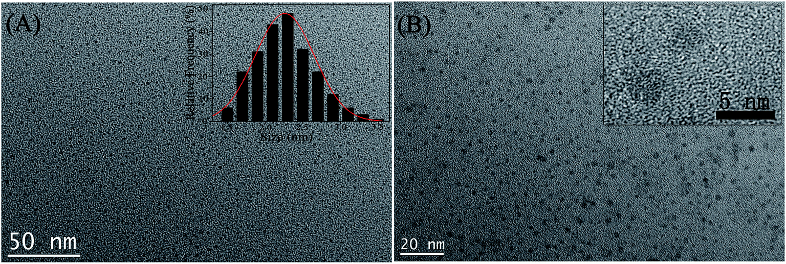 | ||
| Fig. 1 TEM micrographs of the CDs at different magnifications (A) 50 nm and (B) 20 nm. Insets in (A) and (B) show size distribution histograms and high-resolution TEM (HRTEM) images of the CDs, respectively. | ||
The XRD pattern of the CDs has a wide peak around 23.5° (Fig. 2A), revealing an amorphous carbon phase, which was in good agreement with the HRTEM analysis.34 FT-IR spectra were performed to characterize the surface groups of CDs (Fig. 2B). The broad band at 3075–3651 cm−1 is assigned to the stretching vibrations of O–H and N–H. The small band at 2944 cm−1 is ascribed to the stretching vibrations of C–H bonds. The peak at 2072 cm−1 can be identified as the C![[triple bond, length as m-dash]](https://www.rsc.org/images/entities/char_e002.gif) N group.35 The absorption peaks at 1705 cm−1 and 1618 cm−1 indicate the presence of carboxyl functional groups in the surface of CDs.36 In addition, the broad band at 1339–1479 cm−1 is attributed to C–N and N–H groups, and 1025–1155 cm−1 to C
N group.35 The absorption peaks at 1705 cm−1 and 1618 cm−1 indicate the presence of carboxyl functional groups in the surface of CDs.36 In addition, the broad band at 1339–1479 cm−1 is attributed to C–N and N–H groups, and 1025–1155 cm−1 to C![[double bond, length as m-dash]](https://www.rsc.org/images/entities/char_e001.gif) S vibrations.37 These functional groups can improve the stability and hydrophilicity of the CDs in an aqueous system.
S vibrations.37 These functional groups can improve the stability and hydrophilicity of the CDs in an aqueous system.
 | ||
| Fig. 2 The XRD pattern (A) and FT-IR spectrum (B) of the CDs. | ||
XPS was employed to analyze the surface chemical compositions and structures of the CDs. As shown in Fig. 3A, the XPS survey spectra indicate that the CDs mainly contain carbon, oxygen, nitrogen and sulfur elements. The high-resolution XPS spectrum of C1s in Fig. 3B shows five single peaks with binding energies at about 284.2, 284.6, 285.1, 286.0 and 288.6 eV, which are attributed to C–C/CC, C–S, C–N/CN, C–O, and CO/CN, respectively.38,39 The N1s spectrum (Fig. 3C) can be divided into three peaks at 398.1, 399.4, and 400.1 eV, corresponding to C–N–C/CN, N–(C)3, and N–H bonds, respectively.40,41 The S2p spectrum exhibits four main peaks (Fig. 3D). The binding energy peak at 162.0 eV suggests the presence of –SH.42 The peaks at 163.4 and 164.6 eV agree with the S2p2/3 and S2p1/3 positions of the C–S–C covalent bonds due to the spin–orbit couplings.32 The last one located at 168.0 eV can be ascribed to the –C–SO2– species.43
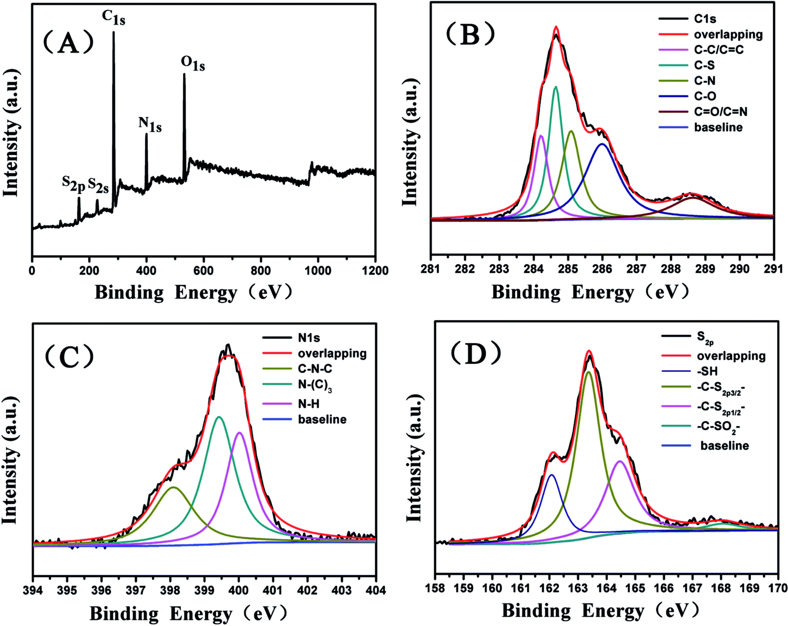 | ||
| Fig. 3 The XPS survey spectrum (A), C1s (B), N1s (C), and S2p (D) spectra of the CDs. | ||
The optical properties of the CDs were analyzed with the UV-vis absorption and fluorescence spectra. The UV-vis absorption spectra of the CDs (Fig. 4A, black line) shows a strong absorption band at 307 nm, which can be ascribed to the n–π* transition of the CO bond.44 The maximum excitation wavelength and maximum emission wavelength of the CDs appear at 360 and 437 nm, respectively (Fig. 4A, red line and blue line). As shown in the inset of Fig. 4A, the CDs suspension exhibits a yellow color under visible light, whereas it is very bright blue luminescence upon excitation with UV light of 365 nm. The quantum yield (QY) of the CDs was measured to be about 32% with quinine sulfate in 0.1 M sulfuric acid solution (QY = 54%) as reference, demonstrating the excellent fluorescence properties of the CDs. Fig. 4B displays the fluorescence emission spectra of the CDs obtained as the excitation wavelength increased from 350 to 420 nm by an increment of 10 nm. The spectra exhibit a typical excitation-wavelength-dependent fluorescence behavior, and the emissive wavelength is red-shifted under excitation with longer wavelength, which is considered to be related to different sizes and a distribution of different surface states of CDs.45
 | ||
| Fig. 4 (A) UV-vis absorption spectrum (black line), fluorescence excitation (red line, λem = 437 nm), and emission spectra (blue line, λex = 360 nm) of the CDs dispersed in water at room temperature. The inserts show the photograph of the CDs in aqueous solutions under visible light (left) and UV light of 365 nm (right). (B) Fluorescence emission spectra of the CDs obtained at different excitation wavelengths with a 10 nm increment from 350 to 420 nm. Inset shows the normalized emission spectra. | ||
The fluorescent stability of the CDs was further investigated to examine their performance. As revealed in Fig. S1A,† only a slight decrease in the fluorescence intensity is observed after continuous irradiation at 365 nm with a UV beam for 200 min, indicating high resistance to photobleaching of the CDs. As demonstrated in Fig. S1B,† the fluorescence intensity of the CDs do not display significant change in the presence of NaCl solutions with different concentrations (up to 1 mol L−1), which verifies that the CDs have excellent stability under high ionic strength environment. In addition, the effect of pH values on the fluorescence intensity of the CDs was also carried out. As shown in Fig. S2,† the fluorescence intensity of the CDs exhibits an irregular pH dependence (pH variation from 2 to 11), which agrees with the previous reports.46
3.2 Detection of Cu2+ based on “turn off” fluorescence of CDs
In order to obtain a high sensitivity for Cu2+ detection, the pH influence on the CDs responding to Cu2+ was first investigated. As shown in Fig. S3A,† the fluorescence intensity ratio F0/F1 (F0 and F1 are the fluorescence intensities of CDs in the absence and presence of Cu2+) increased with pH increase and had a maximum value at pH 6, then decreased as the pH increased above 6. The possible reason for these phenomena was that surface groups of the CDs were protonated and were thus unable to coordinate with Cu2+ in strongly acidic media, while partial hydrolysis of Cu2+ in the alkaline media would impede the coordination between Cu2+ and the functional groups on the surface of the CDs. Thus, pH 6 was selected for the further study. Then, the time for reaction equilibrium between Cu2+ and CDs was also investigated. As shown in Fig. S3B,† the fluorescence intensity of the CDs decreases quickly after addition of Cu2+ into the CDs solution and is stable after 9 min. In order to obtain the maximum quenching effciency, 12 min was chosen as the incubation time between Cu2+ and the CDs before fluorescence measurements.To explore the sensitivity of CDs toward Cu2+, the evolution of the fluorescence intensity of the CDs in the absence and presence of different amounts of Cu2+ was carried out. As shown in Fig. 5A, the fluorescence emission intensity of CDs decreases gradually with the increasing Cu2+ concentration in the solution. The quenching of the fluorescence intensity could be described using the Stern–Volmer equation:
| F0/F1 = 1 + KSVC |
 | ||
| Fig. 5 (A) Fluorescence response of the CDs in the presence of different concentrations of Cu2+ (0–25 μM). Inset: the linear calibration plot of F0/F1 versus Cu2+ concentration. (B) Comparison of fluorescence intensities of the CDs after adding different metal ions with the same concentration of 10 μmol L−1. F1 and F0 represent the fluorescence intensity of CDs in the presence and absence of Cu2+. pH = 6.0. | ||
For a reliable fluorescence probe, high specificity is another significant requirement for analytes detection. To evaluate the specificity of the CDs for Cu2+ detection, we measured the fluorescence changes of the CDs in the presence of various metal ions including Na+, K+, Mg2+, Ca2+, Ba2+, Zn2+, Ag+, Cd2+, Fe3+, Hg2+, Mn2+ and Pb2+ under the same conditions. As shown in Fig. 5B, some metal ions (Na+, K+, Mg2+, Ca2+, Ba2+, Zn2+, Mn2+ and Pb2+) only cause negligible fluorescence change, while some metal ions (Hg2+, Cd2+, Fe3+ and Ag+) induce a little fluorescence quenching. Only Cu2+ can lead to the significant fluorescence quenching effect. The results indicate that the CDs display high selectivity for the determination of Cu2+ over the other metal ions, which can be attributed to that Cu2+ ion has higher thermodynamic affinity and faster chelating process with “N”, “O” and “S” groups on the surface of the CDs than other metal ions.50,51
3.3 Detection of glyphosate based on “turn-off–on” fluorescence of CDs
In general, glyphosate, containing functional groups of carboxyl (–COOH) and phosphonyl (–PO3H2), could be chelate with Cu2+, which might reduce the quenching effect of Cu2+ to the CDs fluorescence, and cause the recovery of fluorescence intensity of quenched CDs. In fact, previous works have reported that glyphosate possesses strong binding affinity to Cu2+.52–54 Thus, the present CDs–Cu2+ system might be a potential candidate for turn-on glyphosate sensing. To illustrate the expected principle for glyphosate detection, the effect of glyphosate on the CDs–Cu2+ system has been investigated thoroughly. As shown in Fig. 6, when excited at 360 nm, CDs show strong fluorescence at 437 nm (curve a). Although a high concentration of glyphosate (50 μg mL−1) was added into the CDs solution, the CDs show no obvious change in fluorescence intensity (curve b), indicating that there is no direct interaction between glyphosate and CDs. The fluorescence of CDs was effectively quenched when Cu2+ was mixed with the CDs solution (curve c). As expected, upon the addition of glyphosate into the CDs–Cu2+ system, the quenched fluorescence of CDs is recovered obviously (curve d) due to competitive affinity to Cu2+ between glyphosate and the functional groups on the surface of CDs, which can be proved once again by the addition of a higher concentration of glyphosate (curve e) into the system. Meanwhile, there was no discernible change in the shape of the emission spectra of CDs, indicating that the recovered emission came from CDs rather than any other newly formed emission centers. Thus, a novel fluorescent strategy for rapid determination of glyphosate was proposed according to the turn-on response of glyphosate to the fluorescence of CDs quenched by Cu2+.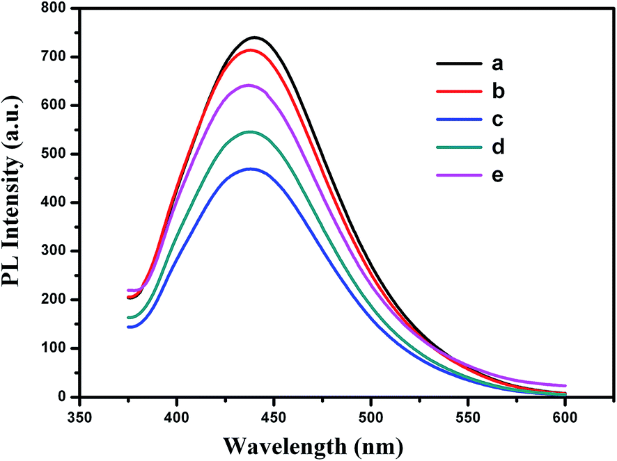 | ||
| Fig. 6 Fluorescence emission spectra: (a) 5 μg mL−1 CDs; (b) a + 50 μg mL−1 glyphosate; (c) a + 25 μM Cu2+; (d) a + 25 μM Cu2+ + 1.5 μg mL−1 glyphosate; (e) a + 25 μM Cu2+ + 7.5 μg mL−1 glyphosate. | ||
To understand the kinetic characteristics of the interaction between CDs–Cu2+ system and glyphosate, we measured the time-dependent fluorescence responses of CDs upon addition of 2 μg mL−1 glyphosate. As shown in Fig. S4,† after the addition of glyphosate to the CDs–Cu2+ system, the fluorescence of the CDs was restored within 10 min and then remained stable. In order to obtain the maximum fluorescence recovery and stable signal, 12 min was chosen as the optimum incubation time for glyphosate detection.
Fig. 7 shows the effects of different concentrations of glyphosate on the fluorescence emission spectra of CDs–Cu2+ system. The fluorescence emission intensity of the system increases gradually with the increment of glyphosate concentration. A good linear relationship between F2/F1 and glyphosate concentration (the inset in Fig. 7) was obtained in the range from 0.03 to 10 μg mL−1 (R2 = 0.996), where F2 and F1 represent the fluorescence intensity of CDs–Cu2+ system in the presence and absence of glyphosate. The limit of detection (LOD, S/N = 3) was estimated to be 16 ng mL−1. The LOD of the proposed method for detecting glyphosate was as good as or better than those of the previously reported methods listed in Table S1.†3,6,55–58
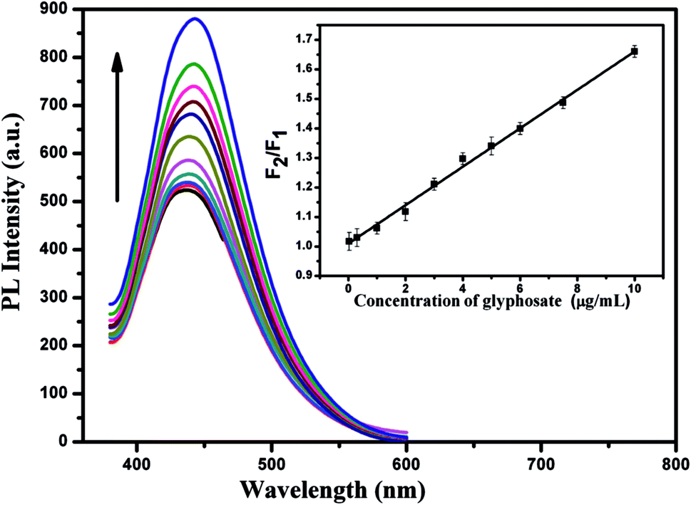 | ||
| Fig. 7 Fluorescence spectra of the CDs–Cu2+ system in the presence of different concentrations of glyphosate (0.03–10 μg mL−1). λex = 360 nm. Cu2+: 25 μM; pH: 6.0. Inset: the linear calibration plot of F2/F1 versus glyphosate concentration. F2 and F1 represent the fluorescence intensity of CDs–Cu2+ system in the presence and absence of glyphosate. | ||
Additionally, the selectivity of the proposed method for glyphosate detection was investigated by examining the fluorescence responses of the CDs–Cu2+ system toward various pesticides, including glufosinate, chlorpyrifos, chlorothalonil, imidacloprid, pymetrozine, carbendazim, chlorfenapyr, dinotefuran, metaflumizone, metsulfuron-methyl and ametryn. As shown in Fig. 8, no obvious fluorescence signal change is observed for these pesticides, whereas the addition of glyphosate results in significant fluorescence recovery, as described previously. The results demonstrated that the CDs–Cu2+ system had a high selectivity for the determination of glyphosate.
 | ||
| Fig. 8 The selectivity of the CDs–Cu2+ system to various pesticides. F2 and F1 are the fluorescence intensity of CDs–Cu2+ system in the presence and absence of pesticides. | ||
3.4 Fluorescence quenching and recovering mechanism analysis
To verify the quenching mechanism, we carried out the time-resolved fluorescence decays of the CDs in the presence and absence of Cu2+, respectively. The data were found to be fit well to a classical double exponential function (Fig. 9A). The average fluorescence lifetime of the CDs was calculated to be 5.6 ns. After addition of Cu2+ into the CDs solution, the average fluorescence lifetime of the CDs show no obvious change (5.2 ns). The results indicate that the fluorescence quenching belongs to static quenching in which a non-fluorescent complex was formed between Cu2+ and the groups on the surface of the CDs, which contributes to the quenched fluorescence of the CDs by Cu2+. | ||
| Fig. 9 (A) Fluorescence decay trace of CDs in the presence and absence of Cu2+ (λex = 360 nm). (B) The UV-vis absorption spectra of (1) glyphosate (1.5 μg mL−1), (2) Cu2+ (20 μM), (3–13) the mixture of Cu2+ in the presence of glyphosate with different concentrations. Concentrations of glyphosate added for spectrum (3)–(13) were 1.5, 2.0, 2.5, 2.8, 3.2, 3.9, 4.5, 5.7, 6.5, 7.8 and 8.3 μg mL−1, respectively. | ||
The formation of a glyphosate–Cu2+ complex was further confirmed by UV-vis absorption spectra of Cu2+ in the presence of glyphosate with different concentrations (Fig. 9B). Compared with glyphosate or Cu2+ solution alone (Fig. 9B, curves 1 and 2), a red shift absorption peak (λmax = 230 nm) was observed in the absorption spectrum of mixture solution of glyphosate and Cu2+. Meanwhile, the absorption intensities (λmax = 230 nm) increased dramatically with increasing concentration of glyphosate with almost the same spectra patterns during evolution (Fig. 9B, curves 3 to 13), indicating the formation of the glyphosate–Cu2+ complex.52,54 This result provides powerful evidence in support of our previously mentioned speculation.
According to the above analysis, Cu2+ was able to quench the fluorescence intensity of CDs through the static quenching process, and the fluorescence intensity of the CDs–Cu2+ system can effectively be recovered by the addition of glyphosate because of its ability to remove Cu2+ from the surface of CDs. Thus, the CDs can be used for Cu2+ and glyphosate detection based on the different fluorescent responses upon the addition of Cu2+ and subsequent glyphosate.
3.5 Detection of glyphosate in environmental water samples
In order to investigate the reliability in practical analysis, the proposed method was applied to the determination of glyphosate in environmental water samples and the results are shown in Table S2.† Among the three real environmental water samples, glyphosate was not detected. The recoveries of glyphosate were studied by adding a certain amount of glyphosate standard solution with concentrations 0.3 and 2.5 μg mL−1 into the water samples. The recoveries in these spiked water samples are in the range of 93.3% to 106.7% and the RSD is lower than 5%. The results indicate the practicability and reliability of the proposed method for glyphosate detection in real water samples.4. Conclusions
In summary, we developed a simple and fast microwave pyrolysis method for the preparation of CDs by using thiourea as the carbon source and diethylene glycol as the reaction medium. The CDs were obtained only within three minutes and the size of the CDs is narrowly distributed in the range of 1.5–3.5 nm. The Cu2+ can quench the fluorescence of CDs through the static quenching process. As expected, the addition of glyphosate into the CDs–Cu2+ system, the Cu2+ can be remove from the surface of the CDs because of the higher affinity of glyphosate for Cu2+ and led to the fluorescence recovery of the CDs. Based on this fluorescence quenching and recovery phenomenon, we developed a new type of CD-based fluorescence probe for “turn-off–on” detection of Cu2+ and glyphosate with high sensitivity and selectivity. Furthermore, the proposed method has been used for glyphosate detection in environmental water samples with satisfactory results, indicating that this fluorescence probe would possess potential applications in environmental fields.Acknowledgements
This work was supported by the Development Program of the Ministry of Science and Technology of Jilin Province, China (Grant number 20150204070GX).Notes and references
- H. Dai, M. Sang, Y. Wang, R. Du, W. Yuan, Z. Jia, Z. Cao and X. Chen, Sens. Actuators, A, 2014, 218, 88–93 CrossRef CAS.
- J. Song, X.-M. Li, A. Figoli, H. Huang, C. Pan, T. He and B. Jiang, Water Res., 2013, 47, 2065–2074 CrossRef CAS PubMed.
- C. F. Coutinho, L. F. Coutinho, L. H. Mazo, S. L. Nixdorf and C. A. Camara, J. Chromatogr. A, 2008, 1208, 246–249 CrossRef CAS PubMed.
- M. V. Khrolenko and P. P. Wieczorek, J. Chromatogr. A, 2005, 1093, 111–117 CrossRef CAS PubMed.
- K. Sato, J.-Y. Jin, T. Takeuchi, T. Miwa, K. Suenami, Y. Takekoshi and S. Kanno, J. Chromatogr. A, 2001, 919, 313–320 CrossRef CAS PubMed.
- K. Qian, T. Tang, T. Shi, F. Wang, J. Li and Y. Cao, Anal. Chim. Acta, 2009, 635, 222–226 CrossRef CAS PubMed.
- M. Malécot, B. Guével, C. Pineau, B. F. Holbech, M. Bormans and C. Wiegand, J. Proteome Res., 2013, 12, 5281–5292 CrossRef PubMed.
- Standardization Administration of China, 2006, GB 5749–2006.
- J. M. Arroyave, C. C. Waiman, G. P. Zanini and M. J. Avena, Chemosphere, 2016, 145, 34–41 CrossRef CAS PubMed.
- E. Avigliano and N. F. Schenone, Microchem. J., 2015, 122, 149–158 CrossRef CAS.
- E. Hogendoorn, F. Ossendrijver, E. Dijkman and R. Baumann, J. Chromatogr. A, 1999, 833, 67–73 CrossRef CAS PubMed.
- A. Botero-Coy, M. Ibáñez, J. Sancho and F. Hernández, J. Chromatogr. A, 2013, 1313, 157–165 CrossRef CAS PubMed.
- Z. H. Kudzin, D. K. Gralak, G. Andrijewski, J. Drabowicz and J. Łuczak, J. Chromatogr. A, 2003, 998, 183–199 CrossRef CAS PubMed.
- M. Motojyuku, T. Saito, K. Akieda, H. Otsuka, I. Yamamoto and S. Inokuchi, J. Chromatogr. B: Anal. Technol. Biomed. Life Sci., 2008, 875, 509–514 CrossRef CAS PubMed.
- Y. Zhu, F. Zhang, C. Tong and W. Liu, J. Chromatogr. A, 1999, 850, 297–301 CrossRef CAS PubMed.
- M. Corbera, M. Hidalgo, V. Salvado and P. Wieczorek, Anal. Chim. Acta, 2005, 540, 3–7 CrossRef CAS.
- M. R. Jan, J. Shah, M. Muhammad and B. Ara, J. Hazard. Mater., 2009, 169, 742–745 CrossRef CAS PubMed.
- Z.-X. Guo, Q. Cai and Z. Yang, J. Chromatogr. A, 2005, 1100, 160–167 CrossRef CAS PubMed.
- Z. Li, H. Yu, T. Bian, Y. Zhao, C. Zhou, L. Shang, Y. Liu, L.-Z. Wu, C.-H. Tung and T. Zhang, J. Mater. Chem. C, 2015, 3, 1922–1928 RSC.
- S. Mandani, B. Sharma, D. Dey and T. K. Sarma, Nanoscale, 2015, 7, 1802–1808 RSC.
- Z. Lin, W. Xue, H. Chen and J. M. Lin, Anal. Chem., 2011, 83, 8245–8251 CrossRef CAS PubMed.
- W. Shi, Q. Wang, Y. Long, Z. Cheng, S. Chen, H. Zheng and Y. Huang, Chem. Commun., 2011, 47, 6695–6697 RSC.
- L. Zhou, Y. Lin, Z. Huang, J. Ren and X. Qu, Chem. Commun., 2012, 48, 1147–1149 RSC.
- H. Li, Y. Zhang, L. Wang, J. Tian and X. Sun, Chem. Commun., 2011, 47, 961–963 RSC.
- Y. Dong, R. Wang, W. Tian, Y. Chi and G. Chen, RSC Adv., 2014, 4, 3685–3689 RSC.
- H. X. Zhao, L. Q. Liu, Z. D. Liu, Y. Wang, X. J. Zhao and C. Z. Huang, Chem. Commun., 2011, 47, 2604–2606 RSC.
- A. Basu, A. Suryawanshi, B. Kumawat, A. Dandia, D. Guin and S. B. Ogale, Analyst, 2015, 140, 1837–1841 RSC.
- C. Yu, X. Li, F. Zeng, F. Zheng and S. Wu, Chem. Commun., 2013, 49, 403–405 RSC.
- X. An, S. Zhuo, P. Zhang and C. Zhu, RSC Adv., 2015, 5, 19853–19858 RSC.
- J. Hou, J. Dong, H. Zhu, X. Teng, S. Ai and M. Mang, Biosens. Bioelectron., 2015, 68, 20–26 CrossRef CAS PubMed.
- S. Li, J. Luo, G. Yin, Z. Xu, Y. Le, X. Wu, N. Wu and Q. Zhang, Sens. Actuators, B, 2015, 206, 14–21 CrossRef CAS.
- Y. Chen, Y. Wu, B. Weng, B. Wang and C. Li, Sens. Actuators, B, 2016, 223, 689–696 CrossRef CAS.
- L. Wang, Y. Bi, J. Hou, H. Li, Y. Xu, B. Wang, H. Ding and L. Ding, Talanta, 2016, 160, 268–275 CrossRef CAS PubMed.
- Y. Wang, Q. Zhuang and Y. Ni, Chem.–Eur. J., 2015, 21, 13004–13011 CrossRef CAS PubMed.
- Y. Wang, C. Lin, X. Ma, Z. Xue, X. Zhu, W. Cao, S. Hu, T. Sheng and X. Wu, Dalton Trans., 2015, 44, 7437–7448 RSC.
- L. Li, B. Yu and T. You, Biosens. Bioelectron., 2015, 74, 263–269 CrossRef CAS PubMed.
- Y.-C. Lu, J. Chen, A.-J. Wang, N. Bao, J.-J. Feng, W. Wang and L. Shao, J. Mater. Chem. C, 2015, 3, 73–78 RSC.
- D. Sun, R. Ban, P.-H. Zhang, G.-H. Wu, J.-R. Zhang and J.-J. Zhu, Carbon, 2013, 64, 424–434 CrossRef CAS.
- A. Majumdar, K. Schröder and R. Hippler, J. Appl. Phys., 2008, 104, 074702 CrossRef.
- S. Zhao, M. Lan, X. Zhu, H. Xue, T.-W. Ng, X. Meng, C.-S. Lee, P. Wang and W. Zhang, ACS Appl. Mater. Interfaces, 2015, 7, 17054–17060 CAS.
- A. Majumdar, J. Schäfer, P. Mishra, D. Ghose, J. Meichsner and R. Hippler, Surf. Coat. Technol., 2007, 201, 6437–6444 CrossRef CAS.
- Y. Su, Y. Zhang, X. Zhuang, S. Li, D. Wu, F. Zhang and X. Feng, Carbon, 2013, 62, 296–301 CrossRef CAS.
- M. Xue, L. Zhang, M. Zou, C. Lan, Z. Zhan and S. Zhao, Sens. Actuators, B, 2015, 219, 50–56 CrossRef CAS.
- Y. Lu, Y. Jiang, W. Wei, H. Wu, M. Liu, L. Niu and W. Chen, J. Mater. Chem., 2012, 22, 2929–2934 RSC.
- S. Zhu, Y. Song, X. Zhao, J. Shao, J. Zhang and B. Yang, Nano Res., 2015, 8, 355–381 CrossRef CAS.
- C. Wang, D. Sun, K. Zhuo, H. Zhang and J. Wang, RSC Adv., 2014, 4, 54060–54065 RSC.
- Q. Li, M. Peng, N. Li, J. Qin and Z. Li, Sens. Actuators, B, 2012, 173, 580–584 CrossRef CAS.
- L. Tang and M. Cai, Sens. Actuators, B, 2012, 173, 862–867 CrossRef CAS.
- J. Zong, X. Yang, A. Trinchi, S. Hardin, I. Cole, Y. Zhu, C. Li, T. Muster and G. Wei, Biosens. Bioelectron., 2014, 51, 330–335 CrossRef CAS PubMed.
- Y. Guo, L. Zhang, S. Zhang, Y. Yang, X. Chen and M. Zhang, Biosens. Bioelectron., 2015, 63, 61–71 CrossRef CAS PubMed.
- S. Liu, J. Tian, L. Wang, Y. Zhang, X. Qin, Y. Luo, A. M. Asiri, A. O. Al-Youbi and X. Sun, Adv. Mater., 2012, 24, 2037–2041 CrossRef CAS PubMed.
- Z. Liu, S. Liu, P. Yin and Y. He, Anal. Chim. Acta, 2012, 745, 78–84 CrossRef CAS PubMed.
- J. Sheals, P. Persson and B. Hedman, Inorg. Chem., 2001, 40, 4302–4309 CrossRef CAS PubMed.
- R. L. Glass, J. Agric. Food Chem., 1984, 32, 1249–1253 CrossRef CAS.
- B. L. Bhaskara and P. Nagaraja, Helv. Chim. Acta, 2006, 89, 2686–2693 CrossRef CAS.
- L. De Almeida, S. Chigome, N. Torto, C. Frost and B. Pletschke, Sens. Actuators, B, 2015, 206, 357–363 CrossRef CAS.
- C. F. Coutinho, L. F. Coutinho, L. H. Mazo, S. L. Nixdorf, C. A. Camara and F. M. Lanças, Anal. Chim. Acta, 2007, 592, 30–35 CrossRef CAS PubMed.
- A. S. da Silva, F. C. B. Fernandes, J. O. Tognolli, L. Pezza and H. R. Pezza, Spectrochim. Acta, Part A, 2011, 79, 1881–1885 CrossRef PubMed.
Footnote |
| † Electronic supplementary information (ESI) available. See DOI: 10.1039/c6ra10115a |
| This journal is © The Royal Society of Chemistry 2016 |