DOI:
10.1039/C6RA09891F
(Paper)
RSC Adv., 2016,
6, 64460-64468
Synthesis of quinoline acetohydrazide-hydrazone derivatives evaluated as DNA gyrase inhibitors and potent antimicrobial agents†
Received
16th April 2016
, Accepted 11th June 2016
First published on 20th June 2016
Abstract
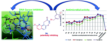
The (E)-N′-(substituted-benzylidene)-2-(7-fluoro-2-methoxyquinolin-8-yl)acetohydrazide-hydrazone derivatives reported in this manuscript represent a new series of antibacterial agents, as well as DNA gyrase inhibitors. Efforts were made to synthesize these quinolone-acetohydrazide-hydrazone derivatives (9a–n) in excellent yields. All the target compounds were evaluated for their in vitro antimicrobial activity against Escherichia coli (MTCC 443) and Pseudomonas aeruginosa (MTCC 424), as specimens of Gram-negative bacteria, and Staphylococcus aureus (MTCC 96) and Staphylococcus pyogenes (MTCC 442), as specimens of Gram-positive bacteria. Excellent antibacterial activity was observed for compounds substituted with R = 3,4,5-trimethoxy, 4-F, 4-OCF3, 4-CF3, and 3-CF3 moieties. Derivatives 9a–n were docked into the active sites of DNA gyrase A and DNA gyrase B in order to obtain more insight into the binding modes of the compounds. Among all the derivatives, 9n showed the lowest binding energy of −91.6 kcal mol−1 with DNA gyrase A. A DNA gyrase enzyme inhibition assay favors compounds 9m and 9n, which were substituted with di-fluorine moieties (R = 2,4-difluoro and 3,4-difluoro) in the quinoline scaffold, as the most potent inhibitors of S. aureus DNA gyrase A (9m, IC50 0.14 mg mL−1 and 9n, IC50 0.19 mg mL−1).
Introduction
Recent reports from the World Health Organization (WHO) state that public health has become a major concern due to the population explosion and an increase in endemic and epidemic diseases. Genomic studies on various microorganisms indicate that the prolonged use of antimicrobials and antibiotics makes these microorganisms more resistant to them.1 The challenge, therefore, for scientists is to design and synthesize novel effective molecules that act against these microorganisms. In this concern, quinoline and its derivatives are well accepted molecules with respect to their antimicrobial properties. In the literature, it is reported that a number of quinoline derivatives possesses antileishmanial properties,1 cytotoxicity,2,3 antituberculosis,4 antimalarial,5 or anti-inflammatory6 properties, or are HIV-1 integrase inhibitors.7 Quinoline derivatives have been developed for the treatment of many diseases, such as malaria,8 HIV,9 tumors,10 and antibacterial infections11 and some of the substituted quinolines have been reported to act as leukotriene D4 receptors,12 5HT3 receptor,13 and antagonists for endothelin14 and NK-3.15 They also function as arachidonate 5-lipoxygenase,16 dihydroorotate dehydrogenase17 and inhibitors of gastric (H+/K+)-ATPase.18 In recent years, hydrazide analogs with azomethine (–CONHN
CH–) functionality were found to exhibit prominent pharmacological and biological activities, such as anticonvulsant, analgesic19,20 and anti-inflammatory properties.21 Additionally, these derivatives were also active against tuberculosis bacteria in guinea pigs.23,24 Reported in vitro metabolism studies suggest that biological systems having hydrazide-hydrazone functionalities can easily undergo hydrolytic reactions, which is an advantage with respect to the treatment of various life-threatening diseases.24,25 Apart from this, hydrazide-hydrazones are the most essential intermediates in the construction of various heterocyclic rings utilizing the hydrogen segment of the –CONHN
CH– azomethine group.26 The fast-growing microbial imperviousness to conventional anti-infectious agents has necessitated a continuing search for new classes of compounds with novel methods of antimicrobial activity.27–29
Our research group is actively involved in the synthesis of N–N bond-bearing molecules and the development of medicinally essential molecules for various disease-causing targets.30 The present study aimed to synthesize novel quinoline acetohydrazide-hydrazone derivatives (9a–n) having a fluorine and a methoxy substituent in the quinoline nucleus and alkoxy, OCF3 and fluorine functionalities on the phenyl moiety as antibacterial agents, as well as DNA gyrase inhibitors. Gyrases present in prokaryotes have different affinities for different molecules and this makes the gyrases a good target for antibiotics. The negative supercoils of bacterial cells, which are necessary for DNA replication, elongation and transcription, will be initiated by the DNA gyrase. Of the two sub-units, DNA gyrase subunit A is selectively inactivated by quinolone-based antibiotics, such as oxolinic and nalidixic acids, and subunit B is selectively inactivated by antibiotics, such as coumermycin A1 and novobiocin. Inhibition of either subunit blocks supertwisting activity and quinolones bind to these enzymes and prevent them from achieving decatenation of replicating DNA.31 Bacterial DNA gyrase and DNA topoisomerase IV (type II topoisomerases), are the main targets against resistance and are mainly present in methicillin-resistant Staphylococcus aureus (MRSA).32 Initially, the synthesized molecules were docked into the active sites of the bacterial DNA gyrase A and DNA gyrase B to ensure the inhibitory abilities of the quinoline acetohydrazide-hydrazone derivatives (9a–n). Autodock is an effective tool for predicting the binding conformations and binding energies of ligands with macromolecular targets and the Lamarckian Genetic Algorithm was used in this study as the primary method for conformational searching.33 Subsequently, to clarify the mechanism by which the acetohydrazide-hydrazone derivatives influence antibacterial activity, the inhibitory activities of compounds 9a–n were evaluated against DNA gyrase A, isolated from S. aureus.
Results and discussion
A synthetic route to prepare (E)-N′-(substituted-benzylidene)-2-(7-fluoro-2-methoxyquinolin-8-yl)acetohydrazide-hydrazone derivatives 9a–n is presented in Scheme 1. Our synthesis commenced with the treatment of 3-fluoro-2-methylbenzenamine 1 with cinnamoyl chloride in the presence of aqueous sodium bicarbonate at room temperature to produce N-(3-fluoro-2-methylphenyl)cinnamamide 2 in 84% yield. Friedel–Crafts acylation of amide 2 resulted in 7-fluoro-8-methylquinolin-2(1H)-one 3 in 82% yield, and 7-fluoro-2-methoxy-8-methylquinoline 4 was obtained in 84% yield from the treatment of compound 3 with methyl iodide in the presence of potassium tert-butoxide in DMSO at 70 °C for 2.5 h.
 |
| Scheme 1 Synthesis of novel quinoline acetohydrazide derivatives, 9a–n. Experimental conditions: (a) cinnamoyl chloride, aq. NaHCO3, isopropyl acetate, room temperature, 30 min; (b) AlCl3, chlorobenzene, 90 °C, 1 h; (c) MeI, KtOBu, DMSO, 70 °C, 2.5 h; (d) NBS, benzoyl peroxide, xylene, 70 °C, 1.5 h; (e) KCN, DMF, 60 °C, 16 h; (f) TMSiCl, MeOH, 70 °C, 2.5 h; (g) NH2–NH2, ethanol, reflux, 20 h; (h) benzaldehydes, a–n, ethanol, reflux, 4 h. | |
Allylic bromination of compound 4 in the presence of NBS and benzoyl peroxide resulted in 8-(bromomethyl)-7-fluoro-2-methoxyquinoline 5 (94% yield), and reaction of 5 with KCN in DMF at 60 °C for 16 h produced 2-(7-fluoro-2-methoxyquinolin-8-yl)acetonitrile 6 in 62% yield. Esterification of 6 in the presence of trimethylsilyl iodide in methanol at 70 °C yielded methyl-2-(7-fluoro-2-methoxyquinolin-8-yl)acetate 7, which upon treatment with hydrazine-hydrate resulted in the key intermediate, 2-(7-fluoro-2-methoxyquinolin-8-yl)acetohydrazide 8 in 82% yield. Syntheses of the intermediates (2–5) were performed as per the reported procedures.30 Condensation of acetohydrazide 8 with various substituted benzaldehydes, a–n, in ethanol, refluxing for 4 h, resulted in a wide variety of desired acetohydrazide derivatives, 9a–n, in quantitative yields. It is important to note that all the synthesized hydrazide-hydrazone derivatives, 9a–n, were found to exist in two rotameric forms in solution,22 e.g. antiperiplanar (ap) and synperiplanar (sp), as indicated by their 1H NMR spectra. As a representative example, the 1H NMR spectral data of compound 9j (R = 4-OCF3) are as follows: the broad singlets at 11.85 ppm (*11.60 ppm) and 8.28 ppm (*8.06 ppm) correspond to the protons in –CO–NH– and –CO–NH–N
CH– groups, respectively. The doublets appearing at 7.80 and 7.46 ppm represent the para-substituted protons attached to the phenyl ring-bearing p-OCF3 substituent. The signals at 8.26 ppm (doublet), 7.90 ppm (triplet), 7.34 ppm (doublet) and 7.0 ppm (doublet) with one proton integration correspond to the quinoline ring skeleton. The protons resonating at 4.50 (*4.08) ppm and 3.94 (*3.88) ppm as singlets correspond to methylene- and methoxy groups, respectively. Mass spectral data ((M + 1) peaks) of all the synthesized compounds represent the expected molecular formulae.
Antibacterial activity
Table 1 (ESI†) represents the antibacterial activity results for 2-(7-fluoro-2-methoxyquinolin-8-yl)acetohydrazide-hydrazone derivatives, 9a–n. Fig. 1a–d depicts some of the representative images showing the antibacterial activity of the synthesized quinolone acetohydrazide-hydrazone derivatives, where the growth of all tested bacterial isolates was inhibited. From Fig. 2, it can be seen that compounds 9a–g showed a moderate antibacterial activity with a zone of inhibition of 8–12 mm against all the tested bacterial strains. Compounds 9h–l exhibit good antibacterial activity with a zone of inhibition ranging from 16–17 mm, and compounds 9m and 9n displayed excellent antibacterial activity with a maximum zone of inhibition ranging from 18–21 mm. The in vivo efficacy of quinoline acetohydrazide derivatives with respect to their antibacterial activity against selected pathogens was tested and compared with existing drugs, i.e. ampicillin, under the established conditions (Table 1, ESI,† entry 15). In terms of structural activity relationships, it is worthwhile mentioning that compounds with R = 2,4-difluoro and 3,4-difluoro in the quinoline scaffold showed potent activity (Table 1, ESI,† entries 13 and 14) and compounds with R = 3,4,5-trimethoxy, 4-F, 4-OCF3, 4-CF3, and 3-CF3 showed good activity (Table 1, ESI,† entries 8 and 12). The remaining compounds in the series with methoxy (–OCH3) substituents showed moderate activity. In conclusion, the antibacterial experimental work suggests that a suitable replacement of the R group in the quinolone scaffold may lead to a promising antibacterial drug candidate. The similar antibacterial activity expression of compounds 9m and 9n suggests that these two molecules may be a suitable alternative to ampicillin, which is an existing and leading antibacterial drug.
 |
| Fig. 1 (a). Excellent activity. (b). Good activity 9h. (c). Moderate activity, 9a. (d). Weak activity, 9c. | |
 |
| Fig. 2 Antibacterial activity of 9a–n against selected pathogens. | |
Molecular docking studies
The ligands 9a–n were docked into the active sites of DNA gyrase A and DNA gyrase B and the results are shown in Table 2. The results less than 2.0 Å in positional root-mean-square deviation (RMSD) were clustered together and represented by the result with the most favorable free energy of binding. The docked poses with the lowest binding energy, hydrogen bonds, and non-covalent interactions such as π–π interactions and π–cation interactions were recorded (Tables 1 & 2) and validated. The expected binding free energy for DNA gyrase A was found to be between −4.25 and −91.6 kcal mol−1 and the DNA gyrase showed a binding free energy between +673.5 and −76.6 kcal mol−1. These free energy values indicated that the newly synthesized compounds showed selectivity towards DNA gyrase A rather than DNA gyrase B.
Table 1 Molecular interaction results for compounds 9a–n with DNA gyrase A (PDB ID: 1ZI0)
Entry |
Binding energy (kcal mol−1) |
Ligand efficiency |
RMSDa |
Inhibitory constant (ki) |
Root mean square deviation. |
9a |
−32.1 |
−1.08 |
6.54 |
2.481 nM |
9b |
−39.1 |
−1.56 |
1.79 |
4.784 nM |
9c |
−31.8 |
−1.02 |
6.07 |
2.181 fM |
9d |
−51.1 |
−1.98 |
80.9 |
214.4 aM |
9e |
−55.1 |
−2.02 |
0 |
91.93 fM |
9f |
−50.3 |
−1.91 |
0 |
118.8 fM |
9g |
−48.5 |
−1.65 |
6.93 |
763.4 fM |
9h |
−75.8 |
−2.62 |
0 |
6.122 zM |
9i |
−42.1 |
−1.76 |
0 |
48.21 zM |
9j |
−53.3 |
−0.47 |
0 |
24.82 fM |
9k |
−64.7 |
−1.29 |
0 |
28.12 fM |
9l |
−68.2 |
−2.53 |
0 |
6.196 zM |
9m |
−82.2 |
−3.29 |
0 |
3.062 zM |
9n |
−91.6 |
−3.67 |
0 |
2.061 zM |
Table 2 Molecular interaction results for compounds 9a–n with DNA gyrase B (PDB ID: 2ZJT)
Entry |
Binding energy (kcal mol−1) |
Ligand efficiency |
RMSDa |
Inhibitory constant (ki) |
Root mean square deviation. |
9a |
−76.6 |
−3.07 |
2.75 |
7.21 zM |
9b |
−5.62 |
−0.27 |
35.7 |
75.64 μM |
9c |
75.8 |
2.8 |
— |
— |
9d |
−45.4 |
−1.69 |
0 |
44.29 fM |
9e |
22.7 |
0.84 |
— |
— |
9f |
2.82 |
0.1 |
— |
— |
9g |
67.5 |
23.3 |
— |
— |
9h |
−59.7 |
−2.06 |
2.2 |
19.12 zM |
9i |
−46.7 |
−1.95 |
1.37 |
44.29 fM |
9j |
3.11 |
0.11 |
— |
— |
9k |
−3.57 |
−0.13 |
0 |
14.95 nM |
9l |
−28.3 |
−1.05 |
0 |
43.99 fM |
9m |
−23.7 |
−0.95 |
0 |
39.12 fM |
9n |
−11.3 |
−0.46 |
0 |
89.12 μM |
Except for 9a (−76.6 kcal mol−1) none of the compounds showed equal or remarkable activity with DNA gyrase B. For DNA gyrase A, the lowest binding energy was found for compound 9n (−91.6 kcal mol−1) and the inhibitory constant was found to be 2.06 zM. The ligand efficiency was −3.67, signifying the potential of compound 9a as an ideal antimicrobial drug, compared to the other compounds. The non-covalent π–π interactions are connected with the contact among the π-orbitals of a molecular organization and the cation–π interactions engross the positive charge of a cation related with the electrons in a π-system of a molecule.
In this study, the presence of hydrogen bonds and non-covalent bonds (π–π interactions and π–cation interactions) were considered with respect to the ligand–receptor interactions. Hydrogen bond interactions were found for all compounds with DNA gyrase A. Compound 9a formed a H-bond with the amino acid residue Glu162, with a length of 2.906 Å (Fig. 3). Compound 9b formed two hydrogen bonds with Leu735 (2.938 Å) and Leu836 (2.798 Å). Compound 9c showed two invisible H-bonds with Ile619 & Leu623, along with a visible H-bond with Leu672 (1.985 Å). Compound 9d formed a H-bond with Arg838 (2.116 Å). Compound 9e showed two invisible and one visible H-bond with Ala780, Leu735, and Arg739 (1.951 Å). Compounds 9f–l and 9n formed a single H-bond (Arg739, 1.786 Å; Leu735, 2.196 Å; Arg580, 2.036 Å; Arg596, 1.234 Å; Arg598, 1.438 Å; Thr632, 2.256 Å; Leu622, 2.173 Å; Glu162, 2.906 Å, respectively), while compound 9m formed the maximum number of four H-bonds, of which two were invisible (Ile674 and Gly597) and two were visible (Gly597, 0.938 Å and Ala673, 0.871 Å).
 |
| Fig. 3 Interaction found with π–cation and hydrogen bonding between DNA gyrase A (PDB ID: 1ZI0) and 9a. | |
Non-covalent interactions (π–π interactions and π–cation interactions) were also found in 9a, 9c, 9h, 9k and 9n. Almost all of the non-covalent interactions were found between the amino acid residues Arg534, Arg580, Lys656, Phe663, and Tyr551 to the Ar-1, Ar-2, and Ar-3 ring systems. DNA gyrase B showed hydrogen bonding only with 9a (Tyr563, 2.134 Å), 9c (Ala570, 1.972 Å and Ser580, Ala570, Ala630 were invisible), 9d (Pro519, 2.072 Å), 9k (Glu540, 2.629 Å; Glu540, 2.639 Å), and 9n (Leu518, invisible). Only one non-covalent interaction was found for 9d to the Ar-1 and Ar-2 ring systems. More hydrogen bonds resulted in greater solubility, whereas more non-covalent interactions resulted in more electromagnetic interactions. These interactions also greatly influence the drug design and are involved in the synthesis of many organic molecules.34,35 DNA gyrase B showed an irregular docking result as well as poor interaction when compared to DNA gyrase A. H-bonds were found only with the compounds 9a (Fig. 5), 9c, 9d, 9l, and 9n. The only non-covalent interaction was found with 9a (Fig. 5).
All ligands were found in the predicted binding pockets of 1ZI0 and 2ZJT (Fig. 4 & 6). This illustrates the potency of the compounds towards ligand–receptor interactions, as well as their inhibitory activity. From the overall results of the docking study, the selective activity on DNA gyrase A by the derivatives 9a–n was understood. In the mechanism, the DNA gyrase A and B subunits attach together to DNA, hydrolyzing ATP and initiating negative supertwists. The A subunit accomplishes breakage-rejoining of DNA, the B subunit introduces negative supercoils, and then the A subunit reseals the strands. Inhibition of either subunit blocks supertwisting activity.36 Fluoroquinolones were observed to bind the A subunit and obstruct its strand-cutting and resealing functions. Remarkably, we could see that compounds 9a–n have the quinoline backbone with –F substituted. This explained why compounds 9a–n were more selective towards DNA gyrase A.
 |
| Fig. 4 Compound 9n in the binding pocket of DNA gyrase A (PDB ID: 1ZI0). | |
 |
| Fig. 5 Interaction found with π–cation and hydrogen bonding between DNA gyrase B (PDB ID: 2ZJT) and 9a. | |
 |
| Fig. 6 Compound 9a in the binding pocket of DNA gyrase B (PDB ID: 2ZJT). | |
DNA gyrase inhibitory assay
The molecular docking results revealed that the docked compounds favor DNA gyrase A over DNA gyrase B. To elucidate the mechanism by which the quinoline acetohydrazide-hydrazone derivatives (9a–n) induce antibacterial activity, the inhibitory activities of the compounds were examined against DNA gyrase isolated from S. aureus. Clorobiocin was used as the standard and was found to have an IC50 of 0.04 (mg mL−1). The IC50 range for compounds 9a–n was 0.14–23.45 mg mL−1 (Table 2, ESI†). Compound 9m, with an effective antibacterial activity, strongly inhibited S. aureus DNA gyrase (IC50, 0.14 mg mL−1), as did 9n (IC50, 0.19 mg mL−1). There was a good correlation between the zone of inhibitions and the IC50, signifying that inhibition of the DNA gyrase by the quinoline acetohydrazide-hydrazone derivatives caused inhibition of bacterial cell growth. Compound 9c showed the least inhibition against the S. aureus DNA gyrase (IC50 = 23.45 mg mL−1). The activity correlation between the antibacterial and anti DNA gyrase was positive, with R2 = 0.8092.
Experimental
Chemistry
All solvents were purified according to standard procedures prior to use and all (commercial) chemicals were used as received. For thin-layer chromatography (TLC) analysis, Merck pre-coated plates (silica gel 60 F254) were used and spots were visualized under UV light. Merck silica gel 60 (230–400 mesh) was used for flash column chromatography and the eluting solvents are indicated in the procedures. Melting point (mp) determinations were performed using Mel-temp apparatus and are uncorrected. The 1H NMR spectra were recorded on a Varian MR-400 MHz instrument, while 13C NMR spectra were recorded on a Bruker-100 MHz instrument. Chemical shifts (δ) are reported in parts per million (ppm) downfield from tetramethylsilane (TMS) as internal standard reference and the signals are reported as s (singlet), d (doublet), dd (doublet of doublet), t (triplet), q (quartet), and m (multiplet); coupling constants are measured in Hz. Mass spectra were recorded on an Agilent ion-trap MS. High resolution mass spectra (HRMS) were recorded on a QTOF micro mass spectrometer. Infrared (IR) spectra were recorded on a Perkin Elmer FT-IR spectrometer.
Preparation of N-(3-fluoro-2-methylphenyl)cinnamamide (2). A pre-mixed solution of cinnamoyl chloride (12.06 g, 72.63 mmol) in isopropyl acetate (48 mL) was added to a mixture of 3-fluoro-2-methyl aniline (9.0 g, 71.90 mmol), saturated aqueous sodium bicarbonate (55 mL) and isopropyl acetate (54.0 mL) over a period of 15 min. The reaction mixture was stirred at room temperature for the next 30 min. After completion of the reaction, the reaction mixture was filtered, washed with water and dried under vacuum to obtain compound 2. White solid, yield: 15.5 g, 84.4%; 1H NMR (400 MHz, CDCl3): δ 9.68 (s, 1H), 7.64 (t, J = 6.8 Hz, 2H), 7.62 (d, J = 15.6 Hz, 1H), 7.48–7.40 (m, 4H), 7.26–7.20 (m, 1H), 7.01 (t, J = 8.4 Hz, 2H), 2.15 (s, 3H); ESI-MS: m/z, 256.2 (M + 1).
Preparation of 7-fluoro-8-methylquinolin-2(1H)-one (3). To a solution of N-(3-fluoro-2-methylphenyl)cinnamamide 2 (4.0 g, 15.70 mmol) in chlorobenzene (20 mL), aluminium trichloride (8.35 g, 6.25 mmol) was added slowly over a period of 30 min at 25 °C. The reaction mixture was then heated to 90 °C for 1 h. Upon completion of the reaction, the reaction mixture was cooled to room temperature, followed by addition of excess ice with vigorous stirring. The resulting precipitate was isolated and washed with water, filtered and dried to obtain compound 3. Light brown solid, yield: 4.6 g, 82.8%; 1H NMR (400 MHz, CDCl3): δ 11.13 (s, 1H), 7.89 (d, J = 9.2 Hz, 1H), 7.58 (t, J = 8.0 Hz, 1H), 7.05 (t, J = 9.2 Hz, 1H), 6.46 (d, J = 10 Hz, 1H), 2.32 (s, 3H); ESI-MS: m/z, 178.2 (M + 1).
Preparation of 7-fluoro-2-methoxy-8-methylquinoline (4). Compound 3, 7-fluoro-8-methylquinolin-2(1H)-one (5.5 g, 57.0 mmol) was suspended in DMSO (35 mL) and treated with potassium t-butoxide (7.03 g, 62.76 mmol) at 25 °C in an argon environment. To the above reaction mixture, methyl iodide (10.52 g, 74.18 mmol) was added over 10 min and heated to 70 °C for 2.5 h. After completion of the reaction, the reaction mixture was diluted with water (250 mL) and extracted with n-hexane (500 mL). The hexane extract was further washed with brine solution, dried over sodium sulphate and concentrated under reduced pressure. The obtained crude product was purified by column chromatography over silica gel (1
:
1 petroleum ether
:
dichloromethane) to obtain compound 4. Pale yellow solid, yield: 4.65 g, 84.4%; 1H NMR (400 MHz, CDCl3): δ 7.91 (d, J = 8.8 Hz, 1H), 7.52 (q, J = 8.8 Hz, 1H), 7.12 (t, J = 8.8 Hz, 1H), 6.83 (d, J = 8.8 Hz, 1H), 4.08 (s, 3H), 2.60 (s, 3H); ESI-MS: m/z, 178.2 (M + 1).
Preparation of 8-(bromomethyl)-7-fluoro-2-methoxyquinoline (5). To a solution of 7-fluoro-2-methoxy-8-methylquinoline 4 (4.65 g, 24.09 mmol) in xylene (150 mL), N-bromosuccinimide (4.67 g, 26.26 mmol) and benzoyl peroxide (0.1 g, 0.38 mmol) were added and heated to 70 °C for 1.5 h. The reaction mixture was cooled to room temperature and filtered, followed by washing with dichloromethane. The combined organic layers were washed with saturated aq. sodium bicarbonate solution, dried over sodium sulphate and concentrated under reduced pressure to obtain compound 5. Pale yellow solid, yield: 3 g, 94%. 1H NMR (400 MHz, CDCl3): δ 7.94 (d, J = 9.2 Hz, 1H), 6.73 (t, J = 8.4 Hz, 1H), 7.14 (t, J = 9.2 Hz, 1H), 6.88 (d, J = 9.2 Hz, 1H), 5.14 (s, 2H), 4.12 (s, 3H); ESI-MS: m/z, 270.1 (M + 1).
Preparation of 2-(7-fluoro-2-methoxyquinolin-8-yl)acetonitrile (6). A solution of 8-(bromomethyl)-7-fluoro-2-methoxyquinoline 5 (6.1 g, 22.60 mmol) in DMF (60 mL) was treated with potassium cyanide (6.82 g, 10.35 mmol) and heated at 60 °C for 16 h. The reaction mixture was then poured into water (500 mL) and extracted with isopropyl acetate (250 mL). The isopropyl acetate layer was washed with brine solution dried over anhydrous sodium sulphate and concentrated under reduced pressure. The obtained crude product was purified by column chromatography over silica gel (10% ethyl acetate and petroleum ether) to obtain compound 6. Brown solid, yield: 3.0 g, 62.5%. 1H NMR (400 MHz, CDCl3): δ 7.98 (q, J = 8.8 Hz, 1H), 7.71 (q, J = 8.8 Hz, 1H), 7.17 (t, J = 8.8 Hz, 1H), 6.94 (t, J = 9.2 Hz, 1H), 4.20 (s, 2H), 4.13 (s, 3H); ESI-MS: m/z, 217.2 (M + 1).
Preparation of methyl-2-(7-fluoro-2-methoxyquinolin-8-yl)acetate (7). A solution of 2-(7-fluoro-2-methoxyquinolin-8-yl)acetonitrile 6 (2.85 g, 13.20 mmol) in dry methanol (40 mL) was treated with trimethylsilyl chloride (4.7 g, 43.55 mmol) and heated to 70 °C for 2.5 h. The reaction mixture was evaporated and diluted with isopropyl acetate (100 mL) and water (50 mL). The combined organic layer was washed with 2 N sodium hydroxide and brine solution, dried over sodium sulphate and evaporated. The crude product was purified by column chromatography over silica gel (100–200 mesh), eluting with dichloromethane, to obtain compound 7. Off-white solid; yield: 4 g, 50%; 1H NMR (400 MHz, CDCl3): δ 7.94 (d, J = 8.4 Hz, 1H), 7.65 (q, J = 8.8 Hz, 1H), 7.18 (t, J = 8.4 Hz, 1H), 6.85 (t, J = 8.8 Hz, 1H), 4.20 (s, 2H), 4.03 (s, 3H), 2.62 (s, 3H); ESI-MS: m/z, 250.1 (M + 1).
Preparation of 2-(7-fluoro-2-methoxyquinolin-8-yl)acetohydrazide (8). A solution of methyl-2-(7-fluoro-2-methoxyquinolin-8-yl)acetate 7 (4 g, 16.06 mmol) in ethanol (60.0 mL) was treated with hydrazine-hydrate (7 equivalents) and refluxed for 20 h. The mixture was evaporated and then slurried with n-hexane, filtered and dried to obtain compound 8. Off-white solid, yield: 3.2 g, 82%; 1H NMR (400 MHz, dmso-d6): δ 9.20 (brs, 1H), 8.24 (d, J = 8.8 Hz, 1H), 7.85 (q, J = 6.4 Hz, 1H), 7.33 (t, J = 9.2 Hz, 1H), 6.98 (d, J = 8.4 Hz, 1H), 4.20 (brs, 2H), 3.99 (s, 3H), 3.89 (s, 2H); ESI-MS: m/z, 250.1 (M + 1).
General procedure for the preparation of hydrazide-hydrazone derivatives (9a–n)
A suspension of compound 8 (100 mg, 0.40 mmol) and the respective benzaldehydes, a–n (0.40 mmol), were taken in ethanol (2 mL) and heated to reflux for 4 h. The obtained products were filtered, washed with ethanol, washed with n-hexane and dried to attain the corresponding hydrazone derivatives, 9a–n. All the derivatives were obtained as solids in good yield and the spectral data of the quinoline acetohydrazide-hydrazone derivatives (9a–n) corresponded with their assigned structures, shown in Scheme 1.
(E)-N′-(4-Methoxybenzylidene)-2-(7-fluoro-2-methoxyquinolin-8-yl)acetohydrazide (9a). White solid; yield: 95% (142 mg); mp: 111–112 °C; 1H NMR (400 MHz, dmso-d6): δ 11.60 (*11.28, s, 1H), 8.28 (d, J = 8.8 Hz, 1H), 8.20 (*8.00, s, 1H), 7.98–7.94 (m, 1H), 7.62 (d, J = 8.6 Hz, 2H), 7.38 (t, J = 9.0 Hz, 1H), 7.06 (d, J = 8.8 Hz, 1H), 7.0 (s, 1H), 4.48 (*4.04, s, 2H), 3.94 (*3.86, s, 3H), 3.80 (s, 3H), (*rotameric peaks); 13C NMR (100 MHz, DMSO): 27.9, 52.9, 55.2, 111.6, 113.1, 114.2, 117.1, 117.8, 121.5, 126.9, 127.7, 128.1, 139.5, 142.3, 145.6, 160.5, 161.9, 162.9, 166.0, 171.4. ESI-MS: m/z, 368.22 (M + 1). HRMS (ESI) m/z (%) calc'd for C20H18FN3O3 (M+): 367.1332, found: 367.1320.
(E)-N′-(2,4-Dimethoxybenzylidene)-2-(7-fluoro-2-methoxyquinolin-8-yl)acetohydrazide (9b). White solid; yield: 95% (151 mg); mp: 119–121 °C; 1H NMR (400 MHz, dmso-d6): δ 11.62 (*11.26, s, 1H), 8.48 (*8.25, s, 1H), 8.28 (s, 1H), 7.91–7.84 (m, 1H), 7.75 (*7.72, d, J = 9.0 Hz, 1H), 7.35 (t, J = 9.0 Hz, 1H), 7.0 (dd, J = 2.1, 8.8 Hz, 1H), 6.58 (td, J = 1.8, 9.9 Hz, 2H), 4.46 (*4.00, s, 2H), 3.94 (*3.86, s, 3H), 3.84 (s, 3H), 3.80 (s, 3H), (*rotameric peaks); ESI-MS: m/z, 398.21 (M + 1).
(E)-N′-(2,5-Dimethoxybenzylidene)-2-(7-fluoro-2-methoxyquinolin-8-yl)acetohydrazide (9c). White solid; yield: 95% (151 mg); mp: 121–124 °C; 1H NMR (400 MHz, dmso-d6): δ 11.58 (*11.49, s, 1H), 8.56 (*8.34, s, 1H), 8.25 (d, J = 9.0 Hz, 1H), 7.92–7.85 (m, 1H), 7.39–7.21 (m, 2H), 7.05–6.97 (m, 3H), 4.48 (*4.03, s, 2H), 3.94 (*3.87, s, 3H), 3.80 (d, J = 4.0 Hz, 3H), 3.71 (s, 3H), (*rotameric peaks); ESI-MS: m/z, 398.16 (M + 1).
(E)-N′-(2,6-Dimethoxybenzylidene)-2-(7-fluoro-2-methoxyquinolin-8-yl)acetohydrazide (9d). White solid; yield: 95% (151 mg); mp: 128–129 °C; 1H NMR (400 MHz, dmso-d6): δ 11.55 (*11.25, s, 1H), 8.35 (*8.28, s, 1H), 8.24 (s, 1H), 7.88 (t, J = 6.0 Hz, 1H), 7.38 (q, J = 8.4 Hz, 2H), 6.97 (d, J = 8.7 Hz, 1H), 6.70 (t, J = 8.0 Hz, 2H), 4.43 (*4.01, s, 2H), 3.98 (*3.89, s, 3H), 3.80 (s, 3H), 3.77 (s, 3H), (*rotameric peaks); ESI-MS: m/z, 398.23 (M + 1).
(E)-N′-(3,4-Dimethoxybenzylidene)-2-(7-fluoro-2-methoxyquinolin-8-yl)acetohydrazide (9e). White solid; yield: 95% (151 mg); mp: 130–131 °C; 1H NMR (400 MHz, dmso-d6): δ 11.60 (*11.38, s, 1H), 9.18 (*8.23, s, 1H), 8.25 (d, J = 8.7 Hz, 1H), 7.95–7.39 (m, 1H), 7.36–7.26 (m, 2H), 7.16 (d, J = 8.4 Hz, 1H), 7.12–6.97 (m, 2H), 4.48 (*4.20, s, 2H), 4.04 (*3.98, s, 3H), 3.88 (s, 3H), 3.76 (s, 3H), (*rotameric peaks); ESI-MS: m/z, 398.12 (M + 1).
(E)-N′-(4-Ethoxy-3-methoxybenzylidene)-2-(7-fluoro-2-methoxy quinolin-8-yl)acetohydrazide (9f). White solid; yield: 95% (156 mg); mp: 98–99 °C; 1H NMR (400 MHz, dmso-d6): δ 11.60 (*11.35, s, 1H), 8.25 (d, J = 8.7 Hz, 1H), 8.15 (*7.95, s, 1H), 7.91–7.85 (m, 1H), 7.36 (t, J = 9.0 Hz, 1H), 7.26 (d, J = 8.4 Hz, 1H), 7.14 (d, J = 8.1 Hz, 1H), 6.99 (t, J = 5.4 Hz, 2H), 4.48 (*4.08, s, 2H), 4.05 (q, J = 6.6 Hz, 2H), 3.95 (*3.88, s, 3H), 3.78 (s, 3H), 1.33 (t, J = 6.8 Hz, 3H), (*rotameric peaks); ESI-MS: m/z, 412.17 (M + 1). HRMS (ESI) m/z (%) calc'd for C22H22FN3O4 (M+): 411.1594, found: 411.1590.
(E)-N′-(3-Methoxy-4-propoxybenzylidene)-2-(7-fluoro-2-methoxy quinolin-8-yl)acetohydrazide (9g). White solid; yield: 95% (161 mg); mp: 87–88 °C; 1H NMR (400 MHz, dmso-d6): δ 11.60 (*11.36, s, 1H), 9.18 (*8.15, s, 1H), 8.28–8.23 (m, 1H), 7.94–7.85 (m, 1H), 7.39–7.26 (m, 2H), 7.14 (dd, J = 1.5, 8.1 Hz, 1H), 6.98 (dd, J = 3.6, 8.4 Hz, 2H), 4.48 (*4.04, s, 2H), 4.04 (q, J = 6.6 Hz, 2H), 3.95 (*3.88, s, 3H), 3.78 (s, 3H), 1.72 (q, J = 6.6 Hz, 2H), 0.97 (t, J = 7.2 Hz, 3H), (*rotameric peaks); ESI-MS: m/z, 426.22 (M + 1). HRMS (ESI) m/z (%) calc'd for C23H24FN3O4 (M+): 425.1751, found: 425.1750.
(E)-N′-(3,4,5-Trimethoxybenzylidene)-2-(7-fluoro-2-methoxy quinolin-8-yl)acetohydrazide (9h). Grey solid; yield: 83% (142 mg); mp: 80–82 °C; 1H NMR (400 MHz, dmso-d6): δ 11.71 (*11.48, s, 1H), 8.25 (d, J = 9.0 Hz, 1H), 8.17 (*7.94, s, 1H), 7.88–7.86 (brm, 1H), 7.36 (t, J = 9.0 Hz, 1H), 7.0 (brs, 3H), 4.48 (*4.05, s, 2H), 3.98 (*3.88, s, 3H), 3.80 (s, 6H), 3.69 (s, 3H), (*rotameric peaks); ESI-MS: m/z, 428.12 (M + 1). HRMS (ESI) m/z (%) calc'd for C22H22FN3O5 (M+): 427.1543, found: 427.1540.
(E)-N′-(4-Fluorobenzylidene)-2-(7-fluoro-2-methoxyquinolin-8-yl)acetohydrazide (9i). White solid; yield: 92% (130 mg); mp: 125–126 °C; IR (KBr): νmax 3421, 3184, 3084, 2972, 2947, 2854, 1672, 1624, 1614, 1589, 1514, 1504, 1481, 1450, 1398, 1350, 1325, 1276, 1255, 1228, 1213, 1130, 1018, 839, 798, 777 cm−1; 1H NMR (400 MHz, dmso-d6): δ 11.78 (*11.58, s, 1H), 8.28 (d, J = 8.6 Hz, 1H), 8.08 (*7.75, s, 1H), 7.98–7.88 (m, 1H), 7.77–7.66 (m, 2H), 7.40 (t, J = 8.0 Hz, 1H), 7.28–7.20 (m, 2H), 6.98 (d, J = 8.2 Hz, 1H), 4.45 (*4.10, s, 2H), 3.92 (*3.88, s, 3H), (*rotameric peaks); ESI-MS: m/z, 356.30 (M + 1). HRMS (ESI) m/z (%) calc'd for C19H15F2N3O2 (M+): 355.1132, found: 355.1130.
(E)-N′-(4-(Trifluoromethoxy)benzylidene)-2-(7-fluoro-2-methoxy quinolin-8-yl)acetohydrazide (9j). White solid; yield: 90% (151 mg); mp: 114–115 °C; IR (KBr): νmax 3421, 3184, 3095, 2972, 2856, 1672, 1624, 1510, 1398, 1348, 1271, 1253, 1228, 1205, 1163, 1128, 1051, 1018, 835, 798 cm−1; 1H NMR (400 MHz, dmso-d6): δ 11.85 (*11.60, s, 1H), 8.28 (d, J = 8.4 Hz, 1H), 8.28 (*8.06, s, 1H), 7.90 (t, J = 8.0 Hz, 1H), 7.80 (d, J = 7.8 Hz, 2H), 7.46 (d, J = 7.8 Hz, 2H), 7.34 (d, J = 8.6 Hz, 1H), 7.0 (d, J = 8.4 Hz, 1H), 4.50 (*4.18, s, 2H), 3.94 (*3.88, s, 3H), (*rotameric peaks); 13C NMR (100 MHz, DMSO): 28.1, 53.4, 112.1, 113.7, 117.5, 118.1, 121.5, 122.0, 128.6, 129.2, 134.1, 140.1, 141.4, 144.6, 146.0, 149.4, 160.7, 162.5, 167.0, 172.2; ESI-MS: m/z, 422.34 (M + 1). HRMS (ESI) m/z (%) calc'd for C20H15F4N3O3 (M+): 421.1050, found: 421.1040.
(E)-N′-(4-(Trifluoromethyl)benzylidene)-2-(7-fluoro-2-methoxy quinolin-8-yl)acetohydrazide (9k). White solid; yield: 96% (155 mg); mp: 92–93 °C; 1H NMR (400 MHz, dmso-d6): δ 11.86 (*11.58, s, 1H), 8.32 (*8.10, s, 1H), 8.28 (d, J = 8.6 Hz, 1H), 7.90 (d, J = 8.0 Hz, 3H), 7.78 (d, J = 7.8 Hz, 1H), 7.36 (t, J = 7.4 Hz, 1H), 6.98 (d, J = 7.4 Hz, 1H), 4.50 (*4.10, s, 2H), 3.92 (*3.88, s, 3H), (*rotameric peaks); 13C NMR (100 MHz, DMSO): 27.6, 52.9, 111.6, 113.1, 117.7, 121.5, 121.5, 125.5, 127.2, 127.8, 129.1, 138.2, 139.6, 140.8, 143.9, 145.6, 159.6, 161.9, 162.8, 166.6, 171.8. ESI-MS: m/z, 406.34 (M + 1). HRMS (ESI) m/z (%) calc'd for C20H15F4N3O2 (M+): 405.1100, found: 405.1100.
(E)-N′-(3-(Trifluoromethyl)benzylidene)-2-(7-fluoro-2-methoxy quinolin-8-yl)acetohydrazide (9l). White solid; yield: 92% (150 mg); mp: 88–89 °C; 1H NMR (400 MHz, dmso-d6): δ 11.88 (*11.68, s, 1H), 8.32 (*8.10, s, 1H), 8.28 (d, J = 8.0 Hz, 1H), 8.06–7.98 (m, 2H), 7.96–7.88 (m, 1H), 7.78–7.58 (m, 3H), 7.38 (t, J = 7.6 Hz, 1H), 6.98 (d, J = 7.6 Hz, 1H), 4.50 (*4.08, s, 2H), 3.92 (*3.88, s, 3H), (*rotameric peaks); 13C NMR (100 MHz, DMSO): 27.7, 52.9, 111.6, 113.1, 117.5, 121.5, 122.7, 126.08, 127.8, 129.4, 135.4, 139.6, 140.8, 143.9, 145.6, 159.5, 161.9, 162.8, 166.5, 171.8. ESI-MS: m/z, 406.36 (M + 1).
(E)-N′-(2,4-Difluorobenzylidene)-2-(7-fluoro-2-methoxyquinolin-8-yl)acetohydrazide (9m). White solid; yield: 90% (135 mg); mp: 126–127 °C; IR (KBr): νmax 3419, 3178, 3113, 3088, 2987, 2966, 2927, 2858, 1670, 1624, 1610, 1589, 1508, 1448, 1386, 1352, 1271, 1249, 1226, 1139, 1091, 968, 840, 788 cm−1; 1H NMR (400 MHz, dmso-d6): δ 11.88 (*11.66, s, 1H), 8.40 (*8.18, s, 1H), 8.28 (d, J = 8.0 Hz, 1H), 7.98–7.82 (m, 2H), 7.38–7.30 (m, 2H), 7.16 (t, J = 7.6 Hz, 1H), 6.98 (d, J = 7.6 Hz, 1H), 4.50 (*4.10, s, 2H), 3.98 (*3.90, s, 3H), (*rotameric peaks); 13C NMR (100 MHz, DMSO): 27.5, 52.9, 104.0, 111.6, 112.3, 113.4, 117.5, 118.6, 121.5, 127.6, 134.5, 139.6, 145.5, 159.6, 162.2, 162.8, 164.5, 166.4, 171.7. ESI-MS: m/z, 374.49 (M + 1). HRMS (ESI) m/z (%) calc'd for C19H14F3N3O2 (M+): 373.1038, found: 373.1030.
(E)-N′-(3,4-Difluorobenzylidene)-2-(7-fluoro-2-methoxyquinolin-8-yl)acetohydrazide (9n). White solid; yield: 95% (142 mg); mp: 116–117 °C; 1H NMR (400 MHz, dmso-d6): δ 11.88 (*11.66, s, 1H), 8.26 (*8.02, s, 1H), 8.28 (d, J = 8.0 Hz, 1H), 7.94–7.68 (m, 2H), 7.58–7.48 (m, 2H), 7.38 (t, J = 7.6 Hz, 1H), 6.98 (d, J = 7.6 Hz, 1H), 4.50 (*4.08, s, 2H), 3.98 (*3.90, s, 3H), (*rotameric peaks); ESI-MS: m/z, 374.49.22 (M + 1).
Antibacterial assay
The antimicrobial activities of the synthesized compounds were determined using an agar well diffusion method.27,28 The compounds were evaluated for antibacterial activity against Escherichia coli (MTCC 443), Pseudomonas aeruginosa (MTCC 424), Staphylococcus aureus (MTCC 96), and Staphylococcus pyogenes (MTCC 442). Commercial antibiotic, ampicillin (250 μg mL−1), was used as reference drug for antibacterial activity. Dimethyl sulphoxide (1%, DMSO) was used as control (without compound). Culture strains of bacteria were maintained on nutrient agar slant at 37 ± 0.5 °C for 24 h. Antibacterial activity was evaluated using a nutrient agar plate seeded with 0.1 mL of the respective bacterial culture strain suspension prepared in sterile saline (0.85%) at 105 CFU per mL dilution. Wells of 6 mm diameter were filled with 0.1 mL of compound solution at a fixed concentration of 250 μg mL−1 separately for each bacterial strain. All the plates were incubated at 37 ± 0.5 °C for 24 h. The zones of inhibition of the compounds in mm were noted.
Molecular docking studies
Autodock version 4.2.6 and Autodock Tools (ADT) version 1.5.6 were used for the docking studies. The ligand structures of compounds 9a–n were drawn using Chemdraw Ultra 10.0 version of Cambridge University. Their 3D atomic coordinates were shaped utilizing the ACD/Labs – Chemsketch 12.0 software. Compound geometries were cleaned and engendered as the corresponding pdb files, using the Arguslab software version 4.0.1. The three-dimensional structures of DNA gyrase A (PDB ID: 1ZI0) and DNA gyrase B (PDB ID: 2ZJT) enzymes were retrieved from the protein data bank (PDB) (source: http://www.rcsb.org/pdb/). The proteins and ligands in the docking tests were treated using the united-atom approximation; only polar hydrogen was added to the protein, and Kollman united-atom partial charges were assigned. Unless stated otherwise, all water was removed.33 The pdbqt files for protein and ligand preparation and grid box creation were completed using Graphical User Interface program, AutoDock Tools (ADT). AutoGrid was used for the preparation of the grid map, using a grid box. The grid size was set to 66 × 66 × 66 xyz points with a grid spacing of 0.385 Å, and the grid center was designated at dimensions (x, y, and z): 1.085, 0.864, and 2.564.
Enzyme inhibition assay
The S. aureus was cultured in medium B (2 g yeast extract, 10 g polypeptone, 1.2 g (NH4)2SO4, 8 g Na2HPO, 2 g KH2PO4, 0.2 g MgSO4, and 4 g glucose in 1 L distilled water). DNA gyrase purification, supercoiling and decatenation were executed as reported by F. Blanche.37
Conclusions
In conclusion, new quinoline-acetohydrazide-hydrazone derivatives are described and evaluated for in vitro antibacterial activity. The majority of the compounds showed reasonably good antibacterial activity towards the tested bacterial strains. In particular, compounds with difluoro substituents in the quinoline scaffold showed excellent activity and had similar activity to the existing drug, i.e. ampicillin, suggesting that these molecules can be alternative drugs to ampicillin. Docking study results give more insight into the binding modes of compounds 9a–n with DNA gyrase A and B. The lowest inhibition constant, 2.06 zM for 9n, was quite remarkable in this study. Furthermore, we anticipate that these molecules may find several other applications in the fields of medicinal chemistry and health care sciences, which need to be investigated.
Acknowledgements
Dr S. R. Reddy and other authors in this study thank the DST-SERB-FAST TRACK SCHEME (SR/FT/CS-93/2011) and DST-SERB (DST/SR/S1/OC-55/2012), Govt. of India for financial support. The authors also thank the DST-FIST and SIF (Sophisticated Instrumentation Facility), VIT University for providing FT-NMR and GC-MS facilities. We would like to thank the IIT Madras (India) for allowing us to record the HRMS data.
Notes and references
- N. Cassir, J. M. Rolain and P. Brouqui, Front. Microbiol., 2014, 5, 551 CrossRef PubMed.
- A. Fournet, A. A. Barrios, V. Munoz, R. Hocquemiller, A. Cave and J. Bruneton, Antimicrob. Agents Chemother., 1993, 37, 859–863 CrossRef CAS PubMed.
- T. Utsugi, K. Aoyagi, T. Asao, S. Okazaki, Y. Aoyagi, M. Sano, K. Wierzba and Y. Yamada, Jpn. J. Cancer Res., 1997, 88, 992 CrossRef CAS PubMed.
- R. S. Upadhayaya, J. K. Vandavasi, N. R. Vasireddy, V. Sharma, S. S. Dixit and J. Chattopadhyaya, Bioorg. Med. Chem., 2009, 17, 2830–2841 CrossRef CAS PubMed.
- R. Vlahov, S. Parushev, J. Vlahov, P. Nickel and G. Snatzke, Pure Appl. Chem., 1990, 62, 1303–1306 CrossRef.
- C. Pellerano, L. Savini, P. Massarelli, G. Bruni and A. I. Fiaschi, Farmaco, 1990, 45, 269–284 CAS.
- Z. G. Luo, C. C. Zeng, F. Wang, H. Q. He, C. X. Wang, H. G. Du and L. M. Hu, Chem. Res. Chin. Univ., 2009, 25, 841–845 CAS.
- R. E. Lutz, P. S. Bailey, M. T. Clark, J. F. Codington, A. J. Deinet, J. A. Freek and J. W. Wilson III, J. Am. Chem. Soc., 1946, 68, 1813–1831 CrossRef PubMed.
- N. Ahmed, K. G. Brahmbhatt, S. Sabde, D. Mitra, I. P. Singh and K. K. Bhutani, Bioorg. Med. Chem., 2010, 18, 2872–2879 CrossRef CAS PubMed.
- G. J. Atwell, B. C. Baguley and W. A. Denny, J. Med. Chem., 1989, 32, 396–401 CrossRef CAS PubMed.
- M. A. Munawar, M. Azad and M. Athar, Chem. Pap., 2008, 62, 288–293 CAS.
- J. Y. Gauthier, T. Jones, E. Champion, L. Charette and R. Dehaven, Med. Chem., 1990, 33, 2841–2845 CrossRef CAS PubMed.
- M. Anzini, A. Cappelli, S. Vomero, G. Giorgi, T. Langer, M. Hamon and J. C. Pinto, J. Med. Chem., 1995, 38, 2692–2704 CrossRef CAS PubMed.
- X. M. Cheng, C. Lee, S. Klutchko, T. Winters, E. E. Reynolds, K. M. Welch and A. M. Doherty, Bioorg. Med. Chem. Lett., 1996, 6, 2999–3002 CrossRef CAS.
- G. A. Giardina, H. M. Sarau, C. Farina, A. D. Medhurst, M. Grugni, L. F. Raveglia and D. W. Hay, J. Med. Chem., 1997, 40, 1794–1807 CrossRef CAS PubMed.
- J. H. Musser, U. R. Chakraborty, S. Sciortino, R. J. Gordon, A. Khandwala, E. S. Neiss and S. M. Coutts, J. Med. Chem., 1987, 30, 96–104 CrossRef CAS PubMed.
- C. Shih-Fong, L. M. Papp, R. J. Ardecky, G. V. Rao, D. P. Hesson, M. Forbes and D. L. Dexter, Biochem. Pharmacol., 1990, 40(4), 709–714 CrossRef.
- R. J. Ife, T. H. Brown, D. J. Keeling, C. A. Leach, M. L. Meeson, M. E. Parsons and K. J. Wiggall, J. Med. Chem., 1992, 35(18), 3413–3422 CrossRef CAS PubMed.
- D. Sriram, P. Yogeeswari and R. V. Devakaram, Bioorg. Med. Chem., 2006, 14, 3113–3118 CrossRef CAS PubMed.
- B. Narasimhan, J. Vikramjeet, N. Rakesh, O. Sucheta and O. Ruchita, Bioorg. Med. Chem. Lett., 2007, 17(21), 5836–5845 CrossRef CAS PubMed.
- C. D. Duarte, J. L. M. Tributino, D. I. Lacerda, M. V. Martins, M. S. Alexandre-Moreira, F. Dutra, E. J. H. Bechara, F. S. De-Paula, M. O. F. Goulart, J. Ferreira, J. B. Calixto, M. P. Nunes, A. L. Bertho, A. L. P. Miranda, E. J. Barreiro and C. A. M. Fraga, Bioorg. Med. Chem., 2007, 15, 2421–2433 CrossRef CAS PubMed.
- A. Nayyar, A. Malde, E. Coutinho and R. Jain, Bioorg. Med. Chem., 2006, 14(21), 7302–7310 CrossRef CAS PubMed.
- B. Ramamurthy, S. B. Rai and T. Ramakrishnan, J. Biosci., 1985, 9, 47–52 CrossRef CAS.
- S. G. Kömürcü, S. Rollas, M. Ülgen, J. W. Gorrod and A. Çevikbas, Boll. Chim. Farm., 1995, 134, 375–379 Search PubMed.
- M. Ülgen, B. B. Durgun, S. Rollas and J. W. Gorrod, Drug Metab. Drug Interact., 1997, 13, 285–294 Search PubMed.
- Z. H. Zhang, J. Drewe, B. Tseng, S. Kasibhatla and S. X. Cai, Bioorg. Med. Chem., 2004, 12, 3649–3655 CrossRef PubMed.
- S. R. Nallamilli, V. Ravi Kumar, V. Hima Bindu, B. Ram and S. R. Alapati, Lett. Drug Des. Discovery, 2011, 8, 626–632 CrossRef CAS.
- D. Shrinivasan, N. Sangeetha, T. Suresh and P. J. Lakshman, J. Ethnopharmacol., 2001, 74, 217 CrossRef.
- N. Subhakara Reddy, V. Ravi Kumar, V. Hima Bindu, B. Ram and A. Srinivas Rao, Lett. Drug Des. Discovery, 2011, 8, 626 CrossRef.
-
(a) C. B. R. S. Reddy, S. R. Reddy and S. Naidu, Catal. Commun., 2014, 56, 50–54 CrossRef CAS;
(b) C. B. R. S. Reddy, S. R. Reddy and S. Naidu, ChemistryOpen, 2015, 4, 107–110 CrossRef CAS PubMed;
(c) C. B. R. S. Reddy, S. Dinesh, T. Itami, S. R. Reddy and R. Sudhakaran, Aquacult. Res., 2015, 1–5 CAS;
(d) C. B. R. S. Reddy, S. R. Reddy, T. Revathy and K. Suthindhiran, Res. J. Pharm., Biol. Chem. Sci., 2014, 5, 873–880 Search PubMed;
(e) A. Manikandan, K. I. Sathiyanarayanan and A. Sivakumar, Int. J. Pharm. Pharm. Sci., 2015, 7, 200 Search PubMed;
(f) A. Manikandan and A. Sivakumar, Pharma Chem., 2015, 7, 210–218 CAS;
(g) A. Manikandan and A. Sivakumar, Pharma Chem., 2016, 8, 315 Search PubMed;
(h) A. Manikandan and A. Sivakumar, Int. J. Pharm. Pharm. Sci., 2016, 8, 339 Search PubMed;
(i) A. Manikandan, S. C. Nemani, V. Sadheeshkumar and S. Arumugam, J. Appl. Pharm. Sci., 2016, 6, 98 CrossRef;
(j) M. Rajesh Kumar, A. Manikandan, A. Sivakumar, N. Sudha Priya, P. T. Perumal and V. Violet Dhayabaran, Pharma Chem., 2016, 8, 315 Search PubMed;
(k) C. B. Rajashekar Reddy, S. Rajashekara Reddy, K. Suthindhiran and A. Sivakumar, Chem.-Biol. Interact., 2016, 246, 69–76 CrossRef CAS PubMed;
(l) S. Sudharsana, C. B. Rajashekara Reddy, S. Dinesh, S. Rajasekhara Reddy, A. Mohanapriya, T. Itami and R. Sudhakaran, J. Fish Dis., 2016 DOI:10.1111/jfd.12454;
(m) R. Jayachandra, R. Lakshmipathy and S. R. Reddy, J. Mol. Liq., 2015, 219, 1172 CrossRef;
(n) R. Jayachandra and S. R. Reddy, Trends Carbohydr. Res., 2015, 7, 1 Search PubMed;
(o) S. Rajasekhara Reddy and J. C. Rayadurgam, RSC Adv., 2016 10.1039/c6ra02792j.
- E. C. Engle, S. H. Manes and K. Drlica, J. Bacteriol., 1982, 149, 92–98 CAS.
-
(a) J. J. Champoux, Annu. Rev. Biochem., 2001, 70, 369–413 CrossRef CAS PubMed;
(b) A. Maxwell and D. M. Lawson, Curr. Top. Med. Chem., 2003, 3, 283–303 CrossRef CAS PubMed.
-
(a) G. M. Morris, D. S. Goodsell, R. Huey and A. J. Olson, J. Comput.-Aided Mol. Des., 1996, 10, 293–304 CrossRef CAS PubMed;
(b) D. S. Goodsell, G. M. Morris and A. J. Olson, J. Mol. Recognit., 1996, 9, 1–5 CrossRef CAS PubMed;
(c) G. M. Morris, D. S. Goodsell, R. S. Halliday, R. Huey, W. E. Hart, R. K. Belew and A. J. Olson, J. Comput. Chem., 1998, 19, 1639–1662 CrossRef CAS;
(d) G. M. Morris, R. Huey, W. Lindstrom, M. F. Sanner, R. K. Belew, D. S. Goodsell and A. J. Olson, J. Comput. Chem., 2009, 30, 2785–2791 CrossRef CAS PubMed.
- A. Eric, Modern Physical Organic Chemistry, University Science, Sausalito, CA, 2004, ISBN 978-1-891389-31-3 Search PubMed.
- L. Cockroft Scott and A. Hunter Christopher, Chem. Soc. Rev., 2007, 36, 172 RSC.
- E. C. Engle, S. H. Manes and K. Drlica, J. Bacteriol., 1982, 149, 92–98 CAS.
- F. Blanche, B. Cameron, F. X. Bernard, L. Maton, B. Manse, L. Ferrero, N. Ratet, C. Lecoq, A. Goniot, D. Bisch and J. Crouzet, Antimicrob. Agents Chemother., 1996, 40, 2714–2720 CAS.
Footnote |
† Electronic supplementary information (ESI) available. See DOI: 10.1039/c6ra09891f |
|
This journal is © The Royal Society of Chemistry 2016 |
Click here to see how this site uses Cookies. View our privacy policy here.