Efficient photocatalytic hydrogen evolution system by assembling earth abundant NixOy nanoclusters in cubic MCM-48 mesoporous materials

Rui Penga, Khadga Shresthaa, Gautam Mishrab, Jonas Baltrusaitisc, Chia-Ming Wua and Ranjit T. Koodali*a
aDepartment of Chemistry, University of South Dakota, Vermillion, South Dakota 57069, USA. E-mail: Ranjit.Koodali@usd.edu
b3M Deutschland GmbH, Carl-Schurz-Str. 1, Neuss 41453, Germany
cDepartment of Chemical Engineering, Lehigh University, Bethlehem, PA 18015, USA

Received 8th April 2016 , Accepted 14th June 2016

First published on 15th June 2016


Abstract

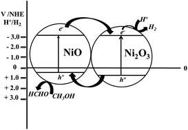
A cubic MCM-48 mesoporous material was employed as a support to encapsulate earth abundant NixOy species (NiO and Ni2O3). The cubic MCM-48 mesoporous support provides an excellent platform to not only effectively disperse NiO and/or Ni2O3 species but also to limit their particle sizes. The presence of Ni2O3 species at an optimal amount seems to enhance the photocatalytic activity of Ni–MCM-48 materials in comparison to a Ni–MCM-48 mesoporous material having only NiO dispersed in it. In addition, the presence of bulk NiO species also seems to be detrimental to the generation of solar hydrogen. The apparent quantum yield (AQY) of the most active material, Ni–MCM-48-2.5% was estimated to be 5.35%. This was over 250 times higher than a bulk, NiO (AQY = 0.02%) under identical experimental conditions. This study indicates that MCM-48 can be used as an effective support to disperse NixOy species.


1. Introduction

Hydrogen production triggered from splitting of water with the assistance of solar energy, as a means of carrying out ultimately clean and renewable energy source, has attracted immense research interest since the ground breaking work that was reported by Fujishima and Honda in 1972.1 During the past few decades, a myriad of semiconductors have been investigated and studied for photocatalytic hydrogen production from the decomposition of water.2–8 However, photocatalytic activities and efficiencies using conventional semiconductor photocatalysts are drastically restrained due to the fast recombination of the photogenerated electron–hole pair that takes place during the photocatalytic reactions in the volume or on the surface of bulk semiconductors. This vital drawback of the bulk semiconductor photocatalysts precludes their wide spread application for a variety of photocatalytic reactions.

In order to minimize the charge-carrier recombination and promote photocatalytic efficiency, an effective strategy has been developed and demonstrated by our group by employing highly ordered, cubic phased MCM-48 mesoporous materials as support for dispersing photoactive semiconductor species such as TiO2, WO3, TiO2 and/or CdS.9–14 MCM-48 is a member of the family of ordered mesoporous materials named M41S. This category of mesoporous materials were first reported by Mobil researchers in 1992.15 Among them, cubic phased, 3-D structured MCM-48 has triggered substantial interest due to its unique pore structure that minimizes pore clogging and provides effective molecular transport of reactant(s) and product(s).16 For instance, MCM-48 mesoporous materials possess large surface area (>1000 m2 g−1) and facilitates incorporation of semiconductor photocatalyst nanoclusters that are spatially isolated and effectively dispersed. High dispersion affords several catalytic sites per unit surface area. Thus, a relatively large number of surface reactive sites can be generated by dispersing spatially isolated and well-dispersed semiconductors on mesoporous materials. In addition, the semiconductor species that are confined in mesoporous materials possess small particle sizes than those of non-supported semiconductors. Small-sized semiconductor particles are also known to be more active than the large ones.17 The smaller is the particle size, the shorter is the distance that the photogenerated charge-carriers need to migrate to the surface. Hence, volume (or bulk) charge-carrier recombination in the smaller-sized semiconductors can be minimized and photocatalytic activity may be enhanced. Also, due to quantization effect, confined semiconductor species in mesoporous supports have larger band gap energies than those of bulk semiconductors. This moves the conduction band and valence band edges to more negative and more positive potentials, respectively. Therefore, the photogenerated electrons and holes in the quantum confined semiconductor species possess higher redox potentials compared with those in bulk semiconductors. Finally, mesoporous support materials can provide a certain extent of protection to semiconductor species, in particular to photocorrosive materials such as CdS. Thus, photocatalysts are more stable in general when dispersed in mesoporous materials such as MCM-48.

NiO, as an earth abundant metal oxide, has been studied as a co-catalyst, dopant, in a variety of photocatalytic reactions and also as a p-type electrode material in dye sensitized solar cells.18–25 In addition, NiO may also function as an “electron sink” when it is deposited on a semiconductor photocatalyst surface. NiO is a p-type semiconductor with a band gap energy ranging within 3.5–4.0 eV.26 The p-type conductivity of NiO results from the presence of Ni3+ ions and cationic vacancies due to the incorporation of chemisorbed oxygen into the NiO lattice. The Ni3+ ions may be considered as positive holes. Because of the relatively low concentration of Ni3+, NiO has a low conductivity. Also, it has been widely suggested that the presence of the p–n or p–p heterojunction structure in the mixed metal oxides materials can tremendously enhance the photocatalytic efficiency by suppressing the photogenerated charge-carrier recombination.27

Due to the above mentioned properties and favorable band edge positions for evolution of hydrogen, NiO itself can be utilized as a photocatalyst for solar hydrogen production from photocatalytic cleavage of water. To the best of our knowledge, this is the first report to incorporate NixOy (in the form of NiO and/or Ni2O3) semiconductor clusters into any mesoporous support (MCM-48 in this work) as a novel photocatalyst in which: (i) UV light generation of H2 in the absence of co-catalyst Pt which is usually indispensable for the production of hydrogen is realized, (ii) the band edges of the composite NixOy species in MCM-48 support have been calculated, (iii) only modest photocorrosion (2.4–11.5%) is observed, and (iv) comprehensive characterization of the photocatalysts using a myriad of techniques (X-ray diffraction (XRD), BET surface area analysis, atomic absorption spectroscopy (AAS), transmission electron microscopy (TEM), UV-vis diffuse reflectance spectra (DRS), and X-ray photoelectron spectroscopy (XPS) and UVPS) have been carried out. The supported photocatalytic system exhibits much higher efficiencies that are 71 to over 250 times (Apparent Quantum Efficiencies (AQY), 1.42 to 5.35%) compared with bulk NiO (AQY of 0.02%) for solar hydrogen production under identical conditions. Our studies also indicate that the presence of an appropriate amount of Ni2O3 in the Ni–MCM-48 material favors the formation of heterojunction structure and promotes the overall photocatalytic hydrogen production rate.

2. Experimental

2.1. Materials

Nickel(ous) nitrate and aqueous ammonia were obtained from Fisher Scientific. Tetraethyl orthosilicate (TEOS, 98%) was obtained from Acros. Cetyltrimethylammonium bromide (CTAB, 98%) was purchased from Alfa Aesar. Ethanol was produced by Pharmco-AAPER. Deionized water was used throughout this study. All these chemicals were used without any further purification.

2.2. Synthesis

2.2.1. Synthesis of Si–MCM-48. Siliceous MCM-48 (Si–MCM-48) mesoporous material was synthesized using a recipe optimized by us and reported previously.28 In brief, 1.2 g (3.3 mmol) of the surfactant, CTAB was added to 50 mL deionized water and stirred vigorously at room temperature until all the surfactant was dissolved. After this, 25 mL of ethanol was added. Then, 6 mL of aqueous NH3 (0.09 mol) was added to the surfactant–ethanol solution. Finally, 1.8 mL (8 mmol) of TEOS was added immediately. This mixture was stirred at 300 rpm for 4 h at room temperature. Then, the product, MCM-48 was recovered by filtration, washed with deionized water, and then dried in an oven at 80–90 °C overnight. The dried powder was ground and calcined at 550 °C for 6 h at a heating rate of 3 °C min−1 to remove the surfactant.
2.2.2. Synthesis of Ni–MCM-48 by impregnation method. In this work, a series of Ni–MCM-48 materials with different molar ratios of Ni contents were prepared by using an impregnation method as described as follows. An appropriate amount of nickel nitrate was first dissolved in 10 mL of deionized water. Then, 0.2 g of the calcined Si–MCM-48 mesoporous was added and the suspension was stirred for 24 h. Afterwards, the beaker was transferred to an oven and dried at 80 °C for overnight. Finally, the dried material was finely ground into a powder and then calcined in air at 550 °C for 6 h at a heating rate of 3 °C min−1 to enable the formation of NixOy nanoclusters in the MCM-48 mesoporous material. In this work, we synthesized 4 materials with different NixOy molar ratios of 1%, 2.5%, 5%, and 10%. Correspondingly, the materials are denoted as Ni–MCM-48-1%, Ni–MCM-48-2.5%, Ni–MCM-48-5%, and Ni–MCM-48-10%.

2.3. Characterizations

The powder XRD measurements were performed at room temperature using a Rigaku Ultima IV X-ray diffractometer with Cu Kα radiation. The diffractometer was operated at 40 kV and 44 mA. The step size for the scans were 0.02° and the scan rate was 0.24° min−1. The textural properties that encompass specific surface area, pore volume, and pore size distribution were analyzed by using N2 physisorption measurements. After the materials were dried for overnight at 80 °C and degassed at 100 °C for sufficient time, N2 isotherms were obtained at 77 K using a NOVA 2200e (Quantachrome Instruments) surface area and pore size analyzer. The specific surface areas were estimated by using the Brunauer–Emmett–Teller (BET) equation within the relative pressure range (P/P0) of 0.05–0.30. The pore volume was determined from the amount of nitrogen adsorbed at the highest relative pressure of P/P0 ≈ 0.99. The pore diameter were calculated by applying the Barrett–Joyner–Halenda (BJH) model to the desorption isotherm. The Ni content in the Ni–MCM-48 materials were measured by using a Varian SpectrAA-200 atomic absorption spectrophotometer equipped with a Ni cathode lamp. The working current was 4 mA and the wavelength of excitation was 232 nm. For AAS studies, 5 mg of the material was digested with known volume of HNO3 and then diluted to 25 mL. The concentration of the Ni2+ ions in the material was determined from a calibration plot. The UV-vis diffuse reflectance spectra were recorded by a Cary 100 Bio UV-visible spectrophotometer with a praying mantis diffuse reflection accessory (Harrick Scientific). The photoluminescence (PL) studies were carried out using a Horiba Fluormax-4 instrument. In the preliminary studies, the emission wavelength was varied in the wavelength increments of 25 nm in the range of 300 to 400 nm and the excitation wavelength were varied accordingly in order to identify the optimal wavelength of excitation and emission. Based on these detailed PL studies, the excitation wavelength was set at 300 nm and the emission was followed in the range of 325–450 nm. The sample was offset at an angle of 30° to the excitation to minimize any excitation beam from entering the emission slit. The optimal slit width for both excitation and emission was found to be 2 nm.

The TEM images were recorded using a Tecnai G2 instrument operating at 120 kV. Prior to TEM analysis, the material was dispersed in ethanol and the suspension was sonicated for 1 h. For each material, one drop of the suspension was placed on a copper grid coated with carbon film, and then allowed to dry overnight. A Kratos Axis Nova DLD X-ray Photoelectron Spectroscopy (XPS) system was used to determine the elemental composition. The surface analysis chamber is equipped with monochromatic radiation at 1486.6 eV from an Al Kα source using a 500 mm Rowland circle silicon single crystal monochromator. The X-ray gun was operated using a 15 mA emission current at an accelerating voltage of 15 kV. Low energy electrons were used for charge compensation to neutralize the material. Survey scans were collected using the following instrument parameters: energy scan range of 1200 to −5 eV; pass energy of 160 eV; step size of 1 eV; dwell time of 200 ms and data was acquired from the material area measuring approximately 700 × 300 μm in size. High resolution spectra were acquired in the region of interest using the following experimental parameters: 20 to 40 eV energy window; pass energy of 20 eV; step size of 0.1 eV and dwell time of 1000 ms. The absolute energy scale was calibrated to the Cu 2p2/3 peak binding energy of 932.6 eV using an etched copper plate. All spectra were calibrated using C 1s peak at 285.0 eV. A Shirley-type background was subtracted from each spectrum to account for inelastically scattered electrons that contribute to the broad background. Casa XPS 2.3.16 software was used to process the XPS data.29 Transmission corrected relative sensitivity factor (RSF) values from the Kratos library were used for elemental quantification.

2.4. Photocatalytic studies

The photocatalytic activities of the Ni–MCM-48 materials were examined in a closed system. A suspension with a catalyst concentration of 1 g L−1 and containing methanol (methanol/water mole ratio of 8) was dispersed in a quartz reactor. Prior to irradiation, the photoreaction system was purged with high purity Ar gas for 30 min to remove air. The light source was a 300 W Xe lamp equipped with an optical cutoff filter (λ > 280 nm). The reaction proceeded under vigorous stirring. The evolved H2 gas was analyzed by sampling the head space of the quartz reactor and using a gas chromatograph SRI 8610C (molecular sieve column, TCD detector, Ar as carrier gas). The amount of hydrogen evolved was calculated from a calibration plot.

3. Results and discussion

3.1. Powder XRD analysis

In order to examine the phases of the support and NiO in the Ni–MCM-48 mesoporous materials, powder X-ray diffraction (XRD) studies were conducted. Fig. 1(A) shows the low-angle XRD patterns of all the calcined Ni–MCM-48 materials along with Si–MCM-48. Si–MCM-48 exhibit typical XRD pattern of highly ordered, cubic phased MCM-48 mesoporous materials that can be evident from the presence of a strong d(211) and a weak d(220) peaks at 2θ values of nearly 2.6 and 3.1°. In addition to these two signature peaks, weak reflections corresponding to d spacings of (321), (400), (420), and (332) belonging to the cubic MCM-48 phase in the 2θ range of 4 to 6° are also observed.28
image file: c6ra09126a-f1.tif
Fig. 1 (A) Low-angle XRD patterns of Ni–MCM-48 materials along with Si–MCM-48; (B) high-angle XRD patterns of Ni–MCM-48 with a bulk NiO material as reference.

The incorporation of NixOy species did not destroy the cubic phase. In addition, one notices that the position of the highest intense peak (due to d(211) diffraction planes) in the nickel containing MCM-48 materials appear at relatively higher angles in comparison to Si–MCM-48. The shift to higher angles is indicative of a decrease in the pore diameter in these four materials. This is validated from nitrogen adsorption studies that are discussed later in this manuscript. Furthermore, the intensity of the reflection peaks in the Ni–MCM-48 materials show a decrease compared with the Si–MCM-48. This is due a decrease in the scattering contrast between the pores and the pore walls due to the presence of NixOy nanoclusters incorporated within the pores of the MCM-48 matrix and is consistent with previous observations in literature.

Fig. 1(B) depicts the high-angle XRD patterns of the Ni–MCM-48 materials and the powder XRD of a bulk NiO material is also shown here as a reference. It can be seen that in the materials with relatively low content of Ni (Ni–MCM-48-1% and Ni–MCM-48-2.5%), the XRD pattern shows a fairly broad peak centered near 2θ = 25°. This peak is due to amorphous SiO2. No other peaks are observed in the high angle scan indicating the NixOy nanoclusters are well dispersed on the relatively high surface area, cubic MCM-48 support in these two materials. With an increase in nickel content, peaks due to crystalline NiO species from (111), (200), (220), (311), and (222) diffraction planes can be seen in the Ni–MCM-48-5% and Ni–MCM-48-10% materials. Hence, it seems that as the nickel content increases, aggregation of NiO species occurs leading to formation of some bulk NiO species in these two materials.

In summary, the low-angle powder XRD results suggest the retention of the cubic phase at all loading levels, whereas the high-angle XRD data indicate the formation of crystalline NiO species at only relatively high loadings.

3.2. N2 physisorption

Nitrogen physisorption analysis was carried out at 77 K in order to probe the textural properties of the mesoporous materials prepared in this work. Fig. 2(A) shows the isotherms. All the materials depict isotherms that belong to type IV isotherm. This isotherm pattern is typical of mesoporous materials. At low relative pressure values (P/P0 < 0.2), nitrogen molecules start to deposit and form a monolayer on the pore walls of MCM-48 mesoporous materials. After this, multilayer adsorption takes place. In accordance, a steep inflection can be seen from the isotherm in the relative pressure range of P/P0 = 0.2 to 0.4. It has been suggested that the sharpness of this steep inflection region in the isotherm is a good indicator for the degree of the periodicity of the pores in mesoporous materials. It can be seen that the slope of the inflection in the Si–MCM-48 is steeper than that in the Ni–MCM-48 materials. This observation suggests that the incorporation of NixOy species, especially at relatively high loadings (5% and 10%), in the MCM-48 may cause some loss in the periodicity of the pores in these two materials. This is supported by powder XRD results which indicate a decrease in the intensity of the peaks due to d[211] and d[220] diffraction planes in these two materials. In addition, the position of the inflection may also provide some clues regarding the pore diameter of the mesoporous materials. It can be noticed that the position of the inflection in the Si–MCM-48 material is shifted slightly to higher relative pressure values compared with that in the Ni–MCM-48 materials. Hence, it can be concluded that the Si–MCM-48 material possesses larger pore diameter than that of Ni–MCM-48 materials, whereas the pore sizes are similar among the Ni–MCM-48 mesoporous materials.
image file: c6ra09126a-f2.tif
Fig. 2 (A) Nitrogen physisorption isotherms of Ni–MCM-48 materials along with Si–MCM-48 material; (B) pore size distribution plot of Ni–MCM-48 materials along with Si–MCM-48 material.

Fig. 2(B) shows the pore size distribution plot of the Ni–MCM-48 materials along with Si–MCM-48 mesoporous materials. As can be seen in the plot, all the materials exhibit a unimodal pore diameter distribution which suggests a highly uniform pore size in all of the materials. Moreover, Si–MCM-48 material presents a peak centered at ∼25 Å, whereas the Ni–MCM-48 materials show peaks located in the range of 19 to 22 Å. This observation is in line with the previous discussion that the pore diameter of Si–MCM-48 is larger than that of Ni–MCM-48 materials. The detailed values of the textural properties of all studied materials, such as surface area, pore volume, and pore size are listed in Table 1. The surface areas and pore volumes of the Ni–MCM-48 materials are lower than that of Si–MCM-48 and in general decrease with increase in Ni loading. However, the surface area and pore volume of Ni–MCM-48-2.5% deviates from this trend and exhibits the lowest surface area and pore volume. This may be due to some minor variability in the impregnation process for this material leading to this discrepancy. The reasons for this are unknown at this time.

Table 1 Textural properties of all studied mesoporous NixOy materials
Material Surface areaa (m2 g−1) Pore volumeb (cm3 g−1) Pore diameterc (nm)
a Determined by applying the Brunauer–Emmett–Teller (BET) equation to a relative pressure (P/P0) range of 0.05–0.30 to the adsorption isotherms.b Calculated from the amount of nitrogen adsorbed at the highest relative pressure (P/P0) of nearly 0.98.c Calculated by applying the Barrett–Joyner–Halenda (BJH) equation to the desorption isotherms.
Si–MCM-48 1262 0.80 2.5
Ni–MCM-48-1% 822 0.47 1.7
Ni–MCM-48-2.5% 596 0.33 2.0
Ni–MCM-48-5% 694 0.39 1.9
Ni–MCM-48-10% 614 0.36 1.9


3.3. XPS studies

XPS studies were conducted in order to discern the oxidation state(s) of the Ni species in the MCM-48 mesoporous materials from the binding energies and the chemical shifts. The high resolution XPS of the Ni 2p region in the various mesoporous materials and that of NiO as well for comparison purposes are shown in Fig. 3. In practice, the XPS of Ni species are fairly complex due to the presence of core level peaks that may be split due to various configurations and also due to the appearance of satellite peaks. Gupta and Sen have provided a method for calculating the multiple components and resolving the peaks of 3d transition metals such as nickel.30 The peaks obtained experimentally were de-convoluted by setting the components corresponding to Ni 2p3/2, Ni 2p3/2 satellite, Ni 2p1/2, and Ni 2p1/2 satellite.30,31 The XPS spectra of bulk NiO in this work indicate profiles similar to previous published report.32 The peak near 854.0 ± 0.1 eV has been previously assigned to Ni 2p3/2 in NiO and the fairly broad peak near 861.0 has been attributed to cd9L (c is metal d-electron state) and the unscreened cd8 final-state configurations (L is a ligand hole) by Carley et al.33 Similarly, Ni 2p1/2 peak appears near 872.5 eV and the associated satellite peak is at 879.2 eV. In addition, to these four peaks, an additional shoulder near 856.1 eV appears for bulk NiO prepared in this study and is invariably found in all NiO samples. This has been attributed to the presence of defect sites.34 The intensity of this peak is dependent on the nature of the sample preparation method and is strongly affected by the presence of defects. The high resolution XPS data of the Ni–MCM-48 materials are indicated in Fig. 3(B–E). The peaks due to Ni 2p1/2 satellite, Ni 2p1/2, Ni 2p3/2 satellite, Ni 2p3/2 appear near the same binding energy values as in NiO, but with small shifts. We could obtain a satisfactory fit only by allowing small variations (±0.1 eV) in the binding energy values. However, it can be seen that the relative intensity of peaks at 854.0 ± 0.1 and near 856.1 eV changes with the Ni concentration in the MCM-48 mesoporous materials. The spectral pattern or the envelope for materials, Ni–MCM-48-5% (Fig. 3(D)) and Ni–MCM-48-10% (Fig. 3(E)) resembles closely to that of bulk NiO as shown in Fig. 3(A). For the materials, Ni–MCM-48-1% and Ni–MCM-48-2.5% the peak at 854.1 eV is of much lower intensity as compared to the peak at 856.1 eV. Literature data suggests that that the higher binding energy peak (856.1 eV) is associated with Ni3+ chemical state.34 Metallic Ni binding energy are generally lower and appears at 852.6 eV.31 Overall it can be postulated that at low loading levels (Ni–MCM-48-1% and Ni–MCM-48-2.5%), Ni is in Ni3+ chemical state predominantly, whereas with an increase in Ni loading, NiO is formed in increasing amounts in Ni–MCM-48-5%, and Ni–MCM-48-10%.
image file: c6ra09126a-f3.tif
Fig. 3 High resolution XPS spectra of Ni 2p (A) NiO; (B) Ni–MCM-48-1%; (C) Ni–MCM-48-2.5%; (D) Ni–MCM-48-5%, and (E) Ni–MCM-48-10%.

Also, by carefully calculating and comparing the intensity of the Ni2+ and Ni3+ peaks in the Ni 2p XPS spectra using pure NiO as reference, the relative ratios of NiO and Ni2O3 in the Ni–MCM-48 mesoporous materials can be determined and the data is shown in Table 2. Table 2 lists all the values of the contents of NiO and Ni2O3 in all studied materials. The elemental quantification from XPS studies is indicated in Table S1 in the ESI section.

Table 2 Relative ratios of NiO and Ni2O3 in the Ni–MCM-48 photocatalysts
Materials % NiO % Ni2O3
NiO 100 0
Ni–MCM-48-1% 15.91 84.09
Ni–MCM-48-2.5% 24.99 75.01
Ni–MCM-48-5% 93.60 6.40
Ni–MCM-48-10% 100 0


The O 1s region of these materials were also probed in our XPS studies. The O 1s peak appears around 529.0 to 533.0 eV. The peaks in bulk Ni–O can be de-convoluted into three peaks appearing near 529.0, 530.6, and 532.3 eV that may be attributed to Ni–O, Ni–OH, and surface oxygen respectively. In the Ni–MCM-48 mesoporous materials, a relatively higher intensity O 1s peak is observed at 532.4 eV, that is due to the Si–O bonds in SiO2.35 In addition, one can see another small peak near 529.0 eV that increases in intensity with Ni loading as one would expect. This peak is due to Ni–O bond. In bulk NiO, and Ni–MCM-48-10%, this peak appears at 529.0 eV, whereas it is shifted to slightly higher values of nearly 529.4 eV in Ni–MCM-48-1% and Ni–MCM-48-2.5%. This shift is indicative of NixOy i.e. Ni3+ species, as per previous literature reports.36,37

In summary, the high resolution XPS results indicate the presence of Ni3+ at low loadings; the relative concentration of Ni3+ progressively decreases with increase in Ni loading in the MCM-48 matrix (Fig. 4).


image file: c6ra09126a-f4.tif
Fig. 4 High resolution XP spectra of O 1s (A) NiO; (B) Ni–MCM-48-1%; (C) Ni–MCM-48-2.5%; (D) Ni–MCM-48-5%, and (E) Ni–MCM-48-10%.

Valence band spectra of bulk NiO and Ni–MCM-48 materials from −1 eV to 10 eV was also acquired and is shown in Fig. 5. The peaks in the 1 to 2 eV region belong to localized Ni 3d orbitals whereas the broad peaks in the 5 to 9 eV region belong to O 2p σ and O 2p π orbitals. The Fermi edge of bulk NiO appears near 0 eV, whereas the valence band edges of the composite NixOy species incorporated onto the mesoporous matrix in Ni–MCM-48-1%, Ni–MCM-48-2.5%, Ni–MCM-48-5%, and Ni–MCM-48-10% are estimated to be 0.8, 0.4, 0.1 and 0.05 eV respectively as per our previous report.13 The absolute numbers should be used with caution because the presence of Ni3+ causes complexity and uncertainty; however the relative trend in the values are consistent with the expectation in that the valence band edges are shifted to less positive values with an increase in Ni loading.


image file: c6ra09126a-f5.tif
Fig. 5 High resolution valence band spectra of bulk NiO and Ni–MCM-48 materials.

3.4. TEM analysis

In order to study the morphology of the mesoporous structure in the Ni–MCM-48 materials, transmission electron microscopy (TEM) analysis was performed in this work. Fig. S1 shows a representative TEM image of Ni–MCM-48-1% material. The TEM image (recorded along the (110) plane) indicates a highly ordered, long-range arrayed, cubic phased mesoporous consistent with our previous studies.9,11,13,14,28

In addition, high resolution TEM studies were also recorded and is shown in Fig. 6. It is worthwhile mentioning here that the inter-penetrating and three-dimensional nature of the pores in MCM-48 poses unique challenges from our experience. In order to observe the NiOx species, a series of under-focus and over-focus studies were carried out. Results from these studies are shown in Fig. 6(A–D). As discussed in the XRD section, the high angle scan of Ni–MCM-48-1% do not indicate the presence of any bulk NixOy (NiO and Ni2O3) species. This indicates that the NixOy species are well dispersed as small clusters (<3 nm) and perhaps amorphous as well. Fig. 6(A) shows the high resolution TEM image of Ni–MCM-48-1%. No lattice fringes due to either NiO or Ni2O3 could be observed despite our best attempts, perhaps indicative of the fact that at this loading the NixOy species are amorphous. The HRTEM of Ni–MCM-48-2.5% shows lattice fringes due to d(111) plane of NiO with spacings of 2.4 Å and this is close to the value of 2.41 Å (Powder Diffraction File, PDF-47-1049) from powder XRD studies. Also, lattice fringes with spacings of 2.8 Å can be observed. This is perhaps due to d(002) plane from Ni2O3 (Powder Diffraction File, PDF-14-0481). This peak typically appears near 2θ = 32°. However, the presence of a fairly broad peak due to the silica support precludes the observation of this peak in our long range powder XRD studies. In addition, one can also notice the formation of some NiO/Ni2O3 heterojunctions in this material in the TEM image (Fig. 6(B)). The TEM images of Ni–MCM-48-5% and Ni–MCM-48-10% indicate the presence of NiO particles with d spacings consistent with powder XRD data.


image file: c6ra09126a-f6.tif
Fig. 6 TEM images of Ni–MCM-48 mesoporous materials.

In summary, the TEM results indicate the presence of well-dispersed NiO and Ni2O3 particles in the some of the mesoporous MCM-48 materials. The particle sizes of the NiOx species appear to be in 4–6 nm range from TEM studies and is larger than the pore sizes of ∼2 nm. In, Ni–MCM-48-1%, the NiOx species seem to be located within the pores because the high angle XRD scan do not indicate the presence of any bulk NiOx species. In Ni–MCM-48-2.5%, a similar situation seems to be prevalent. The particle sizes of NiO and Ni2O3 are larger than the pore sizes and this is perhaps due to the aggregation of NixOy clusters in the three-dimensional pore network of MCM-48 and such phenomena has been observed by us previously for CdS and CdS and TiO2 loaded onto MCM-48 support.12,13 In the case of Ni–MCM-48-5%, and Ni–MCM-48-10%, it is likely that some of the NixOy species are located outside the pores, because the powder XRD results indicate the presence of bulk NiO.

3.5. DRS analysis

In order to evaluate the band gaps of Ni–MCM-48 materials, Diffuse Reflectance Spectroscopy (DRS) studies were carried out. Fig. 7 depicts the DR spectra of all studied materials along with bulk NiO as shown in the inset of Fig. 7. In general, all materials show absorbance in the UV region (<400 nm). Compared with the absorption onset in bulk NiO (∼380 nm), Ni–MCM-48 materials exhibit absorption onsets shifted to lower wavelengths. The blue shift in the absorption onsets can be attributed to size quantization effects due to incorporation of NiOx species into the MCM-48 matrix. This phenomenon is frequently seen in the micro- and mesoporous materials encapsulated semiconductor materials. With a decrease in the loading of Ni in MCM-48, the absorption onset shifts to even lower wavelengths. This is due to the higher dispersion of NixOy in the material at lower NiO loadings. This result is in line with the XRD data, which suggests that some bulk phase NiO was formed in the materials with relatively high loadings of Ni species, whereas the particle sizes of NiO at relatively lower loading Ni–MCM-48 materials are too small (<3 nm) to be monitored by XRD.
image file: c6ra09126a-f7.tif
Fig. 7 DR spectra of Ni–MCM-48 materials; inset: DR spectra of bulk NiO material.

The band gap of Ni–MCM-48-1% is the largest due to the fact that the nanocluster size of the NiOx species is expected to the lowest due to the high dispersion and quantum confinement effects that are more pronounced at lower loadings. In addition, by extrapolating the maximum slope of absorption onset to the X-axis, the band gap energy of the composite NixOy species in the Ni–MCM-48 mesoporous materials can be calculated. We observed similar values when a plot of (αhν)2 versus hν was made (α is the absorption coefficient and is energy in eV) to determine the optical band gap. The values of the band gap energies of the studied materials are listed in Table 3. It should be stated that the band gaps of NiO and Ni2O3 differ by less than 0.1 eV and thus the band gaps estimated in this work are for the composite NixOy species incorporated onto the mesoporous matrix in three of the samples (Ni–MCM-48-1%, 2.5%, and 5%).

Table 3 Band gap energies of all studied materials
Materials Band gap energy (eV)
NiO 3.41
Ni–MCM-48-1% 3.73
Ni–MCM-48-2.5% 3.52
Ni–MCM-48-5% 3.49
Ni–MCM-48-10% 3.46


3.6. Photocatalytic H2 evolution

In order to examine the solar hydrogen generation activity of the prepared Ni–MCM-48 mesoporous materials, photocatalytic reactions were performed in a water/methanol mixed solution under UV light illumination. Fig. 8 shows the photocatalytic hydrogen production from all studied materials as a function of reaction time. It can be observed that the Ni–MCM-48 mesoporous materials exhibit appreciable photocatalytic activity for hydrogen production. A bulk NiO material was also examined in this work under identical experimental conditions. As can be seen from Fig. 8, bulk NiO material generates only a trace amount of hydrogen (∼32 μmol) after 4 hours of irradiation of UV light. The low photocatalytic activity in the bulk NiO material may be due to the relatively large particle size that fastens the charge-carrier recombination process and limits the photocatalytic efficiency.
image file: c6ra09126a-f8.tif
Fig. 8 Photocatalytic hydrogen yields over all studied materials along with a bulk NiO material for comparison.

In the contrast, all of the Ni–MCM-48 mesoporous materials present substantially enhanced photocatalytic activities compared with the bulk NiO photocatalyst. The support itself (MCM-48) does not generate hydrogen under the same experimental conditions. The improved photocatalytic activity of the Ni–MCM-48 mesoporous materials can be ascribed to the utilization of MCM-48 mesoporous support materials that facilitates the high dispersion of NixOy species and also due to the presence of heterojunctions in some of the materials containing NiO and Ni2O3. The highly dispersed and spatially isolated NixOy species result in the presence of a greater amount of surface active sites. In addition, the Ni–MCM-48 materials have conduction band edges of NiO that are relatively more negative compared with bulk NiO. These factors are conducive to the promotion of photocatalytic efficiency by the Ni–MCM-48 materials. Also, by confining the NixOy species within the MCM-48 support materials, the size of the NixOy particles can be restricted. A small particle size has been considered as a positive factor in photocatalytic reactions since the smaller is the size, the shorter is the distance for the photoinduced charge-carriers to migrate to the surface. Hence, the materials with NixOy species encapsulated onto the MCM-48 mesoporous materials exhibit higher photocatalytic activities compared with bulk NiO.

Among the four Ni–MCM-48 mesoporous materials, the photocatalytic hydrogen evolution rate is dependent on the Ni loading in the materials and also on the chemical environment of the NiO species. We also note from DRS results that the absorbance of the NixOy–MCM-48 materials increases with increase in loading of Ni; yet, the photocatalytic activity does not follow the same trend, indicating that the light absorption capability of the NixOy–MCM-48 is not a critical factor in this work and that other factors such as the presence of both NiO and Ni2O3 (discussed in the following paragraphs) are important and impact the photocatalytic activity significantly. In previously published works, we have observed that the photocatalytic activity is dependent on coordination of the transition metal ion, or particle size, and less dependent on the absorption capacity of the photocatalyst.10,12 The highest photocatalytic activity is obtained from the Ni–MCM-48-2.5% material. Further increase in the loading of Ni, causes a decrease in hydrogen production. In the Ni–MCM-48-5% and Ni–MCM-48-10% materials, powder XRD patterns indicate the presence of also bulk NiO. This significantly lowers the activities of these two materials in comparison to Ni–MCM-48-1% and Ni–MCM-48-2.5% (that do not show any bulk NixOy species). However, the NixOy species are well dispersed on the MCM-48 support and, hence, the photocatalytic activities of these two materials are higher than that of bulk NiO as explained previously. It seems that the presence of Ni2O3 is beneficial to production of hydrogen (see discussion later) and, as a result, a more efficient solar hydrogen production can be achieved in Ni–MCM-48-2.5%.

In addition, as suggested by the XPS results, in Ni–MCM-48-10% material, Ni ions are present in the form of Ni2+, whereas in the rest of Ni–MCM-48 materials, Ni ions are present in both Ni2+ and Ni3+. The existence of mixed NiO and Ni2O3 species in the materials could result in the formation of the NiO/Ni2O3 interfaces (as indicated in Fig. 6(B) for Ni–MCM-48-2.5%). The presence of heterojunctions in photocatalytic systems have been recognized as important and as a positive factor in photocatalytic reactions. NiO and Ni2O3 are both p-type semiconductors with almost similar band gap energies varying between 3.4 and 4.0 eV.38 However, due to the different oxidation states in the Ni ions in these two materials, the electron affinity in NiO and Ni2O3 varies.39 From the XPS spectra of O 1s region, it can be observed that the binding energy of Ni2O3 is higher than that of NiO. In other words, Ni3+ species has a higher electron affinity compared with Ni2+. Hence, it is more difficult to remove an electron from the bottom of the conduction band of Ni2O3.40,41 Since the band gap energies and vacuum level of these two semiconductors are very close,42 the conduction band edge of Ni2O3 is located at lower position (less negative) than NiO, whilst the valence band edge of Ni2O3 is also lower (more positive) than NiO.37 This staggered type of band geometry in NiO/Ni2O3 containing mesoporous materials favors separation of the photogenerated charge carriers and enhances the photocatalytic activity in the NiO/Ni2O3 containing mesoporous photocatalysts. As the NiO absorbs the UV light, the electrons on the NiO conduction band can migrate to the conduction band of Ni2O3 whereas the holes on the valence band of NiO cannot migrate to the valence band of Ni2O3. Hence, the photogenerated charge-carrier recombination in such materials can be minimized. In the case of Ni2O3 as a UV light absorbent, the excited electrons on the Ni2O3 conduction band cannot migrate to the NiO conduction band whereas the holes on the valence band of Ni2O3 can be injected into valence band of NiO. Again, the photoinduced charge-carriers can be separated effectively with this type of band structure in the NiO/Ni2O3 containing mesoporous materials.

The photocatalytic activity of the materials with NiO and Ni2O3 (Ni–MCM-48-5%, Ni–MCM-48-2.5%, and Ni–MCM-48-1%) is higher compared with the material containing only NiO (Ni–MCM-48-10%). In Ni–MCM-48-5%, about 94% is NiO and ∼6% is Ni2O3. Due to the relatively low amount of Ni2O3, the photogenerated electrons on the NiO conduction band have only a limited number of outlets to transfer electrons to the conduction band of Ni2O3. Hence, the photocatalytic activity is relatively low in comparison to the other two materials. Ni–MCM-48-2.5% shows the best performance for photocatalytic hydrogen generation. This may be ascribed to the optimum ratio of NiO/Ni2O3 in the material (1[thin space (1/6-em)]:[thin space (1/6-em)]3). The presence of sufficient amount of NiO provides enough outlets for the photogenerated holes on the Ni2O3 valence band. Meanwhile, NiO can also produce photogenerated electrons that can also migrate to the Ni2O3 conduction band. Further increase in the content of Ni2O3 causes a decrease in the hydrogen yield as seen in Ni–MCM-48-1% material which possesses NiO[thin space (1/6-em)]:[thin space (1/6-em)]Ni2O3 ratio of 1[thin space (1/6-em)]:[thin space (1/6-em)]5.3. This may be ascribed to the relatively low amount of NiO that may preclude efficient transfer of holes from Ni2O3 to NiO. Hence, the photocatalytic activity is relatively lower in this material compared to Ni–MCM-48-2.5%. The apparent quantum yield (AQY) of the most active material, Ni–MCM-48-2.5% was estimated to be 5.35%. This was over 250 times higher than a bulk, NiO (AQY = 0.02). In this context it is worth mentioning that Ni3+ has been previously reported to minimize charge-carrier recombination by trapping electrons to form Ni2+, and then the formed, Ni2+ can be trapped by holes to re-form Ni3+.43 Thus, the presence of Ni2O3 (at optimal amounts) in addition to NiO can enhance the efficiency of a photocatalytic reaction.

The mechanism of the photocatalytic reactions taking place at the heterojunctions of the NiO/Ni2O3 mixed oxide materials is illustrated in Scheme 1 for Ni–MCM-48-2.5%. The band gap and valence band edge have been estimated to be 3.52 eV and +0.4 V respectively from DRS and UVPS studies.


image file: c6ra09126a-s1.tif
Scheme 1 Mechanism of photocatalytic reactions in a representative mesoporous material, Ni–MCM-48-2.5%.

In order to better understand the mechanism, photoluminescence (PL) studies were carried out. The PL results are indicated in Fig. S2. As indicated in Fig. S2, a broad emission peak in the UV region centered near 350 nm in observed in these materials. The PL emission intensity varies in the order, Ni–MCM-48-1% > Ni–MCM-48-2.5% > Ni–MCM-48-5% > Ni–MCM-48-10%. This order is consistent with the relative amount of Ni3+ as indicated in Table 3. Thus, it seems that the PL emission is closely related to the presence of Ni2O3. The PL of Ni–MCM-48-10% that only contains NiO is negligible and pure NiO did not show any PL under the same conditions and hence is not plotted in Fig. S2. Our PL results are consistent with a previous report in literature that indicate that the PL emission is primarily dependent on the particle size of the dopant species.44 Thus, the intensity of the PL emission is the highest in Ni–MCM-48-1% since the cluster size of the NixOy species is the least in this composite material.

In order to investigate the stability of the Ni–MCM-48 mesoporous photocatalysts, atomic absorption spectroscopy (AAS) studies of the supernatant of the spent suspension after photocatalytic reaction was performed. Table 4 lists the values of the percentage of nickel ions lost from the Ni–MCM-48 materials after the photocatalytic reactions. It can be seen that all of the Ni–MCM-48 materials exhibit fairly good stability. Only a moderate loss (<5%) of Ni2+ ions from the fresh Ni–MCM-48 materials can be perceived to be leaching out in Ni–MCM-48-10%, Ni–MCM-48-5%, and Ni–MCM-48-2.5%. In the Ni–MCM-48-1% material, there is slight higher loss (11.5%) of nickel ions after photocatalytic reaction. The photostability of the most active catalyst, Ni–MCM-48-2.5% was examined and it was found that the photocatalytic activity was found to decrease by only ∼10% after three cycles indicating good stability.

Table 4 AAS analysis of the suspension after photocatalytic reactions
Materials Percentage of Ni2+ leached from the supernatant after photocatalytic reaction
Ni–MCM-48-1% 11.5%
Ni–MCM-48-2.5% 4.4%
Ni–MCM-48-5% 2.3%
Ni–MCM-48-10% 2.4%
Ni–MCM-48-10% 1.3% (stirred in dark)


4. Conclusions

An efficient and stable photocatalytic system for solar hydrogen production was established by incorporating earth abundant NiOx species onto MCM-48 mesoporous materials. The Ni–MCM-48 materials exhibit significantly enhanced hydrogen evolution compared with bulk NiO. The use of MCM-48 cubic phased mesoporous materials facilitates the high dispersion of NixOy species and is also conducive to restrain the particle size of NiO. The presence of NiO and Ni2O3 species and the formation of the NiO/Ni2O3 heterojunction structure are proven to be positive factors for photocatalytic activity since the photogenerated charge-carrier can be effectively separated. AAS analysis of the spent catalysts suggests that the NixOy species are relatively stable on the MCM-48 mesoporous host materials.

Acknowledgements

Thanks are due to NSF-CHE-0722632, NSF-CHE-0840507, NSF-EPS-0903804, DE-EE0000270, and SD NASA-EPSCOR NNX12AB17G.

References

  1. A. Fujishima and K. Honda, Nature, 1972, 238, 37–38 CrossRef CAS PubMed.
  2. X. Chen, S. Shen, L. Guo and S. S. Mao, Chem. Rev., 2010, 110, 6503–6570 CrossRef CAS PubMed.
  3. A. Kudo and Y. Miseki, Chem. Soc. Rev., 2009, 38, 253–278 RSC.
  4. K. Maeda, J. Photochem. Photobiol., C, 2011, 12, 237–268 CrossRef CAS.
  5. F. E. Osterloh, Chem. Mater., 2007, 20, 35–54 CrossRef.
  6. L. Yuan, C. Han, M. Q. Yang and Y. J. Xu, Int. Rev. Phys. Chem., 2016, 35, 1–36 Search PubMed.
  7. N. Zhang, M.-Q. Yang, S. Liu, Y. Sun and Y.-J. Xu, Chem. Rev., 2015, 115, 10307–10377 CrossRef CAS PubMed.
  8. S. Liu, Z.-R. Tang, Y. Sun, J. C. Colmenares and Y.-J. Xu, Chem. Soc. Rev., 2015, 44, 5053–5075 RSC.
  9. D. Zhao, A. Rodriguez, N. M. Dimitrijevic, T. Rajh and R. T. Koodali, J. Phys. Chem. C, 2010, 114, 15728–15734 CrossRef CAS.
  10. D. Zhao, S. Budhi, A. Rodriguez and R. T. Koodali, Int. J. Hydrogen Energy, 2010, 35, 5276–5283 CrossRef CAS.
  11. R. Peng, D. Zhao, N. M. Dimitrijevic, T. Rajh and R. T. Koodali, J. Phys. Chem. C, 2012, 116, 1605–1613 CrossRef CAS.
  12. R. Peng, D. Zhao, J. Baltrusaitis, C.-M. Wu and R. T. Koodali, RSC Adv., 2012, 2, 5754–5767 RSC.
  13. R. Peng, C. Lin, J. Baltrusaitis, C.-M. Wu, N. M. Dimitrijevic, T. Rajh, S. May and R. T. Koodali, Phys. Chem. Chem. Phys., 2014, 16, 2048–2061 RSC.
  14. R. Peng, C.-M. Wu, J. Baltrusaitis, N. M. Dimitrijevic, T. Rajh and R. T. Koodali, Chem. Commun., 2013, 49, 3221–3223 RSC.
  15. J. S. Beck, J. C. Vartuli, W. J. Roth, M. E. Leonowicz, C. T. Kresge, K. D. Schmitt, C. T. W. Chu, D. H. Olson and E. W. Sheppard, J. Am. Chem. Soc., 1992, 114, 10834–10843 CrossRef CAS.
  16. D. Zhao and Y. Wan, Chem. Rev., 2007, 107, 2821–2860 CrossRef PubMed.
  17. R. Peng, S. Banerjee, G. Sereda and R. T. Koodali, Int. J. Hydrogen Energy, 2012, 37, 17009–17018 CrossRef CAS.
  18. X. Xu, Q. Fu, X. Guo and X. Bao, ACS Catal., 2013, 3, 1810–1818 CrossRef CAS.
  19. S. Lin, L. Shi, H. Yoshida, M. Li and X. Zou, J. Solid State Chem., 2013, 199, 15–20 CrossRef CAS.
  20. Y. Zhang, G. Xiong, S. Sheng and W. Yang, Catal. Today, 2000, 63, 517–522 CrossRef CAS.
  21. M. Antoniadou, P. Panagiotopoulou, D. Kondarides and P. Lianos, J. Appl. Electrochem., 2012, 42, 737–743 CrossRef CAS.
  22. Q. Liu, L. F. Wei, S. Yuan, X. Ren, Y. Zhao, Z. Y. Wang, M. H. Zhang, L. Y. Shi, D. D. Li and A. J. Li, RSC Adv., 2015, 5, 71778–71784 RSC.
  23. J. Zhi, A. Chen, H. L. Cui, Y. Xie and F. Q. Huang, Phys. Chem. Chem. Phys., 2015, 17, 5103–5108 RSC.
  24. Z. J. Huang, G. Natu, Z. Q. Ji, P. Hasin and Y. Y. Wu, J. Phys. Chem. C, 2011, 115, 25109–25114 CrossRef CAS.
  25. Z. Q. Ji, M. F. He, Z. J. Huang, U. Ozkan and Y. Y. Wu, J. Am. Chem. Soc., 2013, 135, 11696–11699 CrossRef CAS PubMed.
  26. M. D. Irwin, D. B. Buchholz, A. W. Hains, R. P. H. Chang and T. J. Marks, Proc. Natl. Acad. Sci. U. S. A., 2008, 105, 2783–2787 CrossRef CAS.
  27. C. Shifu, Z. Sujuan, L. Wei and Z. Wei, J. Hazard. Mater., 2008, 155, 320–326 CrossRef PubMed.
  28. B. Boote, H. Subramanian and K. T. Ranjit, Chem. Commun., 2007, 4543–4545 RSC.
  29. N. Fairley, CasaXPS: Processing Software for XPS, AES, SIMS and More, http://www.casaxps.com/ Search PubMed.
  30. R. P. Gupta and S. K. Sen, Phys. Rev. B: Solid State, 1975, 12, 15–19 CrossRef CAS.
  31. A. P. Grosvenor, M. C. Biesinger, R. S. C. Smart and N. S. McIntyre, Surf. Sci., 2006, 600, 1771–1779 CrossRef CAS.
  32. Z. H. Ibupoto, M. A. Abbasi, X. Liu, M. S. AlSalhi and M. Willander, J. Nanomater., 2014, 6, 928658 Search PubMed.
  33. A. F. Carley, S. D. Jackson, J. N. O'Shea and M. W. Roberts, Surf. Sci., 1999, 440, L868–L874 CrossRef CAS.
  34. M. W. Roberts and R. S. C. Smart, J. Chem. Soc., Faraday Trans. 1, 1984, 80, 2957–2968 RSC.
  35. S. Rasalingam, H. S. Kibombo, C.-M. Wu, S. Budhi, R. Peng, J. Baltrusaitis and R. T. Koodali, Catal. Commun., 2013, 31, 66–70 CrossRef CAS.
  36. J. Knudsen, L. R. Merte, G. Peng, R. T. Vang, A. Resta, E. Lægsgaard, J. N. Andersen, M. Mavrikakis and F. Besenbacher, ACS Nano, 2010, 4, 4380–4387 CrossRef CAS PubMed.
  37. E. L. Ratcliff, J. Meyer, K. X. Steirer, A. Garcia, J. J. Berry, D. S. Ginley, D. C. Olson, A. Kahn and N. R. Armstrong, Chem. Mater., 2011, 23, 4988–5000 CrossRef CAS.
  38. X. Chen, W. Chen, P. Lin, Y. Yang, H. Gao, J. Yuan and W. Shangguan, Catal. Commun., 2013, 36, 104–108 CrossRef CAS.
  39. G. Q. Wang, Y. M. Li and S. P. Zhuo, Int. J. Mater. Res., 2010, 101, 310–315 CrossRef CAS.
  40. Q. C. Du and G. X. Lu, Appl. Surf. Sci., 2014, 305, 235–241 CrossRef CAS.
  41. A. Galtayries and J. Grimblot, J. Electron Spectrosc. Relat. Phenom., 1999, 98, 267–275 CrossRef.
  42. X. Y. Wu, G. C. Xing, S. L. J. Tan, R. D. Webster, T. C. Sum and E. K. L. Yeow, Phys. Chem. Chem. Phys., 2012, 14, 9511–9519 RSC.
  43. S. Z. Hu, F. Y. Li and Z. P. Fan, Bull. Korean Chem. Soc., 2012, 33, 4052–4058 CrossRef CAS.
  44. L. Q. Jing, X. J. Sun, B. F. Xin, B. Q. Wang, W. M. Cai and H. G. Fu, J. Solid State Chem., 2004, 177, 3375–3382 CrossRef CAS.

Footnote

Electronic supplementary information (ESI) available. See DOI: 10.1039/c6ra09126a

This journal is © The Royal Society of Chemistry 2016
Click here to see how this site uses Cookies. View our privacy policy here.