A novel non-viral gene vector for hepatocyte-targeting and in situ monitoring of DNA delivery in single cells†
Yong-Guang Gaoa,
Quan Tanga,
You-Di Shia,
Ying Zhanga,
Ruibing Wangb and
Zhong-Lin Lu*a
aKey Laboratory of Theoretical and Computational Photochemistry, Ministry of Education, College of Chemistry, Beijing Normal University, Xinjiekouwai Street 19, Beijing 100875, China. E-mail: luzl@bnu.edu.cn
bState Key Laboratory of Quality Research in Chinese Medicine, Institute of Chinese Medical Sciences, University of Macau, Taipa, Macau, China
First published on 13th May 2016
Abstract
A macrocyclic polyamine lipid bearing both fluorescent naphthalimide and targeting cholic acid moieties, 1, was designed and synthesized as a novel non-viral gene vector. Plasmid DNA was completely condensed at 10 μM of 1 (N/P = 3.6) in 5 minutes at 37 °C. The process of DNA uptake process by 1 in a single cell was clearly investigated through time-dependent fluorescence microscopy assay. Flow cytometry experiments of liposome 1 clearly proved its targeting activity toward HepG2 cells. Luciferase gene expression transfected by the DNA complex of 1/DOPE gave a higher transfection efficiency in HepG2, A549, HeLa, U2OS and CCC-HPF-1 cells than by lipofectamine 2000. The present work demonstrated that the designed multifunctional organic molecule is able to fluorescently track the gene delivery process at a single-cell level for the first time. This will greatly aid mechanistic studies of gene transfection and further development of new gene vectors for biomedical applications.
Introduction
Gene therapy is a promising treatment modality for both hereditary and acquired diseases, and has attracted significant interest in both fundamental research and clinical applications.1 Viral vectors such as retroviruses, lentiviruses, adenoviruses and adeno-associated viruses (AAVs) have been actively engaged in several clinical applications due to their highly efficient intracellular DNA delivery.2 However, many undesirable complications, such as immunogenicity and relatively low loading capacity, have limited their therapeutic applications. Thus much attention has been paid to non-viral gene vectors because of their low immunogenicity and cytotoxicity, high DNA loading capacity, and their facile production and ability to readily undergo structural modification.3 Accordingly, various non-viral gene vectors such as lipids, functional polymers, metal complexes and nanoparticles have been prepared and examined for gene delivery, and significant progress has been achieved.4In spite of these advancements, there are still some challenges to be overcome before synthetic non-viral gene vectors can be employed in clinical applications. These challenges include limited ability to target specific cells, low transfection efficiency, and high material cost. Moreover, there are currently no tools available to effectively elucidate DNA cellular uptake mechanisms during its delivery, including internalization pathway, and endosomal release.
To track gene delivery processes, fluorescent materials, such as quantum dots and tetranuclear ruthenium complexes, have been incorporated in various non-viral gene vectors.5 However, most of these tracking processes were conducted using model cells that are not those for targeting and treatment. Furthermore, although targeting compounds, such as cholic acid, folate, RGD, and galactose, have been widely used in the development of targeting delivery vehicles,6 those applied in small organic non-viral gene vectors are very rare. To the best of our knowledge, fluorescent non-viral gene vectors with targeting capability have not been reported so far.
Our group has recently developed a series of non-viral gene vectors containing fluorescent moieties such as naphthalimide/coumarin and macrocyclic polyamine [12]aneN3 units.7 Among them, a lipid containing naphthalimide moiety was successfully applied to detect the process of cellular DNA uptake, translocation and release based on real-time fluorescence tracking.7b However, such a tracking process was not based on single cells. Furthermore, the transfection was without targeting property. In this report, a new [12]aneN3 lipid 1 containing both cholic acid and naphthalimide moieties was designed and synthesized, that allowed direct visualization of the processes of cellular DNA uptake, translocation and release in single cells. It also showed hepatocyte-targeting properties and considerably higher transfection efficiency than those of other lipids we have reported7b,c and commercial lipofectamine 2000. This is the first example of the use of a small organic non-viral gene vector for hepatocyte targeting and in situ fluorescent tracking.
Experimental details
Materials and instruments
Anhydrous ethanol, methanol, tetrahydrofuran (THF), dichloro-methane (DCM), dimethylsulfoxide (DMSO) and N,N-dimethyl formamide (DMF) were dried and purified under nitrogen by using standard methods and were distilled immediately before use. 3,5-Bis(azidomethyl)benzoic acid and ditertbutyl 9-(prop-2-ynyl)-1,5,9-triazacyclododecane-1,5-dicarboxylate (7) were prepared according to the literature.14 Electrophoresis grade agarose, 6× loading buffer (30 mM EDTA, 40% glycerol, 0.03% xylene cyanol FF, and 0.05% bromophenol blue), Goldview II, 4,6-diamidino-2-phenylindole (DAPI), 3-(4,5-dimethylthiazol-2-yl)-2,5-diphenyltetrazolium bromide (MTT), plasmid DNA (pUC 18) were purchased from Solarbio Company. Dioleoylphosphatidylethanol amine (DOPE) was from Santa Cruz Biotechnology. Lipofectamine 2000™ with concentration of 1 mg mL−1 and Cy5-labeled double-stranded DNA oligomer 5′-GGTCGGAGTCAAC-GGATTTGGTCG-3′-(Cy5-DNA) were from Invitrogen. Ultrapure milli-Q water (18.25 MΩ) was used in all DNA condensation assays.1H and 13C NMR spectra were obtained on a Bruker Avance III 400 MHz spectrometer at 25 °C. The infrared spectra were taken on a Nicolet 380 spectrometer. Mass spectra were acquired on a Waters Quattro Mocro spectrometer and high resolution mass spectra were acquired on a Waters LCT Premier XE spectrometer. Electrophoresis apparatus was a BG-subMIDI sub marine system (BayGene Biotech Company Limited, Beijing, China). Hydrodynamic diameters were determined using a Brookhaven Zeta Plus Particle Size and Zeta Potential Analyzer. The morphologies of the lipoplexes were observed by SEM (a Hitachi, X650). Fluorescence spectra were measured on a Varian Cary Eclipse spectrometer.
Preparation and characterization of 4
Cholic acid 3 (1.3 g, 3.2 mmol), 1-ethyl-3-(3-dimethylaminopropyl)carbodiimide hydrochloride (EDCI) (0.61 g, 3.2 mmol), N-hydroxybenzotriazole (BtOH) (0.43 g, 3.2 mmol) and N,N-diisopropylethylamine (DIEA) (0.83 g, 6.4 mmol) in dimethyl sulphoxide (DMSO) (30 mL) were stirred for 0.5 h, then compound 2 (1.0 g, 3.2 mmol) was added, and stirring was continued for 4 h. CH2Cl2 (50 mL) was added, the mixture was washed with water (3 × 30 mL). The organic phase was washed with saturated brine, dried over Na2SO4, filtered, and the solvent was evaporated under reduced pressure. The crude material was purified by column chromatography on silica gel (PE/EA = 1/1) to give a yellow solid 1.8 g (79%). 1H NMR (400 MHz, CDCl3) δ 8.66 (d, J = 7.2 Hz, 1H), 8.58 (d, J = 8.5 Hz, 1H), 8.42 (d, J = 7.9 Hz, 1H), 8.06 (d, J = 7.9 Hz, 1H), 7.87 (t, J = 7.9 Hz, 1H), 6.34 (s, 1H), 4.40 (t, J = 5.5 Hz, 2H), 3.87–3.80 (m, 2H), 3.71–3.64 (m, 2H), 3.45 (s, 1H), 2.35–2.13 (m, 6H), 2.08–1.82 (m, 4H), 1.72–1.55 (m, 7H), 1.48–1.25 (m, 6H), 1.20–0.94 (m, 4H), 0.92–0.87 (m, 6H), 0.55 (s, 3H); 13C NMR (101 MHz, CDCl3) δ 174.66, 163.85, 163.80, 133.24, 132.07, 131.21, 131.03, 130.42, 130.29, 128.64, 128.03, 122.50, 121.64, 72.93, 71.80, 68.31, 53.46, 46.45, 46.27, 41.48, 39.76, 39.42, 38.57, 35.31, 34.71, 33.14, 31.57, 30.38, 28.05, 27.40, 26.23, 23.17, 22.43, 17.35, 12.34; IR (KBr, cm−1); ν 3412.08, 2929.87, 2888.78, 2366.66, 2337.72, 1701.22, 1658.78, 1589.34, 1377.17, 1346.31, 1232.51, 1078.21, 1040.56, 783.10, 729.09; MS (ES+): calcd for C38H49BrN2O6 (M + H)+: 709.3, found 709.5.Preparation and characterization of 5
To a stirred solution of compound 4 (1.0 g, 1.4 mmol) in 2-methoxyethanol (30 mL) was added 1,10-diaminodecane (0.24 g, 2.8 mmol). The mixture was refluxed for 24 h and monitored by TLC. After completion of the reaction, the mixture was cooled to room temperature and concentrated under vacuum until most of the solvent was removed. The residue was washed with water and then purified by column chromatography on silica gel (CH2Cl2/MeOH = 5/1) to provide product 0.6 g (54%). 1H NMR (400 MHz, CD3SOCD3) δ 8.70 (d, J = 8.4 Hz, 1H), 8.41 (d, J = 7.3 Hz, 1H), 8.25 (d, J = 8.5 Hz, 1H), 7.78 (d, J = 6.0 Hz, 1H), 7.73 (d, J = 5.2 Hz, 1H), 7.66 (t, J = 7.9 Hz, 1H), 6.75 (d, J = 8.7 Hz, 1H), 4.15–3.94 (m, 4H), 3.70 (s, 1H), 3.59 (s, 1H), 3.31 (s, 2H), 3.27–3.21 (m, 1H), 3.20–3.02 (m, 1H), 2.54 (d, J = 6.8 Hz, 1H), 2.27–2.10 (m, 2H), 2.04–1.91 (m, 2H), 1.88–1.59 (m, 10H), 1.54–1.00 (m, 30H), 0.87 (s, 1H), 0.83 (d, J = 6.2 Hz, 3H), 0.80 (s, 3H), 0.46 (s, 3H); 13C NMR (101 MHz, CD3SOCD3) δ 172.75, 163.95, 163.06, 150.55, 134.10, 130.43, 129.57, 128.50, 123.92, 121.92, 120.12, 107.59, 103.47, 70.98, 70.43, 66.24, 46.05, 45.63, 42.87, 41.53, 41.25, 41.06, 36.65, 35.31, 34.97, 34.86, 34.33, 32.46, 32.01, 31.49, 30.38, 29.04, 28.89, 28.49, 27.86, 27.17, 26.69, 26.34, 26.16, 22.72, 22.53, 17.01, 12.15; IR (KBr, cm−1): ν 3398.57, 2927.94, 2856.58, 1643.35, 1633.71, 1573.91, 1375.25, 1247.96, 1166.93, 775.38; MS (ES+) calcd for C48H72N4O6 (M + H)+: 801.6, found 801.9.Preparation and characterization of 6
3,5-Bis(azidomethyl)benzoic acid (0.097 g, 0.42 mmol), EDCI (0.08 g, 0.42 mmol), BtOH (0.057 g, 0.42 mmol) and DIEA (0.16 g, 0.84 mmol) in DMF (10 mL) were stirred for 0.5 h, then compound 5 (0.3 g, 0.42 mmol) was added, and stirring were continued for 12 h. Water (5 mL) was added and the mixture was extracted with CH2Cl2 (2 × 15 mL). The organic phase was dried (Na2SO4), filtered, and the solvent was evaporated under reduced pressure. The crude material was purified by column chromatography on silica gel (CH2Cl2/MeOH = 5/1) to give a yellow solid 0.22 g (52%). 1H NMR (400 MHz, CDCl3) δ 8.46 (d, J = 7.2 Hz, 1H), 8.35 (d, J = 8.4 Hz, 1H), 8.13 (d, J = 8.3 Hz, 1H), 7.69 (s, 2H), 7.48 (t, J = 7.9 Hz, 1H), 7.38 (s, 1H), 6.62 (d, J = 8.6 Hz, 2H), 6.52 (s, 1H), 4.41 (s, 4H), 4.35 (s, 2H), 3.80 (s, 1H), 3.76 (s, 1H), 3.63 (s, 2H), 3.48–3.30 (m, 6H), 2.38 (s, 2H), 2.25–1.95 (m, 5H), 1.88–1.68 (m, 6H), 1.55–1.21 (m, 27H), 0.98–0.92 (m, 2H), 0.88–0.81 (m, 7H), 0.50 (s, 3H); 13C NMR (101 MHz, CDCl3) δ 174.67, 166.79, 165.37, 164.65, 150.56, 143.51, 136.86, 136.20, 130.23, 129.94, 128.89, 128.79, 126.59, 125.16, 122.13, 120.26, 109.51, 108.64, 85.18, 72.97, 71.91, 68.40, 54.26, 46.68, 46.42, 43.84, 41.64, 40.40, 39.52, 39.20, 35.46, 34.80, 29.79, 29.70, 29.50, 29.41, 29.33, 28.78, 27.27, 27.06, 22.55, 17.44, 12.44; IR (KBr, cm−1): ν 3427.51, 2926.01, 2098.55, 1637.56, 1579.70, 1381.03, 1249.87, 1074.35, 775.38; MS (ES+) calcd for C57H78N10O7 (M + H)+: 1015.6, found 1015.9.Preparation and characterization of 8
Copper sulfate (4.3 mg, 0.021 mmol) and Vc–Na (8.2 mg, 0.42 mmol) were added to a solution of 6 (0.21 g, 0.21 mmol) and 7 (0.18 g, 0.46 mmol) in THF–H2O (10 mL/5 mL) and the mixture was stirred overnight at room temperature. The solvent was removed under reduced pressure. Water (10 mL) was added and the mixture was extracted with CH2Cl2 (2 × 20 mL). The combined organic layer was washed with saturated brine, dried over Na2SO4, filtered, and the solvent was evaporated under reduced pressure. The crude material was purified by column chromatography on silica gel (CH2Cl2/MeOH = 10/1) to give a yellow solid 0.31 g (81%). 1H NMR (400 MHz, CD3OD) δ 8.49 (d, J = 8.3 Hz, 1H), 8.45 (d, J = 7.4 Hz, 1H), 8.31 (d, J = 8.4 Hz, 1H), 7.90 (s, 2H), 7.71 (s, 2H), 7.57 (d, J = 7.4 Hz, 1H), 7.41 (s, 1H), 6.74 (d, J = 8.4 Hz, 1H), 5.62 (s, 4H), 4.27 (s, 2H), 3.73 (s, 4H), 3.57–3.52 (m, 1H), 3.44–3.38 (m, 2H), 3.32–3.29 (m, 16H), 2.38 (t, J = 5.9 Hz, 8H), 2.28–2.09 (m, 3H), 2.03–1.74 (m, 20H), 1.72–1.51 (m, 10H), 1.49–1.39 (m, 48H), 1.32 (s, 10H), 1.06–0.91 (m, 3H), 0.91–0.85 (m, 6H), 0.45 (s, 3H); 13C NMR (101 MHz, CD3OD) δ 176.58, 168.36, 166.08, 165.44, 157.70, 157.56, 152.26, 144.70, 138.20, 137.36, 135.77, 131.99, 131.24, 131.10, 129.17, 127.88, 125.16, 123.07, 121.52, 108.96, 104.82, 81.03, 80.66, 79.61, 79.29, 78.96, 73.69, 72.67, 68.81, 54.09, 50.84, 47.86, 47.26, 46.98, 46.72, 46.54, 44.83, 43.00, 42.72, 41.05, 40.86, 40.33, 36.54, 35.77, 34.18, 32.93, 31.10, 30.59, 30.56, 30.47, 30.39, 30.35, 29.48, 28.83, 28.72, 28.27, 28.02, 27.66, 27.13, 24.11, 23.27, 17.77, 13.00; IR (KBr, cm−1): ν 3448.72, 2929.87, 2366.66, 1687.71, 1641.42, 1581.63, 1365.60, 1047.35, 773.46, 731.02; MS (ES+) calcd for C101H156N16O15 (M + H)+: 1834.2, found 1834.2.Preparation and characterization of 1
Compound 8 (0.31 g, 0.17 mmol) was added to a saturated solution of hydrogen chloride in ethyl acetate (10 mL) and the mixture was stirred for 30 minutes at room temperature. The resulting solid was filtrated off and the solid was washed with ethyl acetate, then dried in vacuum at 60 °C for 24 h, 0.25 g (84%) yellow solid was obtained. 1H NMR (400 MHz, CD3SOCD3) δ 11.27 (s, 1H), 9.53 (s, 9H), 8.73 (d, J = 8.4 Hz, 1H), 8.63 (t, J = 4.9 Hz, 1H), 8.50 (s, 2H), 8.38 (d, J = 7.2 Hz, 1H), 8.22 (d, J = 8.5 Hz, 1H), 7.79 (s, 3H), 7.63 (t, J = 7.8 Hz, 1H), 7.35 (s, 1H), 6.73 (d, J = 8.7 Hz, 1H), 5.69 (s, 4H), 4.40 (s, 2H), 4.08 (s, 4H), 3.72–3.17 (m, 32H), 3.07 (s, 4H), 2.28–1.91 (m, 18H), 1.79–1.15 (m, 36H), 0.92–0.75 (m, 6H), 0.72–0.40 (m, 3H); 13C NMR (101 MHz, CD3SOCD3) δ 173.08, 165.32, 164.07, 163.18, 150.68, 136.53, 135.73, 134.28, 130.65, 129.96, 129.63, 128.78, 128.00, 126.91, 124.08, 121.91, 120.17, 107.50, 103.59, 71.07, 70.50, 66.32, 52.67, 48.30, 46.11, 45.66, 42.90, 41.28, 36.77, 34.99, 34.42, 32.59, 31.44, 30.36, 29.12, 28.95, 28.43, 27.93, 27.17, 26.76, 26.62, 26.16, 22.73, 22.58, 20.32, 19.05, 17.67, 17.07, 12.20; IR (KBr, cm−1): ν 3425.58, 2929.87, 2364.73, 1641.42, 1581.63, 1546.91, 1382.96, 1247.94, 1047.35, 773.46; HRMS (ES+) calcd For C81H124N16O7 (M + H)+: 1833.9839, found 1433.9930.Formation of liposome and lipoplex
The cationic lipid 1 (0.002 mmol) with DOPE in the desired mole ratio was dissolved in anhydrous chloroform (2 mL). Thin films were made by slowly rotary-evaporating the solvent at room temperature. The last trace of organic solvent was removed by keeping these films under vacuum for more than 8 h. The dried films and PBS buffer (10 mM, pH 7.4) were preheated to 70 °C, and then the buffer was added to the films to result in a final lipid concentration of 1 mM. The mixtures were vortexed vigorously until the films were completely re-suspended. Sonication of these suspensions for 20 min in a bath sonicator at 60 °C afforded the corresponding cationic liposomes that were stored at 4 °C.To prepare the liposome 1/pDNA complexes (lipoplexes), various concentrations of cationic liposomes were mixed with a constant amount of DNA (9 μg mL−1), and the mixtures were incubated for 30 min at room temperature.
Agarose gel retardation
Lipoplexes at different concentrations were prepared by adding appropriate volumes of compound 1 solution in the presence or absence of DOPE to 0.9 μL of pUC18 DNA (200 μg mL−1) and 4 μL of HEPES (100 mM, pH 7.2). The obtained complex solution was then diluted to the total volume of 20 μL. After incubation at 37 °C for 5 min, the lipoplexes were electrophoresed on a 0.7% (w/v) agarose gel containing GelRed™ in Tris-acetate (TAE) running buffer at 120 V for 40 min. Then DNA was visualized under an ultraviolet lamp using a Vilber Lourmat imaging system.Dynamic light scattering (DLS)
The liposome/DNA complexes of various concentrations were prepared by adding 0.9 μL of pUC18 DNA (200 μg mL−1) to the appropriate volumes of the liposome solution. Then the complex solution was vortexed for 30 s before being incubated at 37 °C for 5 min and then diluted up to 1 mL by ultrapure water solution prior to be measured. Data were shown as mean ± standard deviation (SD) based on three independent measurements.Scanning electron microscope (SEM) images
0.9 μL of pUC18 DNA (200 μg mL−1) was mixed with the appropriate volume of liposome 1 solution to form complexes, diluted by water to a total volume of 20 μL, and incubated at 37 °C for 5 min. The lipoplexes were added dropwise to a silicon slice. The slice was dried at room temperature at atmospheric pressure for several hours before observation.In vitro transfection experiments
Gene transfection of liposome 1 was investigated in HepG2, A549, HeLa, U2OS and CCC-HPF-1 cells. Cells were seeded in 24-well plates (8 × 10⁴ cells per well) and grown to reach 70–80% cell confluence at 37 °C for 24 h in 5% CO2. Before transfection, the medium was replaced with a serum-free DMEM culture medium containing liposome/DNA (pGL-3) complexes at various concentrations. After 4 h under standard incubator conditions, the medium was replaced with fresh medium containing serum and incubated for another 20 h.Inhibition studies (flow cytometry)
To probe the internalization mechanism of the lipoplex 1, the cellular uptake study was performed at 4 °C or in the presence of various endocytic inhibitors. Briefly, cells were incubated with lipoplex 1 (30 μM) at 4 °C for 4 h, while the energy-dependent endocytosis was not completely blocked. Otherwise, cells were preincubated with various endocytic inhibitors including chlorpromazine (20 μg mL−1), cytochalasin D (10 μg mL−1), genistein (200 μM), and nocodazole (33 μM). Following pretreatment for 30 min, the inhibitor solutions were replaced by the freshly prepared test complexes (Cy5-labeled plasmid DNA) in media containing inhibitors at the same concentrations. After further incubation for 4 h, cells were harvested and analyzed by flow cytometry. In the study, the groups in the presence of test complexes at 37 °C but without inhibitor treatment were used as controls. Results were represented as percentage uptake level of control cells.Cellular uptake of plasmid DNA (flow cytometry)
The cellular uptake of the liposome 1/Cy5-labeled DNA complexes was analyzed by flow cytometry. Briefly, HepG2, A549, HeLa, U2OS and CCC-HPF-1 cells were seeded in 12-well plates (2.0 × 10⁵ cells per well) and allowed to attach and grow for 24 h. The medium was exchanged with serum-free medium. Cells were incubated with complexes containing Cy5-labeled DNA (2 μg of DNA per well) in media for 4 h at 37 °C. Subsequently, the cells were washed with 1× PBS and harvested with 0.25% Trypsin/EDTA and re-suspended in serum-containing DMEM. Cy5-labeled plasmid DNA uptake was analyzed using flow cytometry.Confocal laser scanning microscopy (CLSM)
The cellular uptake of Cy5-labeled dsDNA condensates was observed by fluorescence microscopy. HepG2, A549, HeLa, U2OS and CCC-HPF-1 cells were cultured in DMEM medium supplemented with 10% FBS in a humid atmosphere containing 5% CO2 at 37 °C. The cells were seeded in Glass Bottom Cell Culture Dishes at 10![[thin space (1/6-em)]](https://www.rsc.org/images/entities/char_2009.gif) 000 cells per dish and cultured for 24 h. After being washed three times with DMEM, the cells were treated with freshly prepared Cy5-DNA condensates and the controls (500 μL). The blue fluorescence dye DAPI (5 μg mL−1) was also added to each dish for nuclear staining, after that the cells were cultured for 4 h (or different hours). Finally, the cells were washed 6 times with PBS buffer and observed using a Zeiss Inverted Fluorescence Microscope with a 10× objective and DAPI filter for DAPI (blue), GFP filter for lipid 1 (green), and Rhodamine filter for Cy5 (red), respectively.
000 cells per dish and cultured for 24 h. After being washed three times with DMEM, the cells were treated with freshly prepared Cy5-DNA condensates and the controls (500 μL). The blue fluorescence dye DAPI (5 μg mL−1) was also added to each dish for nuclear staining, after that the cells were cultured for 4 h (or different hours). Finally, the cells were washed 6 times with PBS buffer and observed using a Zeiss Inverted Fluorescence Microscope with a 10× objective and DAPI filter for DAPI (blue), GFP filter for lipid 1 (green), and Rhodamine filter for Cy5 (red), respectively.
Cytotoxicity assay
The cytotoxicity of lipoplex 1 toward HepG2, A549, HeLa, U2OS and CCC-HPF-1 cell lines was tested by MTT assays (MTT = 3-(4,5-dimethylthiazol-2-yl)-2,5-diphenyltetrazolium bromide). The cells were cultured in Dulbecco's modified Eagle's medium (DMEM, Gibco) supplemented with fetal bovine serum (FBS, 10%, v/v) in a humid atmosphere containing 5% CO2 at 37 °C. After 48 h of incubation in the medium, the cells were seeded in 96-well plates at 5000 cells and 100 μL medium per well and cultured for another 24 h. Then the cells were treated with different concentrations of 1 in 100 μL DMEM, 100 μL DMEM with 10% FBS was added to each well 4 h later, and cells were further cultured for 20 h. After that the medium was removed and 20 μL of MTT (5 mg mL−1) was added to the wells, and the cells were incubated for another 4 h. Finally MTT was replaced with 200 μL of DMSO, the plates were oscillated for 10 min to fully dissolve the formazan crystals formed by living cells in the wells. The absorbance of the purple formazan was recorded at 490 nm using a Thermo Scientific Multiskan GO. The relative viability of the cells was calculated based on the data of five parallel tests by comparing to the controls.Results and discussion
Synthesis
Compound 1 was synthesized according to the route shown in Scheme 1. Compound 2 was allowed to react with cholic acid 3 to yield compound 4, which was directly refluxed with 1,10-diaminodecane in 2-methoxyethanol solution to give compound 5. Further reaction of 5 with 3,5-bis(azidomethyl)benzoic acid afforded compound 6. The key intermediate 6 was then allowed to react with N-propargyl [12]aneN3 (7) through copper(I) mediated click cycloaddition to yield compound 8. Finally, the target lipid 1 was obtained by removing the Boc groups under acidic condition. The detailed procedures and characterization of all new compounds are described in the ESI.†Gel retardation assay
Cationic liposome was formed from the combination of cationic lipid 1 with 1,2-dioleoyl-sn-glycero-3-phosphoethanolamine (DOPE) in the molar ratio of 1![[thin space (1/6-em)]](https://www.rsc.org/images/entities/char_2009.gif) :
:![[thin space (1/6-em)]](https://www.rsc.org/images/entities/char_2009.gif) 2. The complexes formed at various liposome 1/DNA concentrations were subjected to agarose electrophoresis to detect the amount required to inhibit the migration of plasmids. As shown in Fig. 1, the electrophoretic mobility of DNA was completely inhibited at 10 μM of 1 (N/P = 3.6) in 5 minutes at 37 °C. The condensation ability is similar to that of the bifunctional compounds that we have reported,7b which is likely attributed to the strong electrostatic interactions between the two triazole-[12]aneN3 units and DNA as well as the hydrophobic interactions from the rigid naphthalimide moiety and the aliphatic chain. In the absence of DOPE, the concentration of 1 to complete the condensation of DNA was found to be 20 μM, which indicated that the presence of DOPE improved the condensation ability of compound 1.
2. The complexes formed at various liposome 1/DNA concentrations were subjected to agarose electrophoresis to detect the amount required to inhibit the migration of plasmids. As shown in Fig. 1, the electrophoretic mobility of DNA was completely inhibited at 10 μM of 1 (N/P = 3.6) in 5 minutes at 37 °C. The condensation ability is similar to that of the bifunctional compounds that we have reported,7b which is likely attributed to the strong electrostatic interactions between the two triazole-[12]aneN3 units and DNA as well as the hydrophobic interactions from the rigid naphthalimide moiety and the aliphatic chain. In the absence of DOPE, the concentration of 1 to complete the condensation of DNA was found to be 20 μM, which indicated that the presence of DOPE improved the condensation ability of compound 1.
Particle size and morphology of the liposome 1/pDNA complexes
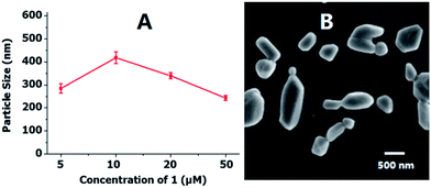
It is well known that the size-distribution and shape of lipoplexes are essential factors for determining their endocytosis pathway, intracellular trafficking and localization behaviors.8 Dynamic light scattering (DLS) was used to characterize the particle size and size distribution of the liposome 1/pDNA aggregates in ultrapure water at different concentrations. As depicted in Fig. 2A, the liposome 1 was able to condense DNA into nano-sized particles (ca. 250 to 450 nm) at concentrations ranging from 5 to 50 μM, which was larger than those of what we reported previously,7b likely due to the presence of the relatively large cholic acid. The size of the nanoparticles was first increased and then decreased with increases in the concentration of liposome 1 within the range, which is consistent with literature reports.9 | ||
| Fig. 2 (A) The effective diameter of liposome 1/DNA (9 μg mL−1) complexes obtained at different concentrations by DLS; (B) SEM images of 1/DNA complexes at concentration of 20 μM (N/P = 7.2). | ||
The morphology of the lipoplexes was further characterized by scanning electron microscope (SEM) at a concentration of 20 μM (N/P = 7.2) in ultrapure water. As shown in Fig. 2B, liposome 1 and DNA could form rhombic nanoparticles with an average particle size of 200–400 nm, which was suitable for cellular endocytosis and subsequent gene transfection.3c,d The results once again indicated that liposome 1 may effectively bind with pDNA and form nano-scale complexes in ultrapure water.
Cytotoxicity assay
As potential gene delivery materials, the cytotoxicity of this carrier is a key index to evaluate its potential for biomedical applications. Thus the cytotoxicity of the cationic lipoplex of 1 was examined at different concentrations in cancer cells (A549, HeLa, HepG2 and U2OS) and normal cells (CCC-HPF-1) using an MTT assay, and compared against lipofectamine 2000. As shown in Fig. 3, the percentages of viable cells when liposome 1 was incubated were generally higher than those involving lipofectamine 2000. | ||
Fig. 3 Cytotoxicity of liposome 1/DOPE (1![[thin space (1/6-em)]](https://www.rsc.org/images/entities/char_2009.gif) : :![[thin space (1/6-em)]](https://www.rsc.org/images/entities/char_2009.gif) 2) with DNA (9 μg mL−1) at different concentrations toward different cell lines, with lipofectamine 2000 (10 μg mL−1) as the control. 2) with DNA (9 μg mL−1) at different concentrations toward different cell lines, with lipofectamine 2000 (10 μg mL−1) as the control. | ||
Gene delivery process study
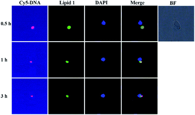
To directly visualize the process of DNA cellular uptake, translocation and release from the lipoplex formed by compound 1, DOPE, and Cy5-labeled dsDNA, time-dependent fluorescence microscopy assay was carried out in single cells (Fig. 4). Bright-field images showed that the cell retained its normal morphology after being incubated with the complex. When the lipoplex of 1 was cultured for 0.5 h at 37 °C, DNA was successfully delivered into the cell. After 1 h, DNA seemed to have surrounded the nucleus. With the incubation time increased to 3 h, DNA was gradually released from the complex and entered into the nucleus. To the best of our knowledge, this is the first time that a gene uptake process was clearly demonstrated in a single cell by a small organic molecule based non-viral gene vector.Cellular uptake mechanism study
The performance of non-viral gene delivery vectors is closely related to their intracellular kinetics, such as cellular uptake level, internalization pathway, and endosomal escape mechanism.10 It is well known that energy-dependent endocytosis is temperature-dependent and can be completely blocked at low temperature (4 °C).11 Chemical inhibitors such as chlorpromazine, cytochalasin D, genistein and nocodazole may inhibit the cellular uptake pathways of clathrin, macropinocytosis, caveolae, and microtubule mediated endocytosis, respectively.12 Thus we performed cellular uptake studies under various conditions. As shown in Fig. 1S,† the cellular uptake level was only inhibited by 25% at 4 °C, suggesting that the majority of DNA entered the cells via an energy-independent and non-endocytosis pathway. For the minor endocytosis of DNA, they were internalized mainly via macropinocytosis or caveolae because the cellular uptake level was reduced by cytochalasin D or genistein by ca. 10%, respectively. Furthermore, chlorpromazine did not inhibit the uptake at all, suggesting that the cellular uptake does not proceed via a clathrin-mediated endocytosis.Targeting ability of liposome 1
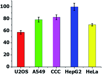
To evaluate the cancer cell targeting ability of liposome 1, flow cytometry was carried out. After incubation of the lipoplexes with HepG2, A549, HeLa, U2OS and CCC-HPF-1 cells for 4 h, the percentage of cells positive for Cy5-labeled pDNA was calculated. As shown in Fig. 5, liposome 1 exhibited excellent cellular uptake in HepG2 cells, and the percentage of uptaken cells was found to be close to 100%. Subsequently, the intracellular distribution of Cy5-labeled DNA (red, 9 μg mL−1) and liposome 1 (green) was detailed by CLSM (Fig. 2S†), and the nuclei were stained with DAPI (blue). The results have shown that almost all of the HepG2 cells were stained by compound 1 (green) and DNA (red), and considerable amounts of DNA (red) were delivered to the perinuclear region of HepG2 cells, which clearly indicated that liposome 1 had a higher cell selectivity toward HepG2 cells than other cell lines, although the selectivity is not excellent. It's worth noting that virtually none of the Cy5-labelled DNA was transferred into the normal cells (CCC-HPF-1). | ||
| Fig. 5 Cellular uptake of 1/DNA complexes (30 μM) toward various cell lines quantified by flow cytometry analysis. | ||
In vitro gene transfection
Although the HepG2 cell line is very important for the study of human liver diseases, metabolism, tumor progression, and toxicity of xenobiotics, it seems more resistant to lipid-mediated transfection than other cell lines.13 The in vitro gene transfection efficiency of liposome 1 was initially tested in HepG2 cells at different concentrations of 1 by using pGL-3 plasmid as a luciferase reporter gene. As shown in Fig. 6A, the liposome of 1 was able to deliver plasmid DNA successfully into HepG2 cells for efficient transfection. The best transfection efficiency of the liposome was obtained at 30 μM (N/P = 10.8) of 1, which was 1.6 times higher than that of commercially available lipofectamine 2000. The mole ratio of lipid 1 against DOPE was also varied (lipid/DOPE ratio of 1![[thin space (1/6-em)]](https://www.rsc.org/images/entities/char_2009.gif) :
:![[thin space (1/6-em)]](https://www.rsc.org/images/entities/char_2009.gif) 1, 1
1, 1![[thin space (1/6-em)]](https://www.rsc.org/images/entities/char_2009.gif) :
:![[thin space (1/6-em)]](https://www.rsc.org/images/entities/char_2009.gif) 2, 1
2, 1![[thin space (1/6-em)]](https://www.rsc.org/images/entities/char_2009.gif) :
:![[thin space (1/6-em)]](https://www.rsc.org/images/entities/char_2009.gif) 3, and 1
3, and 1![[thin space (1/6-em)]](https://www.rsc.org/images/entities/char_2009.gif) :
:![[thin space (1/6-em)]](https://www.rsc.org/images/entities/char_2009.gif) 4) at 30 μM, and the results showed that the lipid 1/DOPE ratio of 1
4) at 30 μM, and the results showed that the lipid 1/DOPE ratio of 1![[thin space (1/6-em)]](https://www.rsc.org/images/entities/char_2009.gif) :
:![[thin space (1/6-em)]](https://www.rsc.org/images/entities/char_2009.gif) 2 (Fig. 3S†) was found to be the best combination for efficient transfection. At the concentration of 30 μM (N/P = 10.8), liposome 1 was applied to the transfection toward other cell lines to further investigate their gene delivery efficiency under the optimized mole ratio of 1
2 (Fig. 3S†) was found to be the best combination for efficient transfection. At the concentration of 30 μM (N/P = 10.8), liposome 1 was applied to the transfection toward other cell lines to further investigate their gene delivery efficiency under the optimized mole ratio of 1![[thin space (1/6-em)]](https://www.rsc.org/images/entities/char_2009.gif) :
:![[thin space (1/6-em)]](https://www.rsc.org/images/entities/char_2009.gif) 2 (1/DOPE). As shown in Fig. 6B, liposome 1 (N/P = 10.8) gave higher transfection efficiency than lipofectamine 2000 in all of the cell lines tested, especially in U2OS cells, which was up to 4 times higher than that of lipofectamine 2000. From the performance of the transfection efficiency shown in Fig. 6B, liposome 1 gave similar results for different cells when compared to those of lipofectamine 2000. Considering the non-targeting property of lipofectamine 2000, the differences can be attributed to the complicated factors affecting the transfection efficiency, which include not only the cellular uptake process, but also the release of DNA from the complex, and the nuclear uptake of DNA.
2 (1/DOPE). As shown in Fig. 6B, liposome 1 (N/P = 10.8) gave higher transfection efficiency than lipofectamine 2000 in all of the cell lines tested, especially in U2OS cells, which was up to 4 times higher than that of lipofectamine 2000. From the performance of the transfection efficiency shown in Fig. 6B, liposome 1 gave similar results for different cells when compared to those of lipofectamine 2000. Considering the non-targeting property of lipofectamine 2000, the differences can be attributed to the complicated factors affecting the transfection efficiency, which include not only the cellular uptake process, but also the release of DNA from the complex, and the nuclear uptake of DNA.
Conclusions
In conclusion, a macrocyclic polyamine lipid containing both cholic acid and naphthalimide moieties, 1, was synthesized and successfully applied as a non-viral gene vector for cellular targeting and in situ monitoring of DNA delivery at a single-cell level. The liposome of 1 showed excellent DNA binding capabilities, low cytotoxicity and outstanding targeting ability toward HepG2 cell lines. In comparison with lipofectamine 2000, 1 gave 4 times higher transfection efficiency in U2OS cells. More importantly, it is the first example of a non-viral gene vector that can facilitate the monitoring of cellular DNA uptake, transportation and release via non-invasive in situ fluorescence imaging in a single cell. These findings may significantly improve our understanding of gene transfection mechanisms and provide important insights for the development of other non-viral gene vectors for targeted DNA delivery.Acknowledgements
The authors gratefully acknowledge the financial assistance provided by the Nature Science Foundation of China (21372032 and 91227109), the Fundamental Research Funds for the Central Universities, Beijing Municipal Commission of Education, the Program for Changjiang Scholars and Innovative Research Team in University, as well as Macau Science and Technology Development Fund (FDCT/020/2015/A1).Notes and references
- (a) I. Guijarro-Munoz, M. Compte, L. Alvarez-Vallina and L. Sanz, Curr. Gene Ther., 2013, 13, 282 CrossRef CAS PubMed; (b) P. Harish, A. Malerba, G. Dickson and H. Bachtarzi, Hum. Gene Ther., 2015, 26, 286 CrossRef CAS PubMed; (c) S. Viswam, J. Chithra and S. A. Surendran, Int. J. Pharm. Sci. Rev. Res., 2015, 31, 14 CAS; (d) I. M. Verma and N. Somia, Nature, 1997, 389, 239 CrossRef CAS PubMed.
- (a) N. Nayerossadat, T. Maedeh and P. Abas Ali, Adv. Biomed. Res., 2012, 1, 1 Search PubMed; (b) R. J. Samulski and N. Muzyczka, Annu. Rev. Virol., 2014, 1, 427 CrossRef PubMed; (c) J. Luo, Y. Luo, J. Sun, Y. Zhou, Y. Zhang and X. Yang, Cancer Lett., 2015, 356, 347 CrossRef CAS PubMed; (d) A. M. Gruntman and T. R. Flotte, Hum. Gene Ther: Methods., 2015, 26, 77 CrossRef CAS PubMed.
- (a) M. A. Mintzer and E. E. Simanek, Chem. Rev., 2009, 109, 259 CrossRef CAS PubMed; (b) U. Laechelt and E. Wagner, Chem. Rev., 2015, 115, 11043 CrossRef CAS PubMed; (c) Y. Shoji-Moskowitz, D. Asai, K. Kodama, Y. Katayama and H. Nakashima, Front. Med. Chem., 2010, 5, 81 CAS; (d) B. Draghici and M. A. Ilies, J. Med. Chem., 2015, 58, 4091 CrossRef CAS PubMed; (e) L. Collins, Methods Mol. Biol., 2006, 333, 201 CAS; (f) C. H. Jones, C.-K. Chen, A. Ravikrishnan, S. Rane and B. A. Pfeifer, Mol. Pharmaceutics, 2013, 10, 4082 CrossRef CAS PubMed; (g) A. Tschiche, S. Malhotra and R. Haag, Nanomedicine, 2014, 9, 667 CrossRef CAS PubMed.
- (a) T. G. Park, J. H. Jeong and S. W. Kim, Adv. Drug Delivery Rev., 2006, 58, 467 CrossRef CAS PubMed; (b) A. Basu, K. R. Kunduru, E. Abtew and A. J. Domb, Bioconjugate Chem., 2015, 26, 1396 CrossRef CAS PubMed; (c) D. Zhi, S. Zhang, S. Cui, Y. Zhao, Y. Wang and D. Zhao, Bioconjugate Chem., 2013, 24, 487 CrossRef CAS PubMed; (d) J. Yang, Q. Zhang, H. Chang and Y. Cheng, Chem. Rev., 2015, 115, 5274 CrossRef CAS PubMed; (e) M. Jaeger, S. Schubert, S. Ochrimenko, D. Fischer and U. S. Schubert, Chem. Soc. Rev., 2012, 41, 4755 RSC; (f) C. O. Mellet, J. M. G. Fernandez and J. M. Benito, Chem. Soc. Rev., 2011, 40, 1586 RSC; (g) G.-Y. Li, R.-L. Guan, L.-N. Ji and H. Chao, Coord. Chem. Rev., 2014, 281, 100 CrossRef CAS; (h) C. Tros de Ilarduya, Y. Sun and N. Duezguenes, Eur. J. Pharm. Sci., 2010, 40, 159 CrossRef CAS PubMed; (i) D. He and E. Wagner, Macromol. Biosci., 2015, 15, 600 CrossRef CAS PubMed; (j) C. R. Safinya, K. K. Ewert, R. N. Majzoub and C. Leal, New J. Chem., 2014, 38, 5164 RSC; (k) H. Tian, J. Chen and X. Chen, Small, 2013, 9, 2034 CrossRef CAS PubMed.
- (a) X. Xu, Y. Jian, Y. Li, X. Zhang, Z. Tu and Z. Gu, ACS Nano, 2014, 8, 9255 CrossRef CAS PubMed; (b) B. He, Y. Chu, M. Yin, K. Müllen, C. An and J. Shen, Adv. Mater., 2013, 25, 4580 CrossRef CAS PubMed; (c) M. Bai, X. Bai and L. Wang, Anal. Chem., 2014, 86, 11196 CrossRef CAS PubMed; (d) B. Yu, C. Ouyang, K. Qiu, J. Zhao, L. Ji and H. Chao, Chem.–Eur. J., 2015, 21, 3691 CrossRef CAS PubMed; (e) B. Yu, Y. Chen, C. Ouyang, H. Huang, L. Ji and H. Chao, Chem. Commun., 2013, 49, 810 RSC; (f) K. Qiu, B. Yu, H. Huang, P. Zhang, L. Ji and H. Chao, Dalton Trans., 2015, 44, 7058 RSC; (g) I. Roy, T. Y. Ohulchanskyy, D. J. Bharali, H. E. Pudavar, R. A. Mistretta, N. Kaur and P. N. Prasad, Proc. Natl. Acad. Sci. U. S. A., 2005, 102, 279 CrossRef CAS PubMed; (h) G. Wang, H. Yin, J. C. Yin Ng, L. Cai, J. Li, B. Z. Tang and B. Liu, Polym. Chem., 2013, 4, 5297 RSC; (i) Y. Su, L. Xu, S. Wang, Z. Wang, Y. Yang, Y. Chen and Y. Que, Sci. Rep., 2015, 5, 10708 CrossRef CAS PubMed.
- (a) W. Tao, X. Zeng, J. Zhang, H. Zhu, D. Chang, X. Zhang, Y. Gao, J. Tang, L. Huang and L. Mei, Biomater. Sci., 2014, 2, 1262 RSC; (b) M. H. Lee, J. H. Han, P. S. Kwon, S. Bhuniya, J. Y. Kim, J. L. Sessler, C. Kang and J. S. Kim, J. Am. Chem. Soc., 2012, 134, 1316 CrossRef CAS PubMed; (c) X. Ma, J. Jia, R. Cao, X. Wang and H. Fei, J. Am. Chem. Soc., 2014, 136, 17734 CrossRef CAS PubMed; (d) Z. Yang, J. H. Lee, H. M. Jeon, J. H. Han, N. Park, Y. He, H. Lee, K. S. Hong, C. Kang and J. S. Kim, J. Am. Chem. Soc., 2013, 135, 11657 CrossRef CAS PubMed.
- (a) H. Yan, Z.-F. Li, Z.-F. Guo, Z.-L. Lu, F. Wang and L.-Z. Wu, Bioorg. Med. Chem., 2012, 20, 801 CrossRef CAS PubMed; (b) Y.-G. Gao, Y.-D. Shi, Y. Zhang, J. Hu, Z.-L. Lu and L. He, Chem. Commun., 2015, 51, 16695 RSC; (c) P. Yue, Y. Zhang, Z.-F. Guo, A.-C. Cao, Z.-L. Lu and Y.-G. Zhai, Org. Biomol. Chem., 2015, 13, 4494 RSC; (d) H. Yan, P. Yue, Z. F. Li, Z. F. Guo and Z. L. Lu, Sci. China: Chem., 2014, 57, 296 CrossRef CAS.
- (a) B. Ruozi, M. Montanari, E. Vighi, G. Tosi, A. Tombesi, R. Battini, C. Restani, E. Leo, F. Forni and M. A. Vandelli, J. Liposome Res., 2009, 19, 241 CrossRef CAS PubMed; (b) S. Schneider and R. Süss, in Liposomes, ed. V. Weissig, Humana Press, 2010, vol. 606, p. 457 Search PubMed; (c) S. E. Gratton, P. A. Ropp, P. D. Pohlhaus, J. C. Luft, V. J. Madden, M. E. Napier and J. M. DeSimone, Proc. Natl. Acad. Sci. U. S. A., 2008, 105, 11613 CrossRef CAS PubMed.
- H.-J. Wang, Y.-H. Liu, J. Zhang, Y. Zhang, Y. Xia and X.-Q. Yu, Biomater. Sci., 2014, 2, 1460 RSC.
- (a) X. Gao and L. Huang, Gene Ther., 1995, 2, 710 CAS; (b) P. L. Felgner and G. M. Ringold, Nature, 1989, 337, 387 CrossRef CAS PubMed.
- (a) N. W. S. Kam, Z. Liu and H. Dai, Angew. Chem., 2006, 45, 577 CrossRef CAS PubMed; (b) Y. Jin, S. Wang, L. Tong and L. Du, Colloids Surf., B, 2015, 126, 257 CrossRef CAS PubMed; (c) K. Kostarelos, L. Lacerda, G. Pastorin, W. Wu, W. Sebastien, J. Luangsivilay, S. Godefroy, D. Pantarotto, J.-P. Briand, S. Muller, M. Prato and A. Bianco, Nat. Nanotechnol., 2007, 2, 108 CrossRef CAS PubMed.
- (a) Q.-F. Zhang, Q.-Y. Yu, Y. Geng, J. Zhang, W.-X. Wu, G. Wang, Z. Gu and X.-Q. Yu, ACS Appl. Mater. Interfaces, 2014, 6, 15733 CrossRef CAS PubMed; (b) Z. Xu, L. Chen, W. Gu, Y. Gao, L. Lin, Z. Zhang, Y. Xi and Y. Li, Biomaterials, 2009, 30, 226 CrossRef CAS PubMed.
- (a) J.-C. Yu, S. Zhu, P.-J. Feng, C.-G. Qian, J. Huang, M.-J. Sun and Q.-D. Shen, Chem. Commun., 2015, 51, 2976 RSC; (b) S. Yamano, J. Dai and A. Moursi, Mol. Biotechnol., 2010, 46, 287 CrossRef CAS PubMed.
- (a) H. Y. Kuchelmeister and C. Schmuck, Eur. J. Org. Chem., 2009, 2009, 4480 CrossRef; (b) Z.-F. Guo, H. Yan, Z.-F. Li and Z.-L. Lu, Org. Biomol. Chem., 2011, 9, 6788 RSC.
Footnote |
| † Electronic supplementary information (ESI) available: Details of synthesis and characterization of all new compounds and properties studies. See DOI: 10.1039/c6ra08935f |
| This journal is © The Royal Society of Chemistry 2016 |


