Magnetic hyperthermia-induced drug release from ureasil-PEO-γ-Fe2O3 nanocomposites

B. L. Caetano*ab, C. Guibertb, R. Finia, J. Fresnaisb, S. H. Pulcinellia, C. Ménagerb and C. V. Santillia
aInstituto de Química, UNESP, Rua Professor Francisco Degni, 55, 14800-900 Araraquara, SP, Brazil. E-mail: caetanobruno@yahoo.com.br
bSorbonne Universités, UPMC Univ Paris 06, CNRS, Laboratoire PHENIX, Case 51, 4 place Jussieu, F-75005 Paris, France

Received 29th March 2016 , Accepted 23rd June 2016

First published on 27th June 2016


Abstract

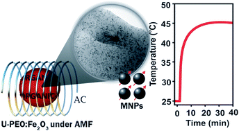
A multifunctional material suitable for cancer therapy, which combines stimuli-responsive properties for drug delivery and magnetic hyperthermia prepared by a one-pot sol–gel synthesis from the conjugation of ureasil cross-linked poly(ethylene oxide) (U-PEO) hybrid materials with superparamagnetic nanoparticles (γ-Fe2O3), is reported in this communication.


Organic–inorganic hybrid (OIH) materials have tremendous potential as a bridge between the organic, mineral, and biological universes, opening new frontiers for the development of systems and devices for human health care, with applications in the areas of dentistry, cosmetics, tissue engineering, therapeutic vectors, and drug delivery.1 Among the OIHs used as drug delivery vehicles, siloxane cross-linked organic macromers are an emerging option.2 The commercially available macromer poly(ethylene oxide) (PEO), otherwise known as poly(ethylene glycol) (PEG), is approved by the FDA for parenteral administration due to its low toxicity and biocompatibility.3 Ureasil-PEO (U-PEO) is a transparent and rubber-like OIH composed of PEO macromers of variable lengths, grafted at both ends to siloxane cross-linked nodes that are conjugated by means of urea bridges. U-PEO presents good mechanical and thermal stability2 and exhibits hydrophilic characteristics, with water uptake resulting in swelling similar to that of hydrogels. Due to these properties, U-PEO has a high capacity to dissolve ionic species and polar molecules, and has been tested as an absorbent for water treatment4 and as a drug delivery system. It offers efficient inclusion of high concentrations of different active molecules such as sodium diclofenac and cisplatin, for which uptake rates of 17 and 5 wt% have been reported, respectively.2,5 Furthermore, U-PEO hybrids prepared by the sol–gel route possess good film-forming properties (skin bioadhesion, workability, and water permeability) and have potential applications in pharmaceutical formulations for transdermal (patches) and implantable (soft tissue) drug carriers.6 In recent work, the PEO polymer has been used in drug targeting and in stimuli-responsive drug delivery systems.7,8 However, there are only a few reports that have focused on the use of U-PEO hybrids as stimuli-responsive systems, and no work concerning drug delivery triggered by an external magnetic field.9

Magnetic nanoparticles (MNP) based on iron oxide (maghemite γ-Fe2O3 or magnetite Fe3O4) are another class of materials highly suitable for use in biomedical applications, due to their biocompatibility and superparamagnetic properties.10,11 Superparamagnetism is crucial for many applications in biomedicine, because when the external magnetic field is removed, superparamagnetic NP do not retain any magnetism. In addition, these MNP can be used as energy transfer mediators by transforming an external magnetic field into heat.12 When MNP are exposed to an alternating magnetic field (AMF), the magnetic energy is dissipated as thermal energy by means of two distinct mechanisms, Néel and Brown relaxation, as the particle returns to its equilibrium state.13 Superparamagnetic NP can therefore be used as a source of hyperthermia to cause the death of cancer cells.14 They can also trigger drug release following application of an AMF, and can provide magnetic guidance of the drug delivery carrier to a specific target.15 This combination of thermo- and chemotherapy can increase efficiency and reduce side effects during the treatment of radiosensitive and radioresistant tumors.16

There are few reports in the literature concerning ureasil-polyether hybrid systems containing MNP. However, the one-pot sol–gel synthesis method has been used to grow ferrihydrite NP in a U-PEO hybrid matrix, resulting in a non-superparamagnetic material.17

In this communication, we propose implantable devices that combine the drug delivery and stimuli-responsive properties of U-PEO with the superparamagnetic properties of γ-Fe2O3 NP, in order to obtain a stimuli-responsive material. To this end, we prepared an innovative multifunctional U-PEO-γ-Fe2O3 nanocomposite loaded with sodium diclofenac (SDCF) as a model drug, of which we were able to control through an external AMF the temperature at an optimum value for hyperthermia, as well as the drug release rate (Scheme 1).


image file: c6ra08127d-s1.tif
Scheme 1 Schematic representation of the synthetic process of UPEO:Fe2O3(x)SDCF nanocomposites. Details presented in the ESI.

The samples were obtained by hydrolysis and condensation of the –Si(OEt)3 groups present in the hybrid precursor (EtO)3Si(CH2)3NHC([double bond, length as m-dash]O)NHCHCH3CH2–(PEO)–CH2CH3CH NH(O[double bond, length as m-dash])CNH(CH2)Si(OEt)3 with PEO1900.18 The hydrolysis was initiated by adding HNO3 solution, followed by addition of γ-Fe2O3 NP dispersed in water at low pH. The MNP were size-sorted at the end of the co-precipitation process in order to retrieve the larger nanoparticles that are more effective in hyperthermia.19,20 In the case of the samples loaded with the model drug, 3 wt% of SDCF was added to 0.5 mL of hybrid precursor, while 5.0, 0.5, 0.05, and 0.005 wt% of γ-Fe2O3 were used in the samples containing MNP. In this work, the ureasil-poly(ethylene oxide) hybrids obtained from the precursor are labelled U-PEO, the OIH nanocomposites containing γ-Fe2O3 are labelled U-PEO:Fe2O3(x), with x being the amount (wt%) of γ-Fe2O3, and the samples loaded with γ-Fe2O3 and SDCF are denoted U-PEO:Fe2O3(x)SDCF. The experimental procedures used for synthesis and characterisation are outlined in the ESI. The γ-Fe2O3 MNP with mean diameter (do) of 12.7 nm and polydispersity (σ) of 4.4 nm are presented in Fig. 1a and b. Cryo-TEM images of the nanocomposite showed the absence of extensive aggregation and a fairly uniform distribution of MNP (do = 12.0 nm, σ = 5.9 nm, Fig. 1c and d). Using Langevin formalism weighted with a log-normal distribution to fit the magnetisation curve (Fig. 1e), do = 10.9 nm and σ = 0.33 were obtained for the MNP in both the aqueous colloid and the OIH matrix. The do value was in good agreement with the average size obtained by TEM, confirming the satisfactory dispersion of the MNP. It is important to note that the good dispersion of bare γ-Fe2O3 MNP was achieved by acid peptisation (pH ∼ 1.7) of the mother suspension. In fact, the sol–gel synthesis of U-PEO:Fe2O3 should be performed at pH below 2.4 in order to maintain good dispersion of the MNP in the OIH matrix. This behaviour can be clearly seen from comparison of the SAXS curves (Fig. 1f) for the nanocomposites prepared at pH ∼ 1.7 (blue line) and pH ∼ 3.7 (dashed line).


image file: c6ra08127d-f1.tif
Fig. 1 (a) TEM image of γ-Fe2O3 colloid; (b) Gaussian size distribution determined from TEM images of γ-Fe2O3 colloid; (c) cryo-TEM image of the U-PEO:Fe2O3(5) nanocomposite, prepared at pH ∼ 1.7; (d) Gaussian size distribution of the U-PEO:Fe2O3(5) nanocomposite; (e) magnetisation curves of the 3.2 wt% γ-Fe2O3 colloid and the U-PEO:Fe2O3(5) nanocomposite, both fitted with the same Langevin function (continuous line); (f) SAXS curves of the U-PEO matrix, the aqueous colloid (3.2 wt% γ-Fe2O3, pH ∼ 1.7), and nanocomposites without (U-PEO:Fe2O3(5)) and with SDCF (U-PEO:Fe2O3(5)SDCF), prepared at pH 1.7 (continuous lines) and pH 3.7 (dashed line).

For the nanocomposite prepared at pH ∼ 3.7 the SAXS curve at low q-range revealed a linear dependence, with a slope value of −1.9, evidencing the growth of fractal aggregates.21 A weak and broad peak centred at ∼0.34 nm−1 was evidence of interaction between the MNP, due to the coexistence of dense aggregates of γ-Fe2O3. In contrast, the plateau at low q-range in the SAXS curve for the nanocomposite prepared at pH ∼ 1.7 was characteristic of a non-interacting set of nanoparticles, reflecting good dispersion of the MNP within the matrix. The presence of well-dispersed MNP in the OIH matrix is essential for hyperthermia applications, because aggregates act to decrease the heating capacity of the MNP.22

After incorporation of the γ-Fe2O3 NP in the hybrid matrix, the characteristic correlation peak of regularly spaced ureasil cross-linking nodes was no longer observed.18 This was due to the greater electronic contrast between γ-Fe2O3 and the U-PEO. It was also observed that the curves for samples containing the MNP prepared at pH ∼ 1.7 presented similar shapes, indicating that the dispersions of MNP in the colloidal suspension and in the U-PEO hybrid with or without SDCF were comparable. The SAXS curves of the γ-Fe2O3 colloidal suspension presented a Gaussian decay in the high q region and a plateau at low q, also known as the Guinier region.

This latter feature is characteristic of scattering by a dilute set of non-interacting particles. The same behaviour could be seen for the U-PEO:Fe2O3 prepared at pH 1.7. At high q, the curves displayed an asymptotic linear trend with a slope of ∼−4, in agreement with Porod's law (I(q) ∝ q−4) and implying that the two-electron density contrast model was satisfied. It also indicated that the interface between the MNP and the matrix was sharp and well-defined, with an absence of heterogeneities inside the MNP, and that each nanoparticle acted as a single object. Under such conditions, the scattering intensity in the low q region (qRg ≈ 1) can be approximated by the Guinier law, enabling determination of the average gyration radius, Rg, of scattering by isolated MNP.23 The average spherical size calculated from Rg was ∼10.5 nm for both the γ-Fe2O3 aqueous colloid and the nanocomposite. These results were in agreement with the TEM images and confirmed the very good dispersion of the MNP in the U-PEO matrix. Magnetic hyperthermia was performed with the γ-Fe2O3 colloids and the U-PEO:Fe2O3 nanocomposite, using the same experimental procedure.

An AMF was applied to samples kept at initial temperature of 25 °C, using maximum amplitude of 14.9 kA m−1 and a frequency of 420 kHz. Fig. 2 shows images of the samples, together with the curves of temperature increase versus the γ-Fe2O3 MNP concentration, with each curve corresponding to a given AMF exposure time.


image file: c6ra08127d-f2.tif
Fig. 2 Isochronous temperature increases after application of an AMF of 14.9 kA m−1 and 420 kHz: (a) aqueous colloid containing different amounts of MNP (3.2, 0.32, 0.032, and 0.0032 wt%); (b) U-PEO:Fe2O3(x) nanocomposite containing different amounts of MNP (x = 5.0, 0.5, 0.05, and 0.005 wt%).

Irrespective of the nature of the sample, the isochronous curves showed a more intense increase of temperature as the concentration of γ-Fe2O3 increased, with this effect being lower for the U-PEO:Fe2O3 nanocomposites. For the aqueous colloid, the onset of heating occurred at a γ-Fe2O3 concentration of ∼0.032 wt%, while for the nanocomposites, the onset of heating occurred at an MNP loading that was 15 times higher (0.5 wt% of γ-Fe2O3).

Fig. 2a and b correspond to different concentrations of γ-Fe2O3 in aqueous colloid (3.2, 0.32, 0.032, and 0.0032 wt%) and in OIH nanocomposite (5.0, 0.5, 0.05, and 0.005 wt%), respectively. The data were obtained from monitoring of the temperature during the application of AMF (ESI: Fig. S1a and S1b).

As shown in Fig. S1a, the colloidal suspension with 3.2 wt% of γ-Fe2O3 exhibited a prompt and continuous temperature increase, reaching 80 °C after 4 min of AMF exposure. Accordingly, for this colloid the isochronous curve displays only the points corresponding to 2 and 4 min. For the ten-fold less concentrated colloid, the temperature increased continuously from 25 °C to an asymptotic value of 53 °C after 50 min of AMF exposure. In the case of the U-PEO:Fe2O3(5) nanocomposite, there was a prompt increase from 25 °C up to a stable plateau at 44 °C. For the ten-fold less concentrated nanocomposite (U-PEO:Fe2O3(0.5)), a constant temperature of 32 °C was achieved after 25 min of AMF exposure. The first 50 min of AMF exposure is shown in Fig. S1b. Experiments were also performed with the same samples at a frequency of 280 kHz (ESI, Fig. S1c and S1d), and smaller ΔT values were observed, as expected.

The temperature plateau indicated that the U-PEO:Fe2O3(5) and U-PEO:Fe2O3(0.5) nanocomposites were able to act as hyperthermia mediators and could be used in cancer therapy. The U-PEO:Fe2O3 nanocomposite prepared without SDCF exhibited a similar temperature increase under an AMF (data not shown). Temperatures between 40 °C and 45 °C lead to inactivation of normal cellular processes, while extensive necrosis occurs above 45 °C.21 In the clinical application of magnetic hyperthermia, local overheating can be avoided by selecting MNP systems with low maximum achievable temperatures, while maintaining the magnetisation that enables efficient local delivery.24 For human applications, where the initial temperature is ∼37 °C, an increase of 4–7 °C, as obtained using U-PEO:Fe2O3(0.5), is suitable for use in cancer therapy.

It is important to highlight that the plateau temperature was dependent on factors including the MNP concentration, MNP–matrix interactions, and the rate of heat transfer to the sample holder tube. In contrast, the initial linear rise in local temperature, dT/dtmax (see Fig. S1 and Table S1), was independent of both heat transfer to the surroundings and sample geometry, and was used to derive the specific loss power (SLP) of the nanocomposites. The SLP (W g−1) is an intrinsic property defined as the amount of energy converted into heat per time and per mass of MNP (mMNP): SLP = C × ms/mMNP × dT/dtmax where C and ms are the specific heat capacity and the mass of the sample, respectively (see values in Table S1).10,14 The SLP was only calculated for the samples that showed appreciable temperature increases, with values of 97, 106, and 110 W g−1 for the 3.2, 0.32, and 0.032 wt% γ-Fe2O3 aqueous colloids, respectively. The minor differences in SLP indicated that the degree of dispersion of the MNP was nearly invariant. In the case of the U-PEO:Fe2O3(0.5) and U-PEO:Fe2O3(5) nanocomposites, an SLP value of 13.3 W g−1 was measured, that is approximately eight-fold smaller than for the aqueous colloids. In the case of superparamagnetic particles only Néel relaxation (rotation of the magnetic moment inside the particle) is involved for heat generation. As a consequence, such superparamagnetic particles are not sensitive to the viscosity of the medium. In the literature, such decrease of the SLP is often explained by the formation of aggregates or strong interactions between the particles.25 However, in our case, SAXS experiments prove that the particles are perfectly dispersed in the matrix. The low SLP value is probably due to the fact that we are not in adiabatic conditions, the measurement of the temperature itself is correct but the temperature elevation rate may be disturbed by temperature gradients inside the U-PEO:Fe2O3 nanocomposite.

The release of SDCF from U-PEO:Fe2O3(5):SDCF was significantly enhanced by heating either in a water bath or using an AMF (Fig. 3). The release of SDCF into pH 7.2 phosphate buffer solution was performed during 8 h, at different isothermal temperatures (0, 25, 37, and 50 °C), and one additional experiment was started at 37 °C under AMF (14.9 kA m−1, 420 kHz). The AMF was applied during ∼45 min and then turned off when the temperature reached 57 °C; the temperature then returned to 37 °C after ∼20 min. This cycle was repeated 8 times (see Fig. S4). Irrespective of the experiment time, the amount of SDCF released followed the order: 0 °C < 25 °C < 37 °C < 50 °C ≅ AMF. This sequence can be explained by the progressive decrease of PEO crystallinity due to the relaxation induced by water uptake and the fusion that occurs near 38 °C (Fig. S5). The former process, which depends on the diffusion of water through the U-PEO matrix, is slower at low temperatures, resulting in less SDCF release. Similar release profiles were observed in experiments performed at 50 °C in a temperature-controlled water bath and under AMF, started at 37 °C. After the first hours, the cumulative drug release under AMF reached twice the value measured at 37 °C. The drug release rate was also significantly higher when the nanocomposites were submitted to an AMF. It could therefore be concluded that the AMF increased the diffusion rate of the drug due to fusion of crystalline domains of the U-PEO matrix following local heating. Taken together, the results supported the use of this system as a combined drug release carrier and hyperthermia source, because fusion of the semi-crystalline U-PEO resulted in a stimuli-responsive effect in the temperature range of biomedical applications.


image file: c6ra08127d-f3.tif
Fig. 3 SDCF release from U-PEO:Fe2O3(5):SDCF at 0, 25, 37, and 50 °C, and under an external AMF started at 37 °C.

Many of the magnetic systems used in biomedical applications rely on the key role played by the surface properties of the nanoparticles, especially in effective interfacing with biological systems, ensuring biocompatibility and specific localisation in proteins, cells, and tissues.26 These systems often use complicated syntheses for modify the surfaces of nanoparticles.27 In the case of the present nanocomposites, the bare MNP were easily inserted in the bulk OIH using the one-pot sol–gel synthesis, and good dispersion was achieved by controlling the pH. Furthermore, the superparamagnetic properties of the nanocomposites were quite stable, because the MNP were not leached and did not aggregate under the effects of the biological medium and the magnetic field. Another advantage of U-PEO is the ability to incorporate high amounts of drugs with different molecular natures, which can be used for specific chemotherapy,2,5 especially when a high dose is required in the first 2 h, after which the nanocomposite can be used continuously as a hyperthermia source. Another important consideration for in vivo applications is that the material exhibits no cytotoxic effects.6

In summary, this work describes the one-pot sol–gel synthesis of a nanocomposite formed by the conjugation of a semi-crystalline ureasil-poly(ethylene oxide) matrix loaded with a model drug (SDCF) and well dispersed γ-Fe2O3 superparamagnetic nanoparticles for remotely triggered therapy. Active control of the drug release rate was achieved using localised hyperthermia induced by exposure to an external AMF. The exposure caused melting of the crystalline PEO at around 38 °C, which favoured the liquid-like diffusion of drug molecules throughout the hybrid network, hence increasing the release rate. These nanocomposites show great potential for use with other drug molecules whose release can be triggered using an external magnetic field. Another very important finding was that the hybrid samples could act as mediators of hyperthermia and could therefore be used in dual cancer therapy, with hyperthermia in the optimal lower-temperature window (between 42 and 45 °C) in order to avoid thermal damage of normal cells, combined with control of the drug release profile.

Acknowledgements

This work was partially supported by the Brazilian National Council for Scientific and Technological Development (CNPq, 482176/2013-0), the Foundation for Research Support of the State of São Paulo (FAPESP, 2011/19253-0 and 2013/10384-0), and the CAPES/COFECUB-767/13 cooperation programs. We acknowledge SOLEIL for the provision of synchrotron radiation facilities, and we would like to thank Dr Javier Perez for assistance in using the beamline SWING.

Notes and references

  1. C. Sanchez, B. Julian, P. Belleville and M. Popall, J. Mater. Chem., 2005, 15, 3559 RSC .
  2. C. V. Santilli, L. A. Chiavacci, L. Lopes, S. H. Pulcinelli and A. G. Oliveira, Chem. Mater., 2009, 21, 463 CrossRef CAS .
  3. D. H. Na and P. P. DeLuca, Pharm. Res., 2005, 22, 736 CrossRef CAS PubMed .
  4. V. Bekiari and P. Lianos, Chem. Mater., 2006, 18, 4142 CrossRef CAS .
  5. E. F. Molina, S. H. Pulcinelli, C. V. Santilli, S. p. Blanchandin and V. r. Briois, J. Phys. Chem. B, 2010, 114, 3461 CrossRef CAS PubMed .
  6. L. K. Souza, C. H. Bruno, L. Lopes, S. H. Pulcinelli, C. V. Santilli and L. A. Chiavacci, Colloids Surf., B, 2013, 101, 156 CrossRef CAS PubMed .
  7. X.-M. Zhang, K. Guo, L.-H. Li, S. Zhang and B.-J. Li, J. Mater. Chem. B, 2015, 3, 6026 RSC .
  8. A. Bin Imran, K. Esaki, H. Gotoh, T. Seki, K. Ito, Y. Sakai and Y. Takeoka, Nat. Commun., 2014, 5, 1 Search PubMed .
  9. E. F. Molina, R. L. T. Parreira, E. H. De Faria, H. W. P. de Carvalho, G. F. Caramori, D. F. Coimbra, E. J. Nassar and K. J. Ciuffi, Langmuir, 2014, 30, 3857 CrossRef CAS PubMed .
  10. N. Lee, D. Yoo, D. Ling, M. H. Cho, T. Hyeon and J. Cheon, Chem. Rev., 2015, 115, 10637 CrossRef CAS PubMed .
  11. A. K. Gupta and M. Gupta, Biomaterials, 2005, 26, 3995 CrossRef CAS PubMed .
  12. R. Hergt, S. Dutz, R. Müller and M. Zeisberger, J. Phys.: Condens. Matter, 2006, 18, 2019 CrossRef .
  13. J.-P. Fortin, C. Wilhelm, J. Servais, C. Ménager, J.-C. Bacri and F. Gazeau, J. Am. Chem. Soc., 2007, 129, 2628 CrossRef CAS PubMed .
  14. R. E. Rosensweig, J. Magn. Magn. Mater., 2002, 252, 370 CrossRef CAS .
  15. V. S. Kalambur, B. Han, B. E. Hammer, T. W. Shield and J. C. Bischof, Nanotechnology, 2005, 16, 1221 CrossRef CAS .
  16. J.-P. Fortin-Ripoche, M. S. Martina, F. Gazeau, C. Ménager, C. Wilhelm, J.-C. Bacri, S. Lesieur and O. Clément, Radiology, 2006, 239, 415 CrossRef PubMed .
  17. N. J. O. Silva, V. S. Amaral, V. de Zea Bermudez, S. C. Nunes, D. Ostrovskii, J. Rocha and L. D. Carlos, J. Mater. Chem., 2005, 15, 484 RSC .
  18. K. Dahmouche, C. V. Santilli, S. H. Pulcinelli and A. F. Craievich, J. Phys. Chem. B, 1999, 103, 4937 CrossRef CAS .
  19. R. Massart, IEEE Trans. Magn., 1981, 17, 1247 CrossRef .
  20. S. Lefebure, E. Dubois, V. Cabuil, S. Neveu and R. Massart, J. Mater. Res., 1998, 13, 2975 CrossRef CAS .
  21. T. A. Witten and L. M. Sander, Phys. Rev. Lett., 1981, 47, 1400 CrossRef CAS .
  22. R. Di Corato, A. Espinosa, L. Lartigue, M. Tharaud, S. Chat, T. Pellegrino, C. Ménager, F. Gazeau and C. Wilhelm, Biomaterials, 2014, 35, 6400 CrossRef CAS PubMed .
  23. A. F. Craievich, in Handbook of Sol-Gel Science and Technology, ed. S. Sakka, Springer US, Norwell, MA, 2005, vol. 2, ch. 8, p. 161 Search PubMed .
  24. A. Jordan, R. Scholz, P. Wust, H. Fähling and F. Roland, J. Magn. Magn. Mater., 1999, 201, 413 CrossRef CAS .
  25. C. Guibert, V. Dupuis, V. Peyre and J. Fresnais, J. Phys. Chem. C, 2015, 119, 28148 CAS .
  26. C. C. Berry, S. Wells, S. Charles, G. Aitchison and A. S. G. Curtis, Biomaterials, 2004, 25, 5405 CrossRef CAS PubMed .
  27. J.-H. Lee, K.-J. Chen, S.-H. Noh, M. A. Garcia, H. Wang, W.-Y. Lin, H. Jeong, B. J. Kong, D. B. Stout, J. Cheon and H.-R. Tseng, Angew. Chem., 2013, 52, 4384 CrossRef CAS PubMed .

Footnote

Electronic supplementary information (ESI) available. See DOI: 10.1039/c6ra08127d

This journal is © The Royal Society of Chemistry 2016
Click here to see how this site uses Cookies. View our privacy policy here.