DOI:
10.1039/C6RA08090A
(Paper)
RSC Adv., 2016,
6, 46696-46701
In situ growth of biocidal AgCl crystals in the top layer of asymmetric polytriazole membranes†
Received
29th March 2016
, Accepted 5th May 2016
First published on 6th May 2016
Abstract
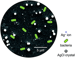
Scalable fabrication strategies to concentrate biocidal materials in only the surface of membranes are highly desirable. In this letter, tight-UF polytriazole membranes with a high concentration of biocide silver chloride (AgCl) crystals dispersed in only their top layer are presented. They were made following a simple dual-bath process that is compatible with current commercial membrane casting facilities. These membranes can achieve a 150-fold increase in their antimicrobial character compared to their silver-free counterpart. Moreover, fine-tuning of their properties is straightforward. A change in the silver concentration in one of the baths is sufficient to tune the permeance, molecular weight cut-off (MWCO) and silver loading of the final membrane.
Introduction
Ultrafiltration (UF), a promising technology for producing safe water in developing countries, can disinfect water and remove its turbidity at relatively low pressures. UF systems have been successfully used in short-term humanitarian and emergency disaster relief missions. However, its wider implementation faces several challenges.1 The principal obstacle is membrane fouling,2,3 and among all the fouling types, biofouling—the “Achilles heel” of membrane processes—is the most challenging one to control.4 Only a small number of microorganisms that manage to survive the pretreatment processes are enough to foul a membrane. They will feed on biodegradable substances present in the water, multiply and colonize the surface of the membrane in the form of a biofilm. This additional layer produces an extra resistance to mass transfer that causes a substantial increase in operational and maintenance costs.5 Effective and efficient methods to control and minimize biofouling are needed.
A well-known and powerful strategy to reduce membrane biofouling is the incorporation of biocidal nano- and micromaterials such as metal/metal oxide nanoparticles (e.g., TiO2 nanoparticles,6 ZnO nanoparticles,7 copper nanoparticles,8 and silver nanoparticles9–18), and carbon-based materials (e.g., graphene oxide nanosheets,19–21 and carbon nanotubes22) into the membrane. The most common and straightforward approach for making such loaded membranes is to disperse the desired material in the membrane casting dope solution.23,24 This strategy offers poor control over the location of the material—it ends up distributed all over the membrane. Biocide materials located far away from the surface will not contribute to the overall antimicrobial character of the membrane, because biofouling formation is a phenomenon that occurs on the surface of the membrane. Development of fabrication strategies to concentrate high amounts of well-dispersed biocidal materials near the surface of the membranes are highly desired. With this in mind, several methods that allow concentration of biocidal materials on or near the surface have been developed.25 While most of them succeed in achieving an efficient control of biofouling, they often relay on multistep modifications to already fabricated membranes. This limits the scalability. Kim et al. reported an approach able to concentrate biocide silver containing polymers on the surface of membranes while maintaining the fabrication simplicity of a phase inversion process.26 Silver incorporated surface modifying macromolecules (SMMs) designed to concentrate at the air interface were added to the membrane casting dope solution. During fabrication of the membranes by non-solvent induced phase separation (NIPS), SMMs migrated to the surface enhancing the resulting membrane antibacterial character. With the fabrication conditions reported by the authors, the silver concentration in the surface was at most 3.4 times higher than in the middle of the membrane. Further improvements to reduce the amount of biocide SMMs in the bulk of the membranes would be of great interest.
We describe a simple process that for the first time incorporates a biocide material in only the top layer of a membrane. We synthesized a polytriazole (PTA) containing 1,2,4-triazole groups in its backbone (Fig. S1†). Triazole is capable of forming complexes with several metal ions and has showed great coordination diversity.27,28 The metal-complexing ability of the synthesized polymer, PTA, allowed us to fabricate membranes via a precipitation process published recently by our group: complexation-induced phase separation (CIPS).29,30 CIPS uses macromolecule–metal intermolecular complexes (coordination-based crosslinks), which precipitate at the surface of a polymer solution film as a thin solid layer made of polymer chains crosslinked by metal ions. Using Ag+ ions to precipitate the top layer and tap water (that contains Cl− ions) to precipitate the porous support made the direct fabrication of polytriazole membranes with a high concentration of AgCl crystals in only their top layer possible. The membranes are ready to use and loaded with AgCl crystals after a simple casting and dual-bath precipitation process. In the first bath the selective top layer is precipitated and loaded with Ag+ ions, and in the second bath a support is formed beneath it, and Ag+ ions react with Cl− ions to precipitate AgCl crystals.
High flux (permeances from 3.4 to 24.4 L m−2 h−1 bar−1), tight-UF polytriazole membranes (MWCO of 1400–2600 Da) with a high concentration of biocide silver chloride crystals dispersed in only their top layer are presented. The low dissolution of these crystals slowly releases Ag+ ions—potent antibacterial agents.31 The membranes containing AgCl crystals showed a great enhancement in the antimicrobial activity compared to silver-free polytriazole membranes.
Materials and methods
Synthesis and characterization of polytriazole
The polytriazole polymer with OH groups was synthesized following a method described before32 with slight modifications to convert most of the oxadiazole to triazole rings (95–98% conversion). Briefly, 2 g of polyphosphoric acid and 37 g (0.3378 mol) of 4-aminophenol were added to 50 g (0.135 mol) of polyoxadiazole precursor33,34 previously dissolved in 333 mL NMP. The reaction mixture was heated to 195 °C in a nitrogen atmosphere for 15 h under constant stirring. The resulting solution was precipitated in a water–methanol mixture at 60 °C and purified by re-precipitation from NMP. The polymer was dried in a vacuum oven at 110 °C and stored. FTIR spectra of the resulting polymer are available in the ESI (Fig. S2†).
Membrane fabrication process
The membranes were prepared by casting a 200 μm film of a polymer solution (20 wt% polytriazole in dimethylacetamide (DMAc)) on a non-woven polyester support and immersing it in two consecutive baths. The first bath consisted of 100–500 mM AgNO3 dissolved in DMAc; all membranes were immersed 5 seconds in this bath. The second bath consisted of either tap water or Milli-Q water; all membranes were immersed for at least 12 hours in this bath. All membranes were coded in the following way: PTA/Ag(“AgNO3 concentration in first bath”, “type of water used in the second bath”). For example, a membrane labeled PTA/Ag(100, tap) means a membrane prepared by immersing the polymer solution film for 5 seconds in a 100 mM AgNO3 bath and then soaking it for at least 12 hours in tap water.
The dense active layer of the membrane is formed in the first bath at the interface of the polymer solution film and the silver-containing DMAc bath. Such dense layer precipitates when sufficient PTA chains are crosslinked by the Ag+ ions present in the DMAc bath. The crosslinking reaction is very fast, the silver-rich layer is formed in less than one second. Once formed, it acts as a barrier delaying further diffusion of Ag+ ions into the bulk of the polymer solution film. Short immersion times, in the order of seconds, guarantee the distribution of silver ions in only the top layer of the membrane. The intermediate product obtained after the first bath is a thin dense silver-rich solid layer floating over the viscous silver-free polymer solution. Careful transition of this intermediary to the second bath, where the porous support is formed, finalizes the fabrication of the membrane. In the second bath, the non-solvent bath, a phase inversion process forms the porous support. Fig. 1 depicts the evolution of the membrane structure throughout the different fabrication steps.
 |
| Fig. 1 Schematic diagram of the membrane fabrication process (top) and the evolution of the membrane structure in each step (bottom). (a) Casting of polymer solution film. (b) Immersion in a bath of AgNO3 in DMAc. (c) Immersion in either a Milli-Q water bath or a tap water bath. | |
Membrane characterization
The morphology of the membranes' surface and cross-section, distribution of AgCl crystals on its surface, and the presence of bacteria on the surface of the membranes were examined via scanning electron microscopy (SEM) imaging. All samples were coated with a thin film of iridium to reduce charging during imaging and imaged either in a FEI Quanta 200 or a FEI Nova Nano. Presence and distribution of AgCl crystals in the top layer were verified by transmission electron microscopy (TEM) imaging of membrane's sections in a FEI Tecnai G12 BIOTWIN operated at 120 kV.
Permeance and molecular weight cut-off (MWCO)
Pure water flux and polyethylene oxide (PEO) rejection measurements were made using a Millipore Amicon 8010 cell (active membrane area of 4.1 cm2), which was connected to a 400 mL feed reservoir. All measurements were made at 4 bar and with an initial feed volume of 10 mL. The measurement procedure for all the membranes was the following: (i) at least 20 mL of water was permeated, (ii) pure water flux was determined, (iii) aqueous solutions containing 0.1 wt% of PEO with a viscosity-average molecular weight (Mv) of either 400, 1000, 1500, 3000, 10
000, or 35
000 were permeated through the membrane. In-between each rejection experiment 10 mL of water was filtered. Rejections were calculated from the concentrations of the particular solute in the feed and permeate—determined using an Agilent 1200 GPC with two columns in series: PL aquagel-OH 60 and PL aquagel-OH 40. The MWCO was approximated by plotting the rejection of solutes versus their molecular weight and interpolating the data to find the molecular weight corresponding to 90% rejection.
Silver content and release
Membranes were digested in 8 mL of concentrated nitric acid using a microwave oven (ETHOS One, Milestone Microwave Systems) at 230 °C for 20 min. The resulting solutions were diluted to 20 mL with Milli-Q water and analyzed with an Agilent Varian 720-ES inductively coupled plasma optical emission spectrometer (ICP-OES) to quantify the total amount of silver in each membrane. Because the silver is located in only the top layer, the silver content was normalized by membrane area instead of by mass. The surface of some membranes was further analyzed with a Kratos Axis Ultra DLD X-ray photoelectron spectrometer (XPS) to determine the silver content in its top layer (Fig. S4†). The presence of AgCl crystals was verified by obtaining the X-ray diffraction (XRD) patterns of the membrane's surface with a Bruker D8 Advance diffractometer (Cu radiation, 40 kV, 40 mA scan range 10–40°). The static leaching of silver was determined by measuring the concentration of silver (ICP-OES) in 20 mL of Milli-Q water after 5.07 cm2 of a membrane were immersed in it and remained there for 30 days. The dynamic leaching was determined right after the static leaching test by permeating Milli-Q water through the membrane and measuring the concentration of silver in the permeate (ICP-OES) at different permeate volumes.
Antimicrobial activity test
The tests were made using cultures of Pseudomonas aeruginosa PAO1 grown in Luria broth medium for 16 h, and diluted with a 0.85% (w/v) NaCl solution to an OD600 nm of 0.06 (cell concentration of approximately 1 × 10⁸ cell per mL). The test consisted of immersing a circle of membrane (diameter of 1.27 cm) in 10 mL of diluted Pseudomonas aeruginosa PAO1 cell suspension, and the suspension was incubated at 37 °C in an orbital incubator shaker for 24 h. After incubation, the cell suspension was diluted 2000 times with a 0.85% (w/v) NaCl solution and stained with an equal volume of 2× LIVE/DEAD BacLight bacterial viability stains for 10 min at 35 °C. The resulting solution was analyzed by flow cytometry on Accuri C6 (BD Biosciences). The membranes were taken out of the solution, treated with a paraformaldehyde solution to fix the cells and imaged using a SEM to look for bacteria adhered to its surface. Each membrane was tested three times. Prior to submitting membranes to this test they were kept for at least three days in Milli-Q water and subjected to a permeation experiment of one liter of Milli-Q water to get rid of weakly bound silver species.
Results and discussion
Concentration of AgNO3 in the first bath determines the permeance, MWCO, and silver loading
The top selective layer of the membranes is formed by crosslinking via formation of intermolecular complexes with silver ions. A higher concentration of Ag+ ions produces thicker selective layers with a higher loading of silver. Thicker selective layers result in membranes with a lower permeance and a lower MWCO. Hence, the permeance and MWCO of the membranes can be tuned—within a certain range—for a desired application by changing this parameter. Fig. 2 and S6† summarize the characteristics of membranes prepared by using three different concentrations of AgNO3 in the first bath. Tap water was used in the second bath for membranes presented in Fig. 2 and Milli-Q water for membranes presented in Fig. S6.† A promising candidate for production of safe water is PTA/Ag(100, tap), a membrane prepared by using 100 mM AgNO3 in the first bath and tap water in the second bath. It presented a high permeance (24.4 L m−2 h−1 bar−1), a MWCO (2600 Da) low enough to reject viruses and bacteria and a large reservoir of silver (28 μg cm−2) in the form of AgCl crystals that can mitigate the formation of a biofilm.
 |
| Fig. 2 Characteristics and performance of PTA/Ag membranes fabricated with different AgNO3 concentrations in the first bath and using tap water for the second bath. | |
Presence of Cl− ions in the second bath is essential to guarantee a long term and controlled release of silver
After several experiments using Milli-Q water for the second precipitation bath, it was evident that the polytriazole–silver complexes are not stable in water. No reproducible loading of silver could be obtained, and the silver content decreased rapidly with the amount of time the membranes spent inside the Milli-Q water bath. To avoid the uncontrolled leaching of the silver ions we used tap water instead of Milli-Q water for the second precipitation bath. The Cl− ions (63.5 mg L−1) in the tap water readily formed complexes with the Ag+ ions and precipitated AgCl crystals in only the top layer of the membrane. Such crystals are stabilized by the polytriazole chains, and become a reservoir of biocidal silver ions to control biofouling during membrane operation. Membranes without the AgCl crystals (Fig. S6†) presented a lower permeance and MWCO than their AgCl-containing counterparts. We postulate that the growth of AgCl crystals inside the selective layer produces extra spaces for the molecules to travel, enhancing the flux and shifting the MWCO to a higher value.
SEM and TEM images (Fig. 3) verified the presence of AgCl crystals in only the surface and top layer of the membranes. When Milli-Q water was used no crystals could be observed in either SEM or TEM. X-ray diffraction patterns of the surface of a membrane prepared using tap water (Fig. S5†) further corroborated the presence of AgCl crystals. Moreover, XPS analysis on the surface of PTA/Ag(100, tap), and PTA/Ag(500, tap) showed in each case the same atomic wt% of Cl and Ag suggesting that the silver in the membranes is predominantly in the form of AgCl crystals. When the polymer solution film is taken out of the first bath and transported to the second bath, a thin layer of solvent with uncomplexed silver ions is carried on top of it. This excess of Ag+ ions near the surface of the membrane produces much larger AgCl crystals on top of the surface compared to the ones located inside the selective layer matrix. PTA/Ag(100, tap) had crystals with a broad size distribution and an average diameter of 390 ± 80 nm in the surface and 60 ± 30 nm inside the matrix (Fig. S3†). The membranes' surface had a high loading of silver; the surface of PTA/Ag(100, tap) and PTA/Ag(500, tap) had a 3.7 and 11.4 wt% of silver respectively (measured by XPS).
 |
| Fig. 3 Surface SEM image (a) and cross-section TEM image (b) of PTA/Ag(100, Milli-Q). Surface SEM image (c) and three cross-section TEM images at different magnifications (d–f) of PTA/Ag(100, tap). PTA/Ag(100, Milli-Q) and PTA/Ag(100, tap) were prepared with a 100 mM concentration of AgNO3 in the first bath and Milli-Q water and tap water respectively in the second bath. AgCl crystals are only present when tap water is used. | |
It is also possible to fix the silver ions loaded in the first bath in the form of silver nanoparticles (AgNPs) by using an aqueous 0.05 M sodium borohydride (NaBH4) solution instead of tap water for the second bath. NaBH4 effectively reduced the silver ions present in the top dense layer to AgNPs. The silver nanoparticles ended up being homogeneously distributed throughout the top selective layer of the final membranes (Fig. S9a†). A drawback of this approach is the formation of defects in the selective layer during the reduction process. The violent release of hydrogen gas during the reduction invariably created defects in the top selective layer (Fig. S9b†). Such defects decreased the performance (primarily the selectivity) of the membranes to unacceptable levels. Future work in this direction could involve the use of milder reducing agents/processes to avoid the formation of defects.
AgCl crystals in situ grown in membrane's top layer strongly enhances the antibacterial activity
Triazole has well-known antimicrobial properties;35,36 and recently reported membranes made of polytriazole-co-polyoxadiazole showed a good antibiofouling character.37 A further—orders of magnitude—improvement in the antibiofouling character is possible by adding biocide AgCl crystals. The antimicrobial activity test results depicted in Fig. 4a show such enhancement. PTA/Ag(100, tap) and PTA/Ag(500, tap) inactivated, respectively, 90 and 150 times more suspended cells than a PTA membrane without the AgCl crystals. Additionally, after the antimicrobial activity test, surface SEM images could not reveal a single bacterial cell attached to the surface of PTA/Ag(100, tap) and PTA/Ag(500, tap) membranes, whereas SEM images of the blank (PTA membrane) reveled that a high amount of cells attached to it (Fig. 5 and S8†). Using tap water to fix the silver in the form of AgCl crystals is necessary to achieve a substantial enhancement in the antibiofouling character of the membranes. The membranes prepared using Milli-Q water showed only a small enhancement in its antimicrobial character as can be observed in Fig. S7†.
 |
| Fig. 4 (a) Growth inhibition of suspended bacteria after exposing the membranes to Pseudomonas aeruginosa PAO1 for 24 h. (b) Leaching of silver from the membrane: silver concentration in the permeate (right) and percentage of silver lost by the membrane (left). The initial value corresponds to the silver lost in the static experiment after 30 days. | |
 |
| Fig. 5 Images of the surface of membranes after exposing them to Pseudomonas aeruginosa PAO1 cells for 24 h. (a) Blank PTA membrane. (b) PTA/Ag(100, Milli-Q). (c) PTA/Ag(100, tap). The scale bars in the insets represent 1 μm. | |
Enough silver is present in the membranes to maintain the antibacterial activity for a long time. PTA/Ag(100, tap) and PTA/Ag(500, tap) only released 1.3 and 2.3 wt% of its silver content in a 30 days static test. A further dynamic test for PTA/Ag(100, tap), depicted in Fig. 4b, shows that initial permeation releases weakly bound silver species followed by a steady release of silver ions. The total loss of silver from both tests combined was less than 4.5 wt% of its content and most of it comes from weakly bound species. A conservative calculation using the steady state leach rate of Ag+ ions and the membrane's silver reservoir forecasts enough silver in the membrane for operating at least 15 years (using a flux of 24.4 L m−2 h−1).
Conclusions
This study shows how a synergistic combination of materials and phenomena allow a straightforward fabrication of tight-UF membranes with a high loading of biocide AgCl crystals in only its top selective layer. CIPS forms the top selective layer and loads it with silver ions, and the right choice of the non-solvent bath simultaneously precipitates AgCl crystals and the porous support beneath the selective layer. The presented polytriazole-AgCl membranes have a good combination of permeance and MWCO and an excellent antibiofouling character.
Acknowledgements
This research was supported by King Abdullah University of Science and Technology (KAUST). The authors thank Mohamed Nejib Hedhili for his assistance with the XPS analysis and Maria Peredo Silva for the illustrations in Fig. 1.
References
- J. Davey and A. I. Schäfer, in Appropriate Technologies for Environmental Protection in the Developing World: Selected Papers from ERTEP 2007, July 17–19 2007, Ghana, Africa, ed. E. K. Yanful, Springer, Netherlands, Dordrecht, 2009, pp. 151–168, DOI:10.1007/978-1-4020-9139-1_16.
- W. Gao, H. Liang, J. Ma, M. Han, Z.-l. Chen, Z.-s. Han and G.-b. Li, Desalination, 2011, 272, 1–8 CrossRef CAS.
- X. Shi, G. Tal, N. P. Hankins and V. Gitis, Journal of Water Process Engineering, 2014, 1, 121–138 CrossRef.
- H. C. Flemming, G. Schaule, T. Griebe, J. Schmitt and A. Tamachkiarowa, Desalination, 1997, 113, 215–225 CrossRef CAS.
- L. Song, J. Membr. Sci., 1998, 139, 183–200 CrossRef CAS.
- A. Rahimpour, S. S. Madaeni, A. H. Taheri and Y. Mansourpanah, J. Membr. Sci., 2008, 313, 158–169 CrossRef CAS.
- A. Ronen, R. Semiat and C. G. Dosoretz, Desalin. Water Treat., 2013, 51, 988–996 CrossRef CAS.
- N. Akar, B. Asar, N. Dizge and I. Koyuncu, J. Membr. Sci., 2013, 437, 216–226 CrossRef CAS.
- M. S. Mauter, Y. Wang, K. C. Okemgbo, C. O. Osuji, E. P. Giannelis and M. Elimelech, ACS Appl. Mater. Interfaces, 2011, 3, 2861–2868 CAS.
- Y. Liu, E. Rosenfield, M. Hu and B. Mi, Water Res., 2013, 47, 2949–2958 CrossRef CAS PubMed.
- F. Diagne, R. Malaisamy, V. Boddie, R. D. Holbrook, B. Eribo and K. L. Jones, Environ. Sci. Technol., 2012, 46, 4025–4033 CrossRef CAS PubMed.
- L. Huang, S. Zhao, Z. Wang, J. Wu, J. Wang and S. Wang, J. Membr. Sci., 2016, 499, 269–281 CrossRef CAS.
- D. Y. Koseoglu-Imer, B. Kose, M. Altinbas and I. Koyuncu, J. Membr. Sci., 2013, 428, 620–628 CrossRef CAS.
- P. Madhavan, P.-Y. Hong, R. Sougrat and S. P. Nunes, ACS Appl. Mater. Interfaces, 2014, 6, 18497–18501 CAS.
- M. Zhang, K. Zhang, B. De Gusseme and W. Verstraete, Water Res., 2012, 46, 2077–2087 CrossRef CAS PubMed.
- X. Zhu, R. Bai, K.-H. Wee, C. Liu and S.-L. Tang, J. Membr. Sci., 2010, 363, 278–286 CrossRef CAS.
- K. Zodrow, L. Brunet, S. Mahendra, D. Li, A. Zhang, Q. Li and P. J. J. Alvarez, Water Res., 2009, 43, 715–723 CrossRef CAS PubMed.
- L. Tang, K. J. T. Livi and K. L. Chen, Environ. Sci. Technol. Lett., 2015, 2, 59–65 CrossRef CAS.
- H.-R. Chae, J. Lee, C.-H. Lee, I.-C. Kim and P.-K. Park, J. Membr. Sci., 2015, 483, 128–135 CrossRef CAS.
- J. Lee, H.-R. Chae, Y. J. Won, K. Lee, C.-H. Lee, H. H. Lee, I.-C. Kim and J.-m. Lee, J. Membr. Sci., 2013, 448, 223–230 CrossRef CAS.
- F. Perreault, M. E. Tousley and M. Elimelech, Environ. Sci. Technol. Lett., 2014, 1, 71–76 CrossRef CAS.
- A. Tiraferri, C. D. Vecitis and M. Elimelech, ACS Appl. Mater. Interfaces, 2011, 3, 2869–2877 CAS.
- L. Y. Ng, A. W. Mohammad, C. P. Leo and N. Hilal, Desalination, 2013, 308, 15–33 CrossRef CAS.
- J. S. Taurozzi, H. Arul, V. Z. Bosak, A. F. Burban, T. C. Voice, M. L. Bruening and V. V. Tarabara, J. Membr. Sci., 2008, 325, 58–68 CrossRef CAS.
- D. Rana and T. Matsuura, Chem. Rev., 2010, 110, 2448–2471 CrossRef CAS PubMed.
- Y. Kim, D. Rana, T. Matsuura and W.-J. Chung, Chem. Commun., 2012, 48, 693–695 RSC.
- G. Aromí, L. A. Barrios, O. Roubeau and P. Gamez, Coord. Chem. Rev., 2011, 255, 485–546 CrossRef.
- J. G. Haasnoot, Coord. Chem. Rev., 2000, 200–202, 131–185 CrossRef CAS.
- L. F. Villalobos, M. Karunakaran and K.-V. Peinemann, Nano Lett., 2015, 15, 3166–3171 CrossRef CAS PubMed.
- L. F. Villalobos, Y. Xie, S. P. Nunes and K.-V. Peinemann, Macromol. Rapid Commun., 2016, 37, 700–704 CrossRef CAS PubMed.
- S. Chernousova and M. Epple, Angew. Chem., Int. Ed., 2013, 52, 1636–1653 CrossRef CAS PubMed.
- S. Chisca, P. H. H. Duong, A. H. Emwas, R. Sougrat and S. P. Nunes, Polym. Chem., 2015, 6, 543–554 RSC.
- D. Gomes, R. Marschall, S. P. Nunes and M. Wark, J. Membr. Sci., 2008, 322, 406–415 CrossRef CAS.
- D. Gomes and S. P. Nunes, J. Membr. Sci., 2008, 321, 114–122 CrossRef CAS.
- N. Singhal, P. Sharma, R. Dudhe and N. Kumar, J. Chem. Pharm. Res., 2011, 3, 126–133 CAS.
- S. Kantheti, R. Narayan and K. V. S. N. Raju, RSC Adv., 2015, 5, 3687–3708 RSC.
- P. H. H. Duong, S. Chisca, P.-Y. Hong, H. Cheng, S. P. Nunes and T.-S. Chung, ACS Appl. Mater. Interfaces, 2015, 7, 3960–3973 CAS.
Footnote |
† Electronic supplementary information (ESI) available: Experimental details; PTA structure and FTIR spectra; membranes' SEM and TEM images; membranes' surface XPS spectroscopy and XRD diffraction patterns; complete characterization including antimicrobial activity test for membranes prepared using Milli-Q water. See DOI: 10.1039/c6ra08090a |
|
This journal is © The Royal Society of Chemistry 2016 |
Click here to see how this site uses Cookies. View our privacy policy here.