Magnetic Co0.5Zn0.5Fe2O4 nanoparticle-modified polymeric g-C3N4 sheets with enhanced photocatalytic performance for chloromycetin degradation

Wei Maab, Long Chenb, Jiangdong Daib, Chunxiang Li*b and Yongsheng Yan*b
aSchool of Chemical and Environmental Engineering, Pingdingshan University, Pingdingshan 467099, China
bSchool of Chemistry and Chemical Engineering, Jiangsu University, Zhenjiang 212013, China. E-mail: 253258398@qq.com; Fax: +86-511-88781119; Tel: +86-511-88789636

Received 27th March 2016 , Accepted 6th May 2016

First published on 6th May 2016


Abstract

The visible-light and heterojunctional photocatalyst Co0.5Zn0.5Fe2O4/g-C3N4 (CN-CZF) was prepared for the first time in a hydrothermal route by adopting Co0.5Zn0.5Fe2O4 and g-C3N4 as monomer. This synthetic method is simple and mild. The activity of the photocatalyst was evaluated by measuring the rate of the degradation of chloromycetin under visible light irradiation. Among the prepared photocatalysts, CN-CZF4 exhibits the highest photocatalytic activity, the reaction rate constant of which is 2.5 times that of pure g-C3N4. The increased photocatalytic activity of CN-CZF composites can be attributed to the formation of a heterojunction between Co0.5Zn0.5Fe2O4 and g-C3N4, which suppresses the recombination of photoinduced electron–hole pairs. Meanwhile, the interface between g-C3N4 and Co0.5Zn0.5Fe2O4 was very important for the photocatalytic activity as determined by comparative tests. A possible photocatalytic mechanism of the CN-CZF composite is proposed.

1. Introduction

Increasing environmental problems and the energy crisis have attracted considerable attention.1,2 The semiconductor photocatalysis technique is a “green” method to completely eliminate most kinds of environmental contaminations.3 From an analysis of the available literature reports,4 there is evidence of potential visible light photocatalytic application of spinel ferrites.

Recently, an inorganic semiconductor, ZnFe2O4, with a relatively narrow band gap of 1.9 eV,5 has been investigated extensively in solar transformation, photocatalysis, and photochemical hydrogen production from water, because of its visible-light response, easy synthesis, low cost, and good photochemical stability.6,7 ZnFe2O4 as an extreme case has an inverse spinel structure.8 Nevertheless, there are still some drawbacks with these magnetic composite materials, such as relatively harsh synthetic conditions, complicated synthesis routes, high cost, and low energy conversion efficiency. Thus, the development of highly efficient photocatalysts by a facile and green method that can be recycled completely from the reaction solution is vitally important and highly desirable.9 Zn2+ substitution in CoFe2O4 may have some distorted spinel structures depending on the concentration of cations. Magnetic Co1−xZnxFe2O4 (x = 0.0–0.5) spinel ferrite nanoparticles were prepared.10 Co(1−x)ZnxFe2O4 nanoparticles with stoichiometric proportion (x) varying from 0.0 to 0.6 were prepared by the chemical coprecipitation method11 and Arulmurugan suggested that substitution of Co2+ with Zn2+ leads to improved magnetic properties of nanocrystalline ferrites.12 Meanwhile, Co0.5Zn0.5Fe2O4 has a mixed spinel type of structure.13 Magnetic properties of ferrites are strictly related to the distribution of cations between octahedral and tetrahedral sites in the spinel structure, and the control of the cation distribution provides a means to tailor their properties. Cation distribution depends on the electronic configuration, valence of ions and nanoparticle size.14

However, as single-phase photocatalysts, their activity is low, because of the fast recombination of charge carriers. Combining two or more semiconductors with appropriate band positions to improve the photocatalytic performance of semiconductors is an established idea. Composites of semiconductors with different band gaps and positions possess a built-in potential gradient at the interface, which facilitates the separation of electron and hole pairs and reduces the chance of recombination.15 For this reason, a magnetic CoFe2O4/g-C3N4 composite photocatalyst was prepared with enhanced visible-light-driven photocatalytic ability.16 ZnFe2O4–C3N4 hybrid was synthesized for photocatalytic degradation of aqueous organic pollutants.17 Therefore, an improvement in the photocatalytic activity of semiconductor photocatalysts can be realized by coupling g-C3N4 with Co0.5Zn0.5Fe2O4.

As a typical stable metal-free inorganic semiconductor, polymeric graphite-like carbon nitride (g-C3N4) with a layered structure similar to graphene has been reported to be a promising candidate for photocatalysis and electrocatalysis owing to its unique structure and electronic properties.18,19 It can be synthesized from a simple precursor via a series of polycondensation reactions without any metal involvement. Due to its high nitrogen content and facile synthesis procedure, g-C3N4 may provide more active reaction sites than other N-carbon materials.20 The two-dimensional planar structure with π-conjugated system benefits the transport of charge carriers.21 It is well known that the narrow band gap of g-C3N4 is about 2.7 eV, which can absorb visible light up to 460 nm. Based on the CB/VB edge potentials, the band gap positions of g-C3N4 were determined as −1.26 and +1.34 eV.22 Furthermore, the CB minimum of g-C3N4 is extremely negative, so photogenerated electrons have a high reduction ability.23

In this paper, easily recycled and highly efficient visible-light-driven heterostructured photocatalysts, g-C3N4/Co0.5Zn0.5Fe2O4 composites, were prepared by a simple hydrothermal method. Chloromycetin was used to evaluate the photocatalytic activities of g-C3N4/Co0.5Zn0.5Fe2O4 under visible light irradiation. The results showed that g-C3N4/Co0.5Zn0.5Fe2O4 had a high photocatalytic activity and such a good photocatalytic property could be maintained at a high level. The novel heterostructure photocatalysts possessed better photocatalytic activity and stability toward the photodegradation of antibiotics under visible light irradiation compared with pure g-C3N4 and Co0.5Zn0.5Fe2O4. Meanwhile, the possible mechanism of g-C3N4/Co0.5Zn0.5Fe2O4 photocatalysis is discussed based on radical trapping and hydroxyl radical detection experiments and the analysis of the results.

2. Experimental section

All chemical reagents were used without further purification in this research.

2.1. Preparation of Co0.5Zn0.5Fe2O4/g-C3N4 nanocomposites

Preparation of g-C3N4 sheets. Graphite-C3N4 sheets were synthesized on the basis of a procedure reported previously.24 Specifically, 5.0 g of melamine was calcined at 550 °C for 4 h with a heating rate of 2 °C min−1 in a muffle furnace. All the experiments were performed under air conditions. The obtained yellow agglomerate was ground into powder.
Preparation of Co0.5Zn0.5Fe2O4. 20 mmol Fe(NO3)3·9H2O, 5 mmol Zn(NO3)2·6H2O and 5 mmol Co(NO3)2·6H2O were dissolved into 30 mL H2O under magnetic stirring for 20 min. Then, 60 mmol of tartaric acid was added into the mixture solution at room temperature. After being stirred vigorously for 30 min, 220 mmol NaOH solid was added. After being magnetically stirred for 30 min, the mixture solution was transferred to a 100 mL Teflon-lined stainless steel autoclave and maintained at 180 °C for 24 h before being cooled down in air. The resulting precipitates were filtered, washed with deionized water and ethanol, and then dried in vacuum at 90 °C.
Preparation of Co0.5Zn0.5Fe2O4/g-C3N4 nanocomposites. The as-prepared Co0.5Zn0.5Fe2O4 nanoparticles (NPs) (300 mg) and g-C3N4 sheets were mixed with 100 mL of deionized water and 100 mL methanol by ultrasonication for 30 min. Then, after carrying out sample boiling reflux for 4 h, the obtained precipitates were centrifuged, washed with deionized ethanol, and then dried in vacuum at 80 °C. The composites with different amounts of g-C3N4 (150 mg, 300 mg, 600 mg and 1200 mg) added were prepared with a similar procedure and named as CN-CZF1, CN-CZF2, CN-CZF4 and CN-CZF8, respectively.

2.2. Characterization

The crystal forms of these materials were investigated by X-ray diffraction (XRD, SMARTLAB, Rigaku, Japan) under the conditions of generator voltage = 45 kV; generator current = 200 mA; scanning range 2θ = 10–70°; and using Cu Kα radiation (λ = 1.5418 Å). The interlayer structure of g-C3N4 and the CN-CZF4 composite microspheres was investigated with transmission electron microscopy (TEM, Tecnai 12, Philips, Netherlands). Fourier transform infrared (FT-IR) spectra of the samples were recorded with Nicolet model Nexus 470 FT-IR equipment using KBr pellets. Surface chemical compositions and chemical status in the as-prepared materials were analyzed by X-ray photoelectron spectroscopy (XPS, VG Scientific ESCA LAB MK II or Thermo ESCALAB 250Xi). UV-visible diffuse reflectance spectra were recorded with a Shimadzu Corporation UV-3600 spectrophotometer. Photoluminescence (PL) spectra were measured at room temperature using an F-7000 fluorescence spectrophotometer (Hitachi, Japan) with an excitation wavelength of 325 nm, equipped with a 150 W Xe lamp at 400 V. The visible light irradiation was obtained from a 300 W xenon lamp (PLS-SXE300, Beijing Trusttech Co. Ltd, China) with a 420 nm cutoff filter.

2.3. Photocatalytic activity test

The photocatalytic activities of the as-prepared g-C3N4, Co0.5Zn0.5Fe2O4 and Co0.5Zn0.5Fe2O4/g-C3N4, and a mixture of g-C3N4 and Co0.5Zn0.5Fe2O4 were evaluated by the degradation of antibiotics in aqueous solution under visible light irradiation. In order to exclude the effect of photosensitivity, phenol as a typical colorless pollutant was also degraded by the as-prepared composites for evaluating the photocatalytic activity. The light source was a 300 W xenon lamp with a UV cutoff filter. In a typical experiment, 0.1 g of the powdered photocatalyst was suspended in 100 mL of 10 mg L−1 chloromycetin aqueous solution. Prior to light irradiation, the dispersion was first sonicated for 10 min and then stirred magnetically for 2 h in the dark to reach the adsorption–desorption equilibrium of chloromycetin molecules on the catalyst. The specified dispersions were pipetted at given time intervals and centrifuged at 10[thin space (1/6-em)]000 rpm for 5 min. The concentration of the chloromycetin aqueous solution was monitored using UV-visible spectroscopy. The absorbance of chloromycetin was determined from the absorption peak at 276 nm, while the change in concentration was recorded as C/C0 (C0 was the initial concentration of chloromycetin aqueous solution and C was the concentration at time t). The direct photolysis experiment of chloromycetin was carried out without the addition of the catalyst under the same conditions.

3. Results and discussion

3.1. Crystal structure

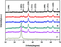
The XRD patterns of the prepared samples are shown in Fig. 1. It is obvious that the pure g-C3N4, Co0.5Zn0.5Fe2O4 NPs and Co0.5Zn0.5Fe2O4 composite microspheres with different compositions exhibit similar diffraction patterns. For pure g-C3N4, a strong peak at 27.3°, corresponding to width of 0.326 nm, can be indexed as (002) diffraction plane, which is well known for the melon networks. Another peak at 13.0°, corresponding to a distance of 0.675 nm, belongs to (100) in-plane ordering of tri-s-triazine units.25 The peaks at around 2θ = 30.3°, 35.5°, 37.1°, 43.2°, 53.6°, 57.3° and 62.8° (marked with “◇”) are attributed to the (111), (220), (311), (222), (400), (511) and (440) crystal planes of mixed spinel in pure Co0.5Zn0.5Fe2O4 NPs, respectively. It should be noted that the typical diffraction peaks of g-C3N4 and Co0.5Zn0.5Fe2O4 appear in the patterns of the CN-CZF composite microspheres.
image file: c6ra07915f-f1.tif
Fig. 1 XRD patterns of (A) Co0.5Zn0.5Fe2O4, (B) CNCZF1, (C) CNCZF2, (D) CNCZF4, (E) CNCZF8, (F) pure g-C3N4.

3.2. Morphology characterization

TEM was used to observe the morphology of the obtained samples. The pure g-C3N4 shows a two-dimensional sheet-like structure consisting of wrinkles (Fig. 2(A)). The average particle size was found to be in the range of 12 nm as measured from the TEM image shown in Fig. 2(B) for the Co0.5Zn0.5Fe2O4 NPs. As depicted in Fig. 2(C), the magnetic Co0.5Zn0.5Fe2O4 NPs were distributed separately on g-C3N4 sheets as nanoislands, which could not only improve the dispersion property of layered materials but also offer more photocatalytic reaction sites, and a heterojunction structure was formed. Therefore, the photocatalytic activity of CN-CZF was improved greatly.
image file: c6ra07915f-f2.tif
Fig. 2 Characterization of g-C3N4, Co0.5Zn0.5Fe2O4 NPs and g-C3N4/Co0.5Zn0.5Fe2O4. TEM images of (A) g-C3N4, (B) Co0.5Zn0.5Fe2O4 NPs and (C) CN-CZF4 composite. The inset is the corresponding SEAD pattern.

3.3. FT-IR analysis

The FT-IR spectra of the pure g-C3N4, Zn0.5Co0.5Fe2O4 and CN-CZF2 composite photocatalyst are shown in Fig. 3. In the FTIR spectrum of the pure g-C3N4, all the IR responses are of aromatic C and N heterocycles. The peak at 808 cm−1 is attributed to the ring-sextant out-of-plane bending vibration characteristic of both triazine and heptazine ring systems,26 and the bands at 1639, 1560, 1462 and 1410 cm−1 were assigned to typical stretching vibration modes of the heptazine-derived repeat units. The bands at 1323 and 1238 cm−1 corresponded to stretching vibrations of connected units of C–N(–C)–C (full condensation) or C–NH–C (partial condensation), which was also supported by the stretching vibration modes of the broad weak band around 3262 cm−1 for hydrogen-bonding interactions.5 The broad band at 3087–3669 cm−1 belonged to the N–H vibration due to partial condensation and adsorbed water molecules. The peaks at 3400 and 1607 cm−1 are attributed to absorbed water molecules on the surface of the Zn0.5Co0.5Fe2O4 sample. The two absorption bands observed at 423 and 581 cm−1 can be assigned to the vibration of metal ions at the octahedral and tetrahedral sites, respectively, involving M–O bonds (M = Zn, Co and Fe) in Zn0.5Co0.5Fe2O4.27 All the characteristic peaks of g-C3N4 and Zn0.5Co0.5Fe2O4 were observed in the spectrum of the CN-CZF2 composite photocatalyst.
image file: c6ra07915f-f3.tif
Fig. 3 FT-IR spectra of pure g-C3N4, CN-CZF4 composite and Co0.5Zn0.5Fe2O4 NPs.

3.4. PL analysis

To understand the trapping, migration and transfer property of electron–hole pairs, PL emission measurements were applied and PL spectra of the samples were recorded.28 As is well known, the intensity of the PL emission spectra depends on the recombination of excited electrons and holes: the lower the PL emission intensity, the less the recombination tendency of the samples.29 Fig. 4 displays the PL spectra of g-C3N4 and CN-CZF2 under an excitation wavelength of 320 nm. It can be observed that the g-C3N4 sample exhibits strong PL at ambient temperature. The PL has a maximum at about 458 nm, which is equivalent to a band gap energy of 2.71 eV. This strong peak is attributed to the band–band PL phenomenon with the energy of light approximately equal to the band gap energy of g-C3N4. In fact, the band–band PL signal is attributed to excitonic PL, which mainly results from the n → π* electronic transition involving lone pairs of nitrogen atoms in g-C3N4.25,30 However, in the PL spectrum of the CN-CZF2 composite, a weaker emission peak in the same position was detected, which indicated that the photogenerated charge recombination rate in CN-CZF2 composite was much lower than that in pure g-C3N4. The recombination of photogenerated electron–hole pairs was greatly inhibited by the introduction of Co0.5Zn0.5Fe2O4, which means that photogenerated electron–hole pair separation efficiency of the CN-CZF2 composite was greater than that of the pure g-C3N4 sample.
image file: c6ra07915f-f4.tif
Fig. 4 Photoluminescence spectra of (a) pure g-C3N4 and (b) CN-CZF4.

3.5. UV-visible diffuse reflectance spectra

The absorption properties of the as-prepared samples were measured using UV-visible diffuse reflectance spectroscopy. Fig. 5 displays the UV-visible diffuse reflectance spectra of the CN-CZF1, CN-CZF2, CN-CZF3 and CN-CZF4 composites, together with those of Co0.5Zn0.5Fe2O4 NPs and pure g-C3N4. For the pure g-C3N4 sheets, the basal absorption edge occurs at about 480 nm. In comparison to the pure g-C3N4, the band edge positions of g-C3N4/Co0.5Zn0.5Fe2O4 composites exhibited a large red shift and the absorption intensity of the composites also became stronger with increasing Co0.5Zn0.5Fe2O4 content. This phenomenon is attributed to the presence of Co0.5Zn0.5Fe2O4 NPs with the red color characteristic as well as a better absorption in the visible light region. The band gap energies of semiconductors can be estimated by the Kubelka–Munk transformation, αhν = A(Eg)n/2, where α represents the absorption coefficient, ν is the light frequency, Eg is the band gap energy, A is a constant and n depends on the characteristics of the transition in a semiconductor.31,32 For g-C3N4 and Co0.5Zn0.5Fe2O4, the values for pure g-C3N4 sheets, Co0.5Zn0.5Fe2O4 NPs and CN-CZF2 composite are calculated to be 2.7, 1.2 and 2.0 eV, respectively. The potentials of VB and CB of a semiconductor material can be estimated according to the following empirical equations:
ECB = χEe − 0.5Eg

EVB = ECB + Eg
where EVB is the valence band edge potential, χ is the electronegativity of the semiconductor, which is the geometric mean of the constituent atoms, Ee is the energy of free electrons on the hydrogen scale (about 4.5 eV vs. NHE) and Eg is the band gap energy of the semiconductor. The χ-values for g-C3N4 and Co0.5Zn0.5Fe2O4 are 4.64 and 4.78 eV, respectively. The ECB values of g-C3N4 and Co0.5Zn0.5Fe2O4 are calculated to be −1.21 and −0.32 eV, respectively. The EVB values of g-C3N4 and Co0.5Zn0.5Fe2O4 are estimated to be +1.49 and +0.88 eV, respectively.

image file: c6ra07915f-f5.tif
Fig. 5 UV-visible diffuse reflectance spectra of pure g-C3N4, CN-CZF composites and Co0.5Zn0.5Fe2O4 NPs.

3.6. XPS analysis

The composition and chemical status of the as-prepared samples were also confirmed using the XPS technique. Fig. 6 displays the XPS survey spectra of the pure Co0.5Zn0.5Fe2O4 and g-C3N4, and the prepared CN-CZF4 composite photocatalyst. Fig. 6(A) shows the survey spectrum of Co0.5Zn0.5Fe2O4 NPs which indicates Zn, Co, Fe, O and C. The survey spectrum of g-C3N4 sheets indicates mainly C and N elements, and the survey spectrum of CN-CZF4 sample indicates Zn, Co, Fe, O, N and C. Carbon of the spectrum of Co0.5Zn0.5Fe2O4 NPs is attributed to the hydrocarbon from the XPS instrument itself. The negligible amount of oxygen element could be ascribed to the tiny amount of O2 adsorbed on the surface of the synthesized product during the polymerization process, which is a common phenomenon for synthetic g-C3N4 materials. As shown in Fig. 6(B), the peaks at 1043.91 eV and 1018.36 eV are attributed to the binding energies of Zn 2p3/2 and Zn 2p1/2 signals in the Zn2+ chemical state. Two binding energies located at 721.17 eV and 710.81 eV are assigned to the distinct Fe 2p3/2 and Fe 2p1/2 of Fe3+ in the spectrum in Fig. 6(C). Furthermore, two peaks of Co 2p located at 799.21 eV and 781.18 eV should be attributed to Co 2p3/2 and Co 2p1/2 in Fig. 6(D). The peak of N 1s appeared in the spectrum of the CN-CZF4 composite located at 398.12 eV (Fig. 6(E)), which had a slight deviation compared with pure g-C3N4 (at 398.13 eV).
image file: c6ra07915f-f6.tif
Fig. 6 XPS spectra of the photocatalyst: (A) XPS survey, (B) Zn 2p XPS spectra, (C) Fe 2p XPS spectra, (D) Co 2p XPS spectra, and (E) N 1s XPS spectra of CN-CZF4 hybrid.

3.7. Safety evaluation of CN-CZF

Due to their favorable magnetic and optical properties, spinel ferrites (MFe2O4, M = Mn, Co, Zn or Ni) nanoparticles are the most attractive iron oxide nanoparticles and have been commonly and widely used in research. Toxicity assays of MFe2O4 have been carried out along with the investigations of their research.33,34 0.1 g of powdered CN-CZF4 was suspended in 100 mL water. We utilized ICP-OES to quantify the Co, Zn, and Fe concentrations of CN-CZF4 at different time points (0 h, 12 h, 24 h, 48 h) as shown in Fig. S2. According to previous reports, Zhao et al. prepared ultra-small magnetite MFe2O4 nanoparticles (<20 nm) and their cytotoxicity was assessed by using the MTT assay in A549 cells. The results indicated that the cytotoxicity of ultra-small MFe2O4 nanoparticles was dose- and time-dependent.35 Wang et al. found that the material modifications can minimize the hazard.36 Podila et al. found that 50 nm NPs exhibited maximum cellular uptake. The results indicated that CN-CZF4 NPs are environmentally friendly after recycling.37

3.8. Photocatalytic performance

In this work, the photocatalytic performances of g-C3N4/Co0.5Zn0.5Fe2O4 hybrids were evaluated by degradation of chloromycetin, an environmentally unfriendly antibiotic pollutant, to test the photodegradation capability. Fig. 7 shows the photocatalytic activity of g-C3N4/Co0.5Zn0.5Fe2O4 composites with different content of magnetic Co0.5Zn0.5Fe2O4 NPs under visible light irradiation. The photodegradation rates of chloromycetin followed the order of CN-CZF4 > CN-CZF8 > CN-CZF2 > CN-CZF1 > CN > CZF > blank. From the blank test, the concentration of chloromycetin remained the same within 240 min, confirming that chloromycetin was very stable and its photolysis could be ignored under visible light irradiation without catalysts. It can be seen from Fig. 7 that Co0.5Zn0.5Fe2O4 and pure g-C3N4 exhibited very low photocatalytic activity; 16% chloromycetin was decomposed by Co0.5Zn0.5Fe2O4 NPs after irradiation for 240 min. Similarly, only 42% chloromycetin was degraded by pure g-C3N4. Surprisingly, the results indicated that the photocatalytic activity of CN-CZF4 composite is much higher than that of Co0.5Zn0.5Fe2O4 and pure g-C3N4, and its catalytic rate is as high as 96%. However, when the content of Co0.5Zn0.5Fe2O4 was too high (such as in CN-CZF1), the photocatalytic activity decreased slightly, but it was still much higher than that of pure g-C3N4 and Co0.5Zn0.5Fe2O4. The reason why excess Co0.5Zn0.5Fe2O decreased the photodegradation efficiency of CN-CZF composites could be as follows: a high content of Co0.5Zn0.5Fe2O4 could significantly affect the dispersion of Co0.5Zn0.5Fe2O4 NPs, which affects the photocatalytic activity. Previous research also indicated the dispersion of nanoparticles dotted on semiconductor surfaces could influence the photodegradation efficiency.38,39 The higher dispersibility of Co0.5Zn0.5Fe2O4 on the g-C3N4 sheets meant higher photocatalytic activity. However, there would be a negative effect when the coverage density was increased. When the coverage density of Co0.5Zn0.5Fe2O4 was too high, the separation efficiency of photogenerated charges was low, leading to a decrease of photodegradation efficiency of g-C3N4/Co0.5Zn0.5Fe2O4. Therefore, only Co0.5Zn0.5Fe2O4 nanoparticles coated on g-C3N4 with the right dispersion can enhance the photocatalytic activity of g-C3N4 composites. For further illustration of the effect and importance of the interface between the components of g-C3N4/Co0.5Zn0.5Fe2O4 heterojunction composites, the photocatalytic activities of CN-CZF4 and PM-CN-CZF4 (physical mixture of g-C3N4 and Co0.5Zn0.5Fe2O4) were compared. Fig. 8 shows that photodegradation efficiency of CN-CZF4 was always much higher than that of PM-CN-CZF4, which indicated definitely that the interface between g-C3N4 and Co0.5Zn0.5Fe2O4 was very important to the photocatalytic activity. The stability of a photocatalyst is also very important from the point of view of its practical application. CN-CZF4 was recycled five times under the same reaction conditions. The sample was separated by using a magnet after each cycle, washed with 0.1 M HNO3 and deionized water several times, and then dried at 80 °C under vacuum. From Fig. 9, the photodegradation efficiency of CN-CZF4 decreased slightly after the five reuse cycles, which indicated that photocatalysts based on CN-CZF composites could be reused completely for wastewater treatment.
image file: c6ra07915f-f7.tif
Fig. 7 Photocatalytic degradation of chloromycetin in aqueous solution over g-C3N4, Co0.5Zn0.5Fe2O4, and CN-CZF composite photocatalysts.

image file: c6ra07915f-f8.tif
Fig. 8 Degradation of chloromycetin under visible light irradiation over different photocatalysts.

image file: c6ra07915f-f9.tif
Fig. 9 Recyclability of the CN-CZF4 photocatalyst in five successive experiments for the photocatalytic degradation of chloromycetin under visible light irradiation.

3.9. Photocatalytic mechanism of the CN-CZF composites

The higher photocatalytic efficiency of the CN-CZF4 sample in comparison with the other samples can be correlated to its greater ability to absorb a larger fraction of visible light, as manifested by the relatively narrow band gap of 2.0 eV. As we know, effective electron–hole pair separation at heterojunction interfaces can significantly improve the photocatalytic performance of composite photocatalysts. Fig. 10 illustrates the photoelectron transfer mechanism in the photocatalytic degradation of chloromycetin over the CN-CZF4 hybrid. The band gap energy of Co0.5Zn0.5Fe2O4 and g-C3N4 was found to be equal to 1.2 and 2.7 eV, respectively. As already stated in the preceding section, the effect of photocatalytic activity is more evident and significant and thus the enhancement in photocatalytic activity can be ascribed to the increased amount of semiconductor and its interaction under visible light irradiation, which is expected to produce h+/e charge carriers. Considering the inner electric field and energy band structure, it is reasonable to conclude that the transfer of electrons between g-C3N4 and Co0.5Zn0.5Fe2O4 is partly restricted, while the transfer of holes can be accelerated. This causes an efficient separation of photogenerated electrons and holes to enhance the photocatalytic activity. As is well known, Co0.5Zn0.5Fe2O4 is a typical p-type semiconductor, while g-C3N4 is n-type. The hypothetical hole h+ in VB is responsible for oxidation and the electron in CB is responsible for reduction.40 As oxidation is the desired route here, the phenomenon is mainly expected with Co0.5Zn0.5Fe2O4 since its band gap is 1.2 eV. In the CN-CZF composites, the photogenerated electrons will have a tendency to transfer from Co0.5Zn0.5Fe2O4 to g-C3N4 and the holes have an opposite transfer because of the inner electric field existed in the p–n junctions.41,42 Thus, under visible light irradiation, both Co0.5Zn0.5Fe2O4 and g-C3N4 can be excited to generate electrons and holes. The holes can also react with surface-adsorbed H2O to produce ˙OH radicals or directly oxidize the adsorbed chloromycetin molecules. Photogenerated electrons (e) play an important role also because they can react at the surface of the photocatalyst with chemisorbed O2 to generate the reactive oxidative species ˙O2−, which combines with H+ from water to form H2O2 under irradiation, and then to produce active ˙OH radicals, which play an important role in degrading and mineralizing the adsorbed molecules of organic peroxides. On the basis of the above results, we propose the possible mechanisms of degradation of chloromycetin as follows:
image file: c6ra07915f-t1.tif

g-C3N4(e + h+)–Co0.5Zn0.5Fe2O4(e + h+) → Co0.5Zn0.5Fe2O4(e) + Co0.5Zn0.5Fe2O4(h+)

Co0.5Zn0.5Fe2O4(h+) + chloromycetin → degradation products

Co0.5Zn0.5Fe2O4(e) + O2 + 4H+ → 2H2O2

OH˙ + chloromycetin → degradation products

image file: c6ra07915f-f10.tif
Fig. 10 Band structure schematic of the CN-CZF4 composite and possibly occurring reaction mechanism of chloromycetin on the surface of the composite.

4. Conclusion

In summary, a magnetic hybrid photocatalyst of g-C3N4/Co0.5Zn0.5Fe2O4 has been successfully constructed via an easily accessible route. During this process, heterostructured g-C3N4/Co0.5Zn0.5Fe2O4 hybrid was first synthesized by a simple hydrothermal synthesis method, which exhibited enhanced photocatalytic activity in the degradation of chloromycetin under visible light irradiation, and its structural and photocatalytic properties were studied by TEM, FT-IR, XRD, diffuse reflectance spectroscopy, and XPS techniques. Experimental results indicate that the close interfacial connections between Co0.5Zn0.5Fe2O4 and g-C3N4, where induced photoelectrons and holes were efficiently separated in space, were conductive to retarding the charge recombination and improving the photoactivity. On the basis of the results of this study, the present approach for hybrid photocatalyst preparation and the detailed mechanism discussion in this work can provide valuable knowledge on the development of MnX1−nFe2O4-based highly efficient photocatalysts and magnetic photocatalysts in environmental pollution cleanup.

Acknowledgements

This work was financially supported by the National Natural Science Foundation of China (no. 21107037, no. 21176107), National Postdoctoral Science Foundation (no. 2013M530240), Postdoctoral Science Foundation funded Project of Jiangsu Province (no. 1202002B) and Programs of Senior Talent Foundation of Jiangsu University (no. 12JDG090), Key Scientific Programs of Higher Education of Henan Province of China (no. 15A150069), Youth Foundation of Pingdingshan University (no. 2012004), and the National Scientific Research Project Cultivating Foundation of Pingdingshan University (no. PXY-PYJJ2016005).

Notes and references

  1. S. Cao and J. Yu, g-C3N4-based photocatalysts for hydrogen generation, J. Phys. Chem. Lett., 2014, 5(12), 2101–2107 CrossRef CAS PubMed.
  2. F. Sastre, A. V. Puga and L. Liu, et al., Complete Photocatalytic Reduction of CO2 to Methane by H2 under Solar Light Irradiation, J. Am. Chem. Soc., 2014, 136(19), 6798–6801 CrossRef CAS PubMed.
  3. C. Chen, W. Ma and J. Zhao, Semiconductor-mediated photodegradation of pollutants under visible-light irradiation, Chem. Soc. Rev., 2010, 39(11), 4206–4219 RSC.
  4. J. S. Jang, S. J. Hong and J. S. Lee, et al., Synthesis of zinc ferrite and its photocatalytic application under visible light, J. Korean Phys. Soc., 2009, 54, 204 CrossRef CAS.
  5. S. Zhang, J. Li and M. Zeng, et al., In situ synthesis of water-soluble magnetic graphitic carbon nitride photocatalyst and its synergistic catalytic performance, ACS Appl. Mater. Interfaces, 2013, 5(23), 12735–12743 CAS.
  6. K. Aranishi, H. L. Jiang and T. Akita, et al., One-step synthesis of magnetically recyclable Au/Co/Fe triple-layered core–shell nanoparticles as highly efficient catalysts for the hydrolytic dehydrogenation of ammonia borane, Nano Res., 2011, 4(12), 1233–1241 CrossRef CAS.
  7. H. Lv, L. Ma and P. Zeng, et al., Synthesis of floriated ZnFe2O4 with porous nanorod structures and its photocatalytic hydrogen production under visible light, J. Mater. Chem., 2010, 20(18), 3665–3672 RSC.
  8. J. Wu, N. Li and J. Xu, et al., Partially inverse spinel ZnFe2O4 with high saturation magnetization synthesized via a molten salt route, Appl. Phys. Lett., 2011, 99(20), 202505 CrossRef.
  9. Y. Yao, J. Qin and H. Chen, et al., One-pot approach for synthesis of N-doped TiO2/ZnFe2O4 hybrid as an efficient photocatalyst for degradation of aqueous organic pollutants, J. Hazard. Mater., 2015, 291, 28–37 CrossRef CAS PubMed.
  10. R. S. Yadav, J. Havlica and M. Hnatko, et al., Magnetic properties of Co1−xZnxFe2O4 spinel ferrite nanoparticles synthesized by starch-assisted sol–gel autocombustion method and its ball milling, J. Magn. Magn. Mater., 2015, 378, 190–199 CrossRef CAS.
  11. I. H. Gul, A. Z. Abbasi and F. Amin, et al., Structural, magnetic and electrical properties of Co1−xZnxFe2O4 synthesized by co-precipitation method, J. Magn. Magn. Mater., 2007, 311(2), 494–499 CrossRef CAS.
  12. R. Arulmurugan, B. Jeyadevan and G. Vaidyanathan, et al., Effect of zinc substitution on Co–Zn and Mn–Zn ferrite nanoparticles prepared by co-precipitation, J. Magn. Magn. Mater., 2005, 288, 470–477 CrossRef CAS.
  13. A. Manikandan, L. J. Kennedy and M. Bououdina, et al., Synthesis, optical and magnetic properties of pure and Co-doped ZnFe2O4 nanoparticles by microwave combustion method, J. Magn. Magn. Mater., 2014, 349, 249–258 CrossRef CAS.
  14. M. Veverka, Z. Jirák and O. Kaman, et al., Distribution of cations in nanosize and bulk Co–Zn ferrites, Nanotechnology, 2011, 22(34), 345701 CrossRef CAS PubMed.
  15. J. Li, Z. Liu and Z. Zhu, Magnetically separable ternary hybrid of ZnFe2O4–Fe2O3–Bi2WO6 hollow nanospheres with enhanced visible photocatalytic property, Appl. Surf. Sci., 2014, 320, 146–153 CrossRef CAS.
  16. S. Huang, Y. Xu and M. Xie, et al., Synthesis of magnetic CoFe2O4/g-C3N4 composite and its enhancement of photocatalytic ability under visible-light, Colloids Surf., A, 2015, 478, 71–80 CrossRef CAS.
  17. Y. Yao, Y. Cai and F. Lu, et al., Magnetic ZnFe2O4–C3N4 hybrid for photocatalytic degradation of aqueous organic pollutants by visible light, Ind. Eng. Chem. Res., 2014, 53(44), 17294–17302 CrossRef CAS.
  18. X. Wang, K. Maeda and A. Thomas, et al., A metal-free polymeric photocatalyst for hydrogen production from water under visible light, Nat. Mater., 2009, 8(1), 76–80 CrossRef CAS PubMed.
  19. X. Wang, K. Maeda and A. Thomas, et al., A metal-free polymeric photocatalyst for hydrogen production from water under visible light, Nat. Mater., 2009, 8(1), 76–80 CrossRef CAS PubMed.
  20. Y. Zheng, Y. Jiao and J. Chen, et al., Nanoporous graphitic-C3N4@carbon metal-free electrocatalysts for highly efficient oxygen reduction, J. Am. Chem. Soc., 2011, 133(50), 20116–20119 CrossRef CAS PubMed.
  21. Q. Sun, K. Lv and Z. Zhang, et al., Effect of contact interface between TiO2 and g-C3N4 on the photoreactivity of g-C3N4/TiO2 photocatalyst: (001) vs. (101) facets of TiO2, Appl. Catal., B, 2015, 164, 420–427 CrossRef.
  22. F. Dong, Y. Sun and L. Wu, et al., Facile transformation of low cost thiourea into nitrogen-rich graphitic carbon nitride nanocatalyst with high visible light photocatalytic performance, Catal. Sci. Technol., 2012, 2(7), 1332–1335 CAS.
  23. H. Katsumata, T. Sakai and T. Suzuki, et al., Highly efficient photocatalytic activity of g-C3N4/Ag3PO4 hybrid photocatalysts through Z-scheme photocatalytic mechanism under visible light, Ind. Eng. Chem. Res., 2014, 53(19), 8018–8025 CrossRef CAS.
  24. S. C. Yan, Z. S. Li and Z. G. Zou, Photodegradation performance of g-C3N4 fabricated by directly heating melamine, Langmuir, 2009, 25(17), 10397–10401 CrossRef CAS PubMed.
  25. Y. Yao, Y. Cai and F. Lu, et al., Magnetic ZnFe2O4–C3N4 hybrid for photocatalytic degradation of aqueous organic pollutants by visible light, Ind. Eng. Chem. Res., 2014, 53(44), 17294–17302 CrossRef CAS.
  26. D. R. Miller, D. C. Swenson and E. G. Gillan, Synthesis and structure of 2,5,8-triazido-s-heptazine: an energetic and luminescent precursor to nitrogen-rich carbon nitrides, J. Am. Chem. Soc., 2004, 126(17), 5372–5373 CrossRef CAS PubMed.
  27. R. D. Waldron, Infrared spectra of ferrites, Phys. Rev., 1955, 99(6), 1727 CrossRef CAS.
  28. J. Wang and W. D. Zhang, Modification of TiO2 nanorod arrays by graphite-like C3N4 with high visible light photoelectrochemical activity, Electrochim. Acta, 2012, 71, 10–16 CrossRef CAS.
  29. S. Zhou, Y. Liu and J. Li, et al., Facile in situ synthesis of graphitic carbon nitride (gC3N4)-N-TiO2 heterojunction as an efficient photocatalyst for the selective photoreduction of CO2 to CO, Appl. Catal., B, 2014, 158, 20–29 CrossRef.
  30. V. N. Khabashesku, J. L. Zimmerman and J. L. Margrave, Powder synthesis and characterization of amorphous carbon nitride, Chem. Mater., 2000, 12(11), 3264–3270 CrossRef CAS.
  31. S. I. In, D. D. Vaughn and R. E. Schaak, Hybrid CuO–TiO2xNx Hollow Nanocubes for Photocatalytic Conversion of CO2 into Methane under Solar Irradiation, Angew. Chem., Int. Ed., 2012, 51(16), 3915–3918 CrossRef CAS PubMed.
  32. J. Tauc, Absorption edge and internal electric fields in amorphous semiconductors, Mater. Res. Bull., 1970, 5(8), 721–729 CrossRef CAS.
  33. L. C. Wehmas, C. Anders and J. Chess, et al., Comparative metal oxide nanoparticle toxicity using embryonic zebrafish, Toxicol. Rep., 2015, 2, 702–715 CrossRef CAS PubMed.
  34. S. L. Harper, J. L. Carriere and J. M. Miller, et al., Systematic evaluation of nanomaterial toxicity: utility of standardized materials and rapid assays, ACS Nano, 2011, 5(6), 4688–4697 CrossRef CAS PubMed.
  35. H. Zhao, Z. Chen and L. Tao, et al., In vitro toxicity evaluation of ultra-small MFe2O4 (M = Fe, Mn, Co) nanoparticles using A549 cells, RSC Adv., 2015, 5(84), 68454–68460 RSC.
  36. Z. Wang, D. Xie and H. Liu, et al., Toxicity assessment of precise engineered gold nanoparticles with different shapes in zebrafish embryos, RSC Adv., 2016, 6(39), 33009–33013 RSC.
  37. R. Podila and J. Mbrown, Toxicity of Engineered Nanomaterials: A Physicochemical Perspective, J. Biochem. Mol. Toxicol., 2013, 27(1), 50–55 CrossRef CAS PubMed.
  38. M. Murdoch, G. I. N. Waterhouse and M. A. Nadeem, et al., The effect of gold loading and particle size on photocatalytic hydrogen production from ethanol over Au/TiO2 nanoparticles, Nat. Chem., 2011, 3(6), 489–492 CAS.
  39. D. Jassby, J. Farner Budarz and M. Wiesner, Impact of aggregate size and structure on the photocatalytic properties of TiO2 and ZnO nanoparticles, Environ. Sci. Technol., 2012, 46(13), 6934–6941 CrossRef CAS PubMed.
  40. I. O. Ali and A. G. Mostafa, Photocatalytic reduction of chromate oxyanions on MMnFe2O4 (M = Zn, Cd) nanoparticles, Mater. Sci. Semicond. Process., 2015, 33, 189–198 CrossRef.
  41. L. Li, L. Xu and W. Shi, et al., Facile preparation and size-dependent photocatalytic activity of Cu2O nanocrystals modified titania for hydrogen evolution, Int. J. Hydrogen Energy, 2013, 38(2), 816–822 CrossRef CAS.
  42. M. Xu, L. Han and S. Dong, Facile fabrication of highly efficient g-C3N4/Ag2O heterostructured photocatalysts with enhanced visible-light photocatalytic activity, ACS Appl. Mater. Interfaces, 2013, 5(23), 12533–12540 CAS.

Footnote

Electronic supplementary information (ESI) available. See DOI: 10.1039/c6ra07915f

This journal is © The Royal Society of Chemistry 2016
Click here to see how this site uses Cookies. View our privacy policy here.