Danieli C. Rodrigues*a,
Sathyanarayanan Sridhara,
Izabelle M. Gindria,
Danyal A. Siddiquia,
Pilar Valderramab,
Thomas G. Wilson Jr.b,
Kwok-Hung Chungc and
Chandur Wadhwanic
aDepartment of Bioengineering, University of Texas at Dallas, Richardson, TX, USA. E-mail: danieli@utdallas.edu
bPrivate Practice of Periodontics, Dallas, TX, USA
cDepartment of Restorative Dentistry, University of Washington, Seattle, WA, USA
First published on 12th May 2016
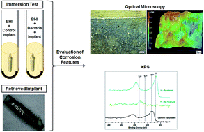
The surface morphology and chemical composition of commercially pure titanium dental implants and healing abutments exposed in vitro or in vivo to oral bacteria were studied. Implants exposed in vitro to media containing S. mutans and healing abutment retrievals were used to evaluate the effect of early bacterial colonizers. Another dental implant retrieval that failed due to peri-implantitis and implants exposed to an in vitro culture of P. gingivalis were analyzed to assess the effect of late stage colonizing bacteria. Using optical microscopy and X-ray photoelectron spectroscopy, it was determined that both S. mutans and P. gingivalis adversely affected the implant surface, with the early colonizing S. mutans creating conditions leading to more severe corrosion features being observed, including pitting and surface discoloration. Overall, this study demonstrated the ability of bacteria in the oral environment to solely induce changes in the oxidation states of titanium and subsequent corrosion of the implant surface, even in the absence of mechanical loads.
Implant failures can be induced by a number of individual or synergistic events.12–16 Typically, these failure mechanisms are classified as either early or late stage failures.17,18 Early stage failure occurs when the establishment of osseointegration is interrupted mainly due to bacterial contamination, premature overloading, impaired healing and excessive surgical trauma.17,18 Late stage failures involve breakdown of the established osseointegration, having major etiological factors associated with bacteria-induced marginal bone loss (peri-implantitis) and excessive occlusal stresses.18 Bacteria is clearly involved in the early and late stages of implant failure, inducing surface changes that can trigger inflammatory reactions in soft tissues.17–19 According to Zhao et al., the performance of a dental implant surface is dependent on a “race-for-the-surface” between human gingival fibroblasts and bacterial biofilm adhesion.2 Early failure can be initiated by surface adhesion of early bacterial colonizers such as Streptococci and Actinomyces species, which upon formation of a biofilm will prevent sealing of epithelial tissues on the implant surface.20,21 Failure to achieve soft tissue integration on implant surfaces will enable the first colonizers to infiltrate and interrupt the early stages of osseointegration.22 Attachment of these early colonizers will create the necessary anaerobic conditions for pathogenic late colonizers such as Aggregatibacter actinomycetemcomitans, Porphyromonas gingivalis and Fusobacterium nucleatum.20,21,23 These periodontal pathogens can ultimately lead to bone loss and are considered causative agents of peri-implantitis.24 Inflammation due to bacterial biofilm is then assumed as the primary etiological factor for peri-implantitis.25 However, other factors including, excessive mechanical loads,26 corrosion,4,27 excess cement28 and previous periodontal disease history29 have also been reported to play an important role in the inflammatory process.
Although, bacteria is a major cause for both early and late stage failures, there are only a few studies that discuss the ability of bacteria to create unfavorable conditions leading to implant surface damage.30–32 We demonstrated in a recent in vitro study that immersion of dental implants in medium containing Streptococcus mutans for 60 days led to significant changes in surface morphology with evidence of corrosion and generation of metal ions in solution.33 Wilson et al. reported histopathological findings in soft tissue biopsies of implants with peri-implantitis, demonstrating that the predominant foreign bodies found were titanium and dental cement surrounded by inflammatory cells.34 Retrospective studies of the surface of dental implants retrieved from patients with peri-implantitis also illustrate the degree of surface damage that can be imparted by late colonizers. Morphological features such as pitting, surface etching, discoloration, cracking, scratches and fractures were observed on the surface of these implants.35–37
Bacterial biofilms are assumed to change the electrochemical environment surrounding an implant, which can lead to disruption of the surface oxide layer and induce osseointegration loss.34,38 This is hypothesized based on two mechanisms: (1) contact and adhesion of early-colonizing planktonic bacteria on surfaces that release lactic acid during glycolysis, thereby decreasing the pH of the oral environment. Acidic environments can result in changes in the oxidation states of titanium and can lead to metal ion dissolution;30,39,40 and (2) biofilm formation on implant surfaces, leading to the development of localized crevice environments on the implant surface. Oxygen depletion within these crevices will induce a significant decrease in local pH and consequent metal surface attack.41–43 Implant micromotion, resulting from occlusal loading, in the presence of a stagnant acidic environment can lead to permanent disruption of the oxide layer through a mechanism of fretting-crevice corrosion.44 Besides leading to active metal ion and particle generation, this process can result in disruption of osseointegrated and sealed surfaces, exacerbating inflammatory reactions.45
Hence, it has been suggested that metal ions and wear release can affect the biological response of cells that are vital to attain implant integration with soft and bone tissues.37,46 The possibility of implant corrosion mediated by bacteria requires a better understanding of the role of early and late colonizers on the surface of titanium dental implants. The primary goal of this study was to evaluate whether bacteria found in the oral cavity are able to change the surface topography and surface composition of dental implants and abutments exposed to them.
Sample | Brand | Dimension | Morphology and surface treatment | History | Retrieval stage |
---|---|---|---|---|---|
a Not available.b No exposure to medium containing bacteria.c Used as control against implants or components analyzed as early or late stage failures. | |||||
Implant (IC) | Straumann LLC., (Andover, MA) | 4.1 × 10 mm | Regular Neck (RN), Sand-blasted, Large-grit and Acid-etched (SLA) | NEb | Nonec |
Implant 1 (I1) | Immersed in medium containing Streptococcus mutans (in vitro) | Early | |||
Implant 2 (I2) | Immersed in medium containing Porphyromonas gingivalis (in vitro) | Late | |||
Implant 3 (I3) | |||||
Implant 4 (I4) | |||||
Implant 5 (I5) | |||||
Implant 6 (I6) | NAa | 4.1 × 8 mm | Unknown | Retrieved implant (in vivo) | |
Healing Abutment Control (HAC) | Straumann LLC., (Andover, MA) | 4.5 × 6 mm | Regular connection (RC), conical emergence profile, machined surface | NEb | Nonec |
(HA1) | Retrieved healing abutments (in vivo) | Early | |||
(HA2) | |||||
(HA3) |
A retrieved implant removed from a patient presenting a case of peri-implantitis and three healing abutments (HA) were received from a private periodontal practice (Dallas, TX). Patients provided consent as according to the guidelines of the Helsinki Declaration to donate their retrieved implants and abutments for research. Upon retrieval and storage, there were no identifiers that could be linked to the patient who donated the implant/abutment. Therefore, information like time elapsed from implant placement to implant removal and surface treatment were not available.
A visual inspection was initially performed to detect particular areas of interest and gross features and to verify the severity of corrosion present (discoloration, cracking and debris). Specimens were then analyzed with a digital optical microscope (OM) for identification of surface features and failure mechanisms. Areas of interest were further analyzed with X-ray photoelectron spectroscopy (XPS) to evaluate the elemental composition and the nature of chemical interactions on the surface. After surface characterization, all specimens were subjected to a cleaning protocol for complete removal of biological deposits that could have covered important surface features. They were first cleaned in an aqueous solution with soap powder, followed by cleaning in deionized water and finally 70% ethanol. For this protocol, specimens were fully immersed in each of the solutions and subjected to sonication for 1 hour. Microscopy and XPS were then repeated.
Experiments were performed with a PHI 5000 Versa Probe II X-ray Photoelectron Spectrometer (Chanhassen, MN). Measurements were taken at an angle of 45° in relation to the sample surface. Areas scanned in each analysis had dimensions of approximately 200 μm2. A monochromatic Al Ka source of 1486.6 eV was used. The survey spectra were acquired using 187.850 eV pass energy and 0.8 eV step size, while the high resolution scans were obtained using 23.5 eV pass energy and 0.200 eV step size. The pressure in the analysis chamber was maintained below 10−8 Torr. The data was analyzed and normalized using CasaXPS 2.3.16 Software.
XPS allowed for identification of chemical elements present in the samples by measuring the binding energy spectra of emitted X-ray photoelectrons. A typical XPS spectrum is a plot of X-ray intensity versus binding energy (BE). Before a more detailed discussion of XPS data, some general information is provided regarding the XPS profiles of oxygen (O1s) and titanium (Ti2p), which are the main elements investigated in this study. The XPS spectrum of oxygen consists of a singlet peak and its BE varies according to the chemical environment in which oxygen molecules are located. For example, it is already established that when bound to titanium in the oxide layer (TiO2), the BE of oxygen is around 529.70 ± 0.10 eV.48 However, this BE can be shifted to higher values (530.99 ± 0.15 eV) when oxygen is in its hydroxyl form (OH), which can occur when either linked to Ti or bound in water.48 The titanium spectrum is characterized by doublet peaks of Ti2p, which are denominated Ti2p3/2 and Ti2p1/2. To avoid dubiety in the discussion of results, only the binding energy of Ti2p3/2 in the Ti spectrum will be investigated and discussed throughout this paper. XPS can detect four distinct electronic states of Ti2p (Ti2p3/2) with BE values of 453.74 ± 0.32 eV (Ti0, metallic state), 455.34 ± 0.39 eV (Ti2+, TiO), 457.13 ± 0.35 eV (Ti3+, Ti2O3) and 458.66 ± 0.22 eV (Ti4+, TiO2).48 These different values of BE for both oxygen and titanium are important for probing the chemical environment in which these elements are located as well as their oxidation states. Moreover, it can be a powerful strategy to evaluate corrosion processes on the surface of dental implants, which is mainly characterized by damage of the oxide layer (TiO2) and changes in the oxidation states of Ti.
Microscopic analysis of the surface of healing abutments did not show significant surface deformation or corrosion features when compared with the control abutment (Fig. 2D). Fig. 2F illustrates the features present on the surface of the retrieved healing abutment HA2. There were discolorations and scratches (black arrows) observed on the surface of HA2 (Fig. 2F). However, since the abutments were color coded, it was difficult to visualize discoloration with optical microscopy. Also, the height profile (Fig. 2G) of scratches observed in HA2 did not show any significant change in the surface morphology in comparison to the control (Fig. 2E).
Surface analysis of the retrieved dental implant I6 from a patient with peri-implantitis revealed severe corrosion with the characteristic purple-yellow discoloration observed in several regions of the rough implant surface (Fig. 3D). Fig. 3C shows the bottom rough part of the failed implant while Fig. 3E shows a 3D mapping of the surface with no indication of roughness variation (absence of peaks and valleys).
The XPS results obtained for all healing abutments (HA) were, in general, similar with the spectrum obtained for the control abutment (HAC). Two different areas of each HA were scanned. The first area was the screw, which is in direct contact with the implant placed in maxillar/mandibular bone. The second area was the upper section, which is in contact with soft tissues. Each of these areas was analyzed as received and after sputtering. On the screw area, titanium was observed only in its Ti4+ state before and after sputtering in all samples, with the exception of specimen HA3 where this element was not detected (Fig. 5). The other elements detected were oxygen, carbon, nitrogen and calcium. The BEs found for O1s (as received) are associated both with hydroxyl groups and TiO2. However, after sputtering, only O1s related to TiO2 was observed. The elements detected in the HA upper body as received were oxygen, titanium, nitrogen, calcium, carbon and silicon. The BE related to titanium was associated with Ti4+ (TiO2). The oxygen spectra are in agreement with this result, in which both oxygen in its hydroxyl and TiO2 forms are observed. Moreover, the amount of titanium detected in HA3 was lower than those observed on other samples, which indicates a thicker layer of organic contaminants from the oral environment (Fig. 5a). After sputtering, the BE related to Ti4+, Ti3+, Ti2+ and Ti0 (metallic titanium) was observed in all samples (Fig. 5b), showing that the oxide layer in the upper section of the HAs was thinner.
![]() | ||
Fig. 6 Ti2p spectra of (a) I5 before and after sputtering; (b) implant control (IC) and I2–I5 after sputtering; (c) IC and I6 after sputtering. |
The elements observed for the retrieved implant (I6) as received, were oxygen, carbon and titanium. The O1s spectrum consisted of two peaks that have binding energies associated both with hydroxyl form and also with TiO2. Titanium was observed in trace quantity and associated with its Ti4+ (TiO2) form. This result was expected since the implant was maintained in the oral environment. After sputtering the surface, both Ti2+ and Ti0 were detected (Fig. 6c).
Topographical analysis of the control implant specimen surface showed no evidence of deformation, as expected, in any of the rough and smooth collar areas of the implant (Fig. 1). The Ti spectrum of the sample as received showed a characteristic peak for Ti4+, which represents the native oxide layer (TiO2) that is spontaneously formed on the surface of titanium when in contact with air.52,53 Even after sputtering, the oxide layer could be detected on the surface of the control implant by XPS, despite having different oxidation states (Ti2+ and Ti3+) as shown in Fig. 4. This indicates that the oxide layer on the surface of the control implant was thick and stable, which can be also inferred from the absence of the metallic bulk state Ti0 peak. Previous depth profile studies have shown that the oxide layer may comprise of all oxidation species (Ti4+, Ti3+, Ti2+). Ti4+ is increased at the surface relative to the sub-oxides layers, while the concentration of Ti3+ and Ti2+ is enhanced closer to the metal-oxide interface.52,53 According to Hashimoto and Tanaka,54 after sputtering, Ti and O atoms are not able to recombine in the same coordination and concentration of TiO2, and as a result, new oxidation forms can be detected in the XPS spectrum. Thus, it is reasonable to expect that titanium surfaces covered by a stable and thick oxide layer will exhibit peaks for these oxidation states (Ti4+ → Ti2+) upon sputtering due to removal of surface layers. In contrast, a surface with damaged or depleted oxide layers will exhibit the Ti0 peak, indicating bulk exposure upon removal of surface layers.
Microscopic analysis of the surface of the implant immersed in bacterial culture of S. mutans (I1, Fig. 2) showed significant morphological changes in relation to the control (Fig. 1). I1 displayed features that suggested an electrochemical attack. Areas of the smooth collar-rough junction interfaces of the implant showed the characteristic purplish and yellowish discoloration associated with the oxidation states of Ti3+ and Ti2+, respectively.49 Pitting attack was also observed, indicating a mechanism of metal dissolution induced by breakdown of the oxide layer and active corrosion. The direct contact of the implant with the bacterial medium provided a worst case scenario for the surface, simulating a situation of high bacteria concentration such as in the presence of a biofilm. In the first area of I1 analyzed, high levels of carbon and oxygen were detected which can be associated with environmental contamination and, in this particular case, attributed to bacterial products.4 After sputtering, iron and other elements in trace concentrations were detected with no peaks for titanium, which indicated a thick layer of contaminants. Iron is one of the essential micronutrients for S. mutans growth, which can be correlated with previous exposure of the implant surface to bacteria.55 In the second area evaluated, Ti4+ (TiO2) was detected among other elements. After sputtering, Ti2+ and Ti0 were detected, suggesting that the oxide layer was almost completely removed after sputtering. Compared to the control specimen (Fig. 4), it can be inferred that I1 had a thinner oxide layer. It is well documented that S. mutans produces an acidic environment due to the generation of organic acids during its sugar metabolism,56 which can produce the effects observed in the morphological and spectroscopy results. These results are supported by a previous investigation showing that S. mutans affected the corrosion behavior of commercially pure titanium by reducing its open circuit potential (OCP) and corrosion potential (Ecorr), thereby accelerating surface corrosion processes.30
Microscopic analysis of the three retrieved HAs did not reveal any features that could indicate surface damage (Fig. 2). The color of the HAs surfaces (deep purple) indicated that the implants were subjected to an anodization process which increases the thickness of the oxide layer and corrosion resistance.57 The XPS results obtained for all HAs were, in general, similar with the spectrum obtained for the control abutment (Fig. 5). On the screw area, titanium was observed in its Ti4+ state before and after sputtering in all samples with the exception of specimen HA3, which was covered in a thick layer of biological materials. This reveals that minimal corrosion occurred as the oxide layer of HAs was much thicker than that of the implants analyzed, considering the same sputtering treatment was performed on both. On the cap region, the BE was associated with Ti4+ (TiO2). After sputtering, Ti4+, Ti3+, Ti2+ and Ti0 was observed in all samples, including the control, showing that the oxide layer in the upper section of HA was thinner than that of the screw. The distinct surface chemistries of these two regions can also be related to the different environments in which those areas were located. The screw is seated inside and in direct contact with the implant while the upper section is maintained in contact with soft tissue and is therefore exposed to higher loads of bacteria.
HAs are not subjected to the same mechanical loading conditions as observed in implant–abutment–crown systems.58 Therefore, any damage that may be present on the surface of retrieved HAs must have been a result of chemical attack due to the presence of early colonizing bacteria. This situation would be similar to the conditions imposed in the in vitro implant immersion testing in S. mutans. Studies on HAs are particularly scarce, probably due to the low probability of usage of these devices and their short life span in vivo.59 Comparing the surface composition and morphology of I1 and HAs, it was observed that the HA surfaces were significantly less damaged. This can be due to the fact that the implant immersed in vitro in a culture of S. mutans was exposed to a higher bacterial load, which was supplemented by nutrients that provided ideal conditions for bacterial growth. However, in vivo (as in the case of HAs), a lower potential for bacterial growth exists due to the natural immune system assisting in the host defenses against bacterial contamination.60 Furthermore, it can be assumed that the anodization process contributed to forming thicker and stable oxide layers, as indicated by the intense Ti4+ peaks that may have assisted in producing surfaces more resistant to the acidic environment produced by early colonizing bacteria.
Morphological analysis of the implants immersed in P. gingivalis (I2–I5) culture did not reveal signs of surface oxidation or corrosion features such as pitting attack or changes in surface roughness on the smooth and rough implant interfaces with the exception of one sample (I5) (Fig. 3). I5 in particular displayed discoloration, which was milder in comparison to the attack observed for the implant immersed in a culture of S. mutans (I1, Fig. 2). The discoloration suggested the possibility of damage due to the release of lipopolysaccharides (LPS), which is one the main metabolic products of P. gingivalis. Previous studies have reported that LPS negatively affects the stability of the oxide layer and corrosion behavior of Ti alloys.61,62 Color formation on titanium has been reported to be a result of shifts in surface potential in the presence of acidic environments.57 Interestingly, Ti4+ as well as Ti2+ and Ti0 were detected on I5 as received (Fig. 6a). This suggests that the oxide layer of I5 was possibly thinner than that of I2–I4 which only displayed Ti4+ before sputtering, corroborating with the corrosion features observed. The presence of Ti2+ and Ti0 after sputtering samples I2–I5 demonstrates that, as observed for I1, the oxide layer was thinner in comparison to the control implant. The morphological and XPS results indicate that immersion in a culture of P. gingivalis was less damaging to the implant surface in comparison to the effects of immersion in S. mutans. The difference in immersion time for the two group of specimens is not considered as a factor for the differences observed since we have demonstrated in a previous study that implant damage occurred as early as 3 days when immersed in S. mutans.33 Similarly, a previous study reported that implants immersed in bacterial broth containing a mixture of early and late colonizers exhibited signs of pitting corrosion and surface damage after one month of exposure to in vitro culture.63
Surface analysis of the retrieved implant from a patient with peri-implantitis (I6) revealed features that indicated severe corrosion and mechanical failure. The surface features and characteristic titanium discoloration were similar to those features observed for implants I1 and I2–I5, which were immersed in cultures of S. mutans and P. gingivalis, respectively. The 3D surface profile of I6 indicated smoother surfaces, possibly an indication that the surface layers were removed as a result of delamination caused by corrosion and wear processes. The XPS results for this implant were similar to those obtained for the implants immersed in S. mutans and P. gingivalis, with peaks detected for Ti4+ as received and Ti2+ and Ti0 after sputtering, indicating a thinner oxide layer structure in comparison to the control specimen.
The results from the morphological and spectroscopy studies revealed that oxide layer damage and corrosion can be induced by early and late stage colonizers on the surface of dental implants, even in the absence of mechanical loading. This is due to the creation of unfavorable acidic environments, which result in surface potential shifts that facilitate metal ion dissolution. This was particularly pronounced when the implant was exposed to a culture of S. mutans. Results from the in vivo retrieved implant illustrated synergistic effects of bacteria and occlusal loads on the surface of the implant with discoloration and delamination features, which were corroborated by XPS results demonstrating thinner oxide layer structure. The oxidation state of titanium may interfere with tissue integration, and the results of this study clearly illustrate the changes in oxidation states induced by bacteria in the presence and absence of mechanical loading.
Loss of passivity and corrosion of dental implants is a concerning issue. Metallic ions and particles resulting from these processes can deposit in soft tissues and induce macrophage accumulation at the implant site, which causes destruction of bone tissues and loss of integration.4 Features observed in this study, such as surface pitting and delamination, indicate processes of metal ion and wear generation. According to Mouhyi et al., corrosion can therefore be considered much more than a triggering factor and instead is a phenomenon underlying osseointegration and peri-implantitis.4
In summary this study showed that bacteria can create unfavorable electrochemical conditions that can disrupt the stability of the surface oxide layer. One of the limitations of this study was the use of monoculture in the in vitro studies instead of poly-microbial culture, which better resembles clinical conditions experienced by dental implants in vivo. However, understanding of the effects of individual bacterial strains on the implant surface is also important to provide information on the surface features induced by different species and their respective metabolites. In this study, S. mutans created an acidic electrochemical environment due to lactic acid metabolite whereas LPS releasing P. gingivalis resulted in a basic immersion medium. It is important to note that deleterious effects on the surface oxide layer were witnessed in both scenarios. Another limitation of this study was the lack of including electrochemical techniques as a direct means to characterize and quantify corrosion. Although other factors can play a major role in inducing failure, it is important to understand the individual contribution of each of these events. Therefore, future studies will thoroughly evaluate the role of a series of early and late colonizing bacterial strains in changing the morphology and surface composition of different implant materials under conditions of immersion and mechanical loading.
This journal is © The Royal Society of Chemistry 2016 |