A. P. Subramaniana,
S. K. Jaganathan*a,
A. Manikandanb,
K. N. Pandiarajc,
Gomathi Nd and
E. Supriyantoa
aIJN-UTM Cardiovascular Engineering Centre, Faculty of Biosciences and Medical Engineering, Universiti Teknologi Malaysia, Johor Bahru 81310, Malaysia. E-mail: jaganathaniitkgp@gmail.com; Fax: +60-7-5558553; Tel: +60-7-5558548
bDepartment of Chemistry, Bharath University, Chennai 600073, Tamil Nadu, India
cSurface Engineering Laboratory, Department of Physics, Sri Shakthi Institute of Engineering and Technology, L&T by pass, Chinniyam Palayam, Coimbatore-641062, India
dDepartment of Chemistry, Indian Institute of Space Science and Technology, Thiruvananthapuram, Kerala 695547, India
First published on 4th May 2016
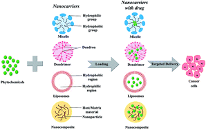
The advent of nanotechnology has revolutionized various scientific inventions, out of which the debut of nanomedicine is outstanding. Especially, research has embarked on nano-drug delivery for treating cancer. Natural compounds present in plants, namely phytochemicals, have been extensively exploited for their anticancer properties. Despite their excellent anticancer abilities, phytochemicals are limited by their low water solubility and poor bioavailability. However, the field of nanotechnology has overcome these limitations. This review focusses on various methods of nano-drug delivery of phytochemicals against the killer disease, cancer. Common carriers that were employed ranged from micelles, with a polymeric base, to dendrimers, liposomes and nanoparticles. The phytochemicals were found to become more soluble when delivered by the nanocarriers and exhibited a remarkable effect on the cancer cells, compared to their free form. More interestingly, the half-maximal dose of the phytochemical was reduced significantly when it was delivered by the nanocarrier. On the whole, this review encourages the idea of “cancer-nanotechnology” after in-depth clinical studies on these phytochemical-loaded nanocarriers. Moreover, it will epitomize the nanocarriers as a crusader in improving cancer chemotherapy by reducing undesired effects and will invigorate site-specific drug delivery.
Several population studies have shown that there are a few phytochemicals from fruits, vegetables and whole grains that are active against cardiovascular disease, diabetes and neurodegeneration. Apart from this, there is much literature evidence to show the various health benefits of phytochemicals present in plants.3–5 Among these, the potential ability of natural compounds against the killer disease, cancer, is significant. Regardless of these biological benefits possessed by the bioactive plant components, their usage in the medicinal field has a long way to go.6 Some of the factors that influence this are low water solubility, poor bioavailability and the requirement for high doses. Furthermore, many investigations have shown that these issues can be easily overcome by the nano-based delivery of these phytochemicals. Nano-based formulations have been found to improve solubility, bioavailability, and specific targeting, while reducing the doses and achieving steady-state therapeutic levels in cancer treatment.7
In general, all the biological processes that happen inside the body, including the origin and prognosis of cancer, are said to occur at the nano-level.8 Due to this, the application of nanotechnology in the field of medicine has flourished. Nanotechnology is one of the advanced multidisciplinary technologies that typically involves nano-level (10−9 m) investigation.9 Cancer is a major threat to mankind and is claimed to be the second leading cause of death worldwide. According to a survey, about 595
690 Americans are expected to die of cancer and 1
685
210 new cancer cases are expected to be diagnosed in the US during the year 2016.10 The available treatments for cancer vary from chemotherapy to targeted radiotherapy. Regardless of these different treatment procedures, there is no single medical procedure to treat cancer. Hence, the continuing threat of cancer deaths motivates scientists to steadily concentrate on the field of oncology. One such more advanced mode of treatment is cancer-nanotechnology, which combines nanomedicine and cancer.11
The application of nanotechnology in the field of oncology offers different approaches to making significant advances in both cancer diagnosis and treatment. These renewed approaches mainly deal with nanoparticles that are designed to specifically target and treat cancer by increasing the solubility and bioavailability of encapsulated drugs.12 Although there is a large number of studies to show the anticancer properties of phytochemicals, this review mainly deals with the nanotechnology-based drug delivery of several naturally available chemical compounds in the treatment of various types of cancer. Before leading into this, a brief introduction to chemotherapy is provided.
Some of the commercially available chemotherapeutic drugs include 5-fluorouracil, doxorubicin, paclitaxel, oxaliplatin, cisplatin, and epirubicin. All these chemotherapeutic drugs affect the cancer cells and induce apoptosis.16 Apoptosis is a programmed cell death in multicellular organisms involving a series of biochemical changes as well as morphological changes, finally leading to the death of the cell.17 A schematic representation of the action of anti-cancer drugs on cancer cells is given in Fig. 1. Chemotherapeutic drugs are generally consumed orally. However, not all drugs can be orally administered and hence some are injected subcutaneously, intra-muscularly or intravenously. Another major complexity of chemotherapy is the dosage at which the chemotherapeutic drugs can be administered. The dose is prescribed depending upon the weight and height of a person and also based on the type of drug.18 This is mainly because, if the dosage is low, then the treatment becomes ineffective, whereas if the dosage is high, the chemotherapeutic drugs cause side effects.
Adverse effects or side-effects of chemotherapy are a foremost concern of physicians and cancer patients around the globe. A wide range of side-effects are exhibited, depending on the type of medication used. As chemotherapeutic drugs affect fast-growing cells, other growing cells, such as hair cells, blood cells, bone marrow cells and the cells lining the mouth, stomach, and intestines, are also affected. Due to this, pregnant women are advised to undergo abortion before chemotherapy.19 Besides this, chemotherapy leads to immunosuppression, infertility, myelosuppression, gastrointestinal distress, typhlitis, anemia, fatigue, peripheral neuropathy, nausea and vomiting.20 All these undesirable effects of chemotherapeutic drugs trigger research to search for a harmless substance with significant anticancer properties for cancer treatment. In addition to new anticancer drugs, there is also much research on administration methods that can individually target cancer cells.
Phytochemicals are more abundant in the fruit and vegetable extracts, chocolate, and tea, which are consumed in day-to-day life. These compounds are usually described as non-essential nutrients as they minimally contribute to the growth, development and well-being of the individual. They have long remained unnoticed by healthy eaters or dietitians due to the fact that they are not required to sustain life. However, much research has been focused on these plant-derived bioactive compounds and their biological benefits. Phytochemicals, when consumed along with the diet, have proven effective in fighting many chronic diseases, especially cardiovascular disorders and cancer.26 Phytochemicals are found to be a great source of antioxidants. Most phytochemicals are found to excellently inhibit the proliferation of cells as well as angiogenesis, the two main trademark characteristics of cancer.27 Investigations of the anti-carcinogenic effects of phytochemicals under laboratory conditions and in various animal models have been conducted. The results obtained showed an improvement in the excretion of carcinogens, inhibition of mitosis, subdual of inflammatory processes, such as cyclooxygenase-2 expression, and induction of apoptosis at different stages of cancer.28 Phytochemicals are proven to fight against various types of cancers, such as lung cancer, prostate cancer, oral cancer, melanoma, leukemia, lymphoma, colon cancer and breast cancer.29 Before embarking on various studies related to the anticancer properties of phytochemicals, understanding their bioavailability is more important.
The bioavailability of the phytochemicals present in the food we consume is a critical factor that is extensively researched. Manach et al. found that the bioavailability of dietary polyphenols is influenced by the absorption in the gut, the microbiota metabolism, glucuronide excretion to the intestinal lumen, plasma kinetics, liver and gut metabolism, accumulation in tissues and bile, urinary excretion and a variety of metabolites in the bloodstream, bonding to albumin, cell assimilation and metabolism.32 The plasma concentrations of the various phytochemicals were calculated to study their complex bioavailability. For example, catechins are absorbed rapidly and are supposed to be absorbed in the small intestine, yet are affected by dimerization. Compounds such as epigallocatechin gallate and other catechin monomers are found to have similar properties. Meanwhile, the flavanones are slowly absorbed due to the attached disaccharides. The highest bioavailability was seen in the isoflavones subclass. In the case of aglycones and glucosides, the absorption was relaxed, suggesting absorption from the colon. Anthocyanins are quite rapidly absorbed, but their bioavailability seems to be the lowest of all flavonoids.33 Similarly, proanthocyanidins and hydroxycinnamic acids, which are abundant in the human diet, were also not absorbed. The plasma concentrations of the isoflavones were found to be 5 μmol L−1, whilst the plasma concentrations for proanthocyanidins did not exceed 1 μmol L−1. The bioavailability of phytochemicals is also influenced by the esterification process.34
For the most part, the phytochemicals present in plant foods are poorly absorbed by human subjects, in which they are rapidly metabolized and excreted. This is one of the major issues to be faced before implementing these phytochemicals in the war against cancer.
However, drug delivery involving nanocarriers has proven to increase the bioavailability of these drugs. Manzoor et al. studied the bioavailability of doxorubicin when delivered with nano-liposomes. The bioavailability of doxorubicin was improved by thermally sensitive liposomes released inside the tumor vasculature. The maximum penetration of free doxorubicin was previously limited to 34 μm, while this study depicted a diffusion distance of about 78 μm in both sides of the capillary bed of the tumor.35,36 Hence, the nano-drug delivery of phytochemicals is thought to increase the possibility of anticancer activity. The improved bioavailability during nanodrug delivery is related to the route by which the phytochemicals from food and the nanocarrier reach cancer cells. This is shown diagrammatically in Fig. 3. The anticancer effects of the nano-drug delivery of phytochemicals against various types of cancer is discussed in further chapters.
A particular study was carried out to overcome this limitation by Manzoor et al. The bioavailability of doxorubicin was improved by the release of thermally sensitive liposomes inside the tumor vasculature. The maximum penetration of doxorubicin was previously limited to 34 μm, while this study depicted a diffusion distance of about 78 μm in both sides of the capillary bed of the tumor.25,26 Therefore, the nano-drug delivery of the phytochemicals is thought to increase the possibility of anticancer activity. The anticancer effect of the nano-drug delivery of phytochemicals against various types of cancer is discussed in further chapters.
All these nanocarriers allow the delivery of hydrophobic and hydrophilic drugs throughout the body. As most of the human body contains water, this ability of the nanocarrier contributes to the therapeutic efficiency, while another is the targeted delivery of the drug.44 The drugs carried are harmful to the normal cells present in the human body. The nanocarriers carefully deliver the drugs to the specific site due to their site-specificity and smaller size. The four types of targeting characteristics of nanocarriers are passive, active, pH-sensitive and temperature-sensitive. The nanocarrier targets the delivery based on pH and temperature changes in cancer cells, and it may also be cloaked to match the circulation time by coating materials such as PEG. In the case of active targeting, the nanocarrier is provided with a cell-specific ligand.45 The foremost application of these nanocarriers is focused on cancer chemotherapy. This may be because of the lower pH, the higher temperature, the hydrophobic nature of the anticancer drugs and the potential need for specific targeting ability.46 Another major advantage of nanocarriers in cancer is the enhanced permeability and retention (EPR) effect. This involves the aggregation of macromolecules or nanoparticles in the extravasation in the tumor tissue during angiogenesis or the formation of new blood vessels. All the above factors make the application of nanotechnology in cancer beneficial.47 However, the major literature on the nano-drug delivery of various phytochemicals against major cancers is enumerated in the following subdivisions.
The novel anticancer drug, β-lapachone (β-lap), which is bioactivated by NAD(P)H:quinone oxidoreductase-1 (NQO1), an enzyme found specifically overexpressed in non-small-cell lung cancer (NSCLC), was delivered using nanoparticles. The β-lap was incorporated into poly(ethylene glycol)-co-poly(D,L-lactic acid) (PEG–PLA) polymer micelles using a film sonication procedure. The prepared micelles had core–shell architecture and were about 30 nm in size. The β-lap micelles were injected via the tail vein or caudal vein of mice with subcutaneous A549 lung tumors and the biodistribution was studied. The results showed a prolonged blood circulation and increased accumulation of β-lap. In addition, the in vitro administration of the micelles to LLC tumors led to DNA damage and PARP-1 hyperactivation.51 The basic principal involved in the formation of polymeric micelles is illustrated in Fig. 5. Zhang et al. examined the combinational effect of β-lapachone and paclitaxel micelles against A549 non-small-cell lung cancer (NSCLC) cells. The co-encapsulation of β-lapachone and paclitaxel in the PEG–PLA micelles had an encapsulation efficiency of 100.7 ± 2.2% and a drug-loading efficiency of about 100.3 ± 3.0%. The combinational β-lapachone and paclitaxel micelle was found to have an improved effect compared to the β-lapachone micelle and paclitaxel micelle alone. The combinational micelle had a significant antiproliferative effect at an IC50 of 0.16 μM, while the individual IC50 s were 4.5 μM and 0.32 μM for the β-lapachone micelle and the paclitaxel micelle, respectively. Thus, the two compounds were supposed to exhibit a synergistic effect against lung cancer.52
The flavonoid, luteolin, was encapsulated into a nanocarrier and tested against H292 lung cancer cells. The nanocarrier had a polymeric base, made up of polylactic acid and polyethylene glycol (PLA–PEG). The nanoparticle formed had a mean size of about 115 nm. The luteolin-loaded nanoparticle and free luteolin both showed antiproliferative activity against H292 cells. The IC50 of nano-luteolin was significantly less than that of free luteolin, showing that the nanosystem contributes to improved bioavailability. Similar results were observed in a colony formation assay.53
A pH-sensitive liposome was used to deliver ursolic acid, a triterpenoid compound, to MDA-MB-231 breast cancer cells. The pH-sensitive liposomes were prepared by the lipid hydration method. The liposomes had a mean diameter of 191.1 ± 6.4 nm and long-term stability. The liposomes were predominantly of a vesicle size less than 100 nm, promising good drug-loading efficiency. The MDA-MB cells were exposed to the pH-sensitive ursolic acid liposomes. The IC50 value of the ursolic acid liposomes was much lower than that of the free ursolic acid, indicating the improved anticancer activity of the nano-liposomes.56 Odeh et al. prepared two different kinds of liposomes, thymoquinone-loaded liposomes (TQ-LP) and thymoquinone loaded in liposomes modified with Triton X-100 (XLP). Thymoquinone is an herbal-derived phytochemical that has excellent chemopreventive properties and is hydrophobic in nature. Both the nanoparticles had a diameter of about 100 nm, while the entrapment efficiency was more than 90% for TQ-LP and 49.6% for XLP. Their biological activity was studied using both MCF-7 cancer cells and fibroblast cells. However, the TQ-LP effectively suppressed the proliferation of MCF-7 cells and exerted very low toxicity on normal periodontal ligament fibroblasts.57
The inhibitory effect of silibinin and D-α-tocopheryl polyethylene glycol 1000 succinate (TPGS) on breast cancer cells was recorded. Silibinin-loaded lipid nanoparticles containing TPGS and phosphatidylcholine were designed and prepared by a thin-film hydration method. The nanoparticles had an average size of 45 nm and the encapsulation efficiency of silibinin in the nanoparticle was 98.63 ± 0.30%. Cellular uptake studies showed that the drug content in MDA-MB-231 breast cancer cells after silibinin nanoparticle treatment for 24 h was about twice as much as that after free silibinin treatment. Corresponding results were observed for cell viability, invasion and migration assays. However, the silibinin-loaded nanoparticles strongly suppressed the invasive and migratory capabilities of MDA-MB-231 cells at a concentration of 20 μg mL−1 through the downregulation of the MMP-9 and Snail pathways. Thus, it was concluded that the silibinin-loaded TPGS nanoparticles could be used as a novel therapeutic agent against breast cancer.58 Sharma et al. used dendrimers to deliver the phenolic phytochemical, gallic acid, for inhibiting breast cancer cells. The dendrimers were made up of polyamidoamine (PAMAM) using Tomalia's divergent growth approach. The dendrimers provided a high degree of surface functionality and versatility for the gallic acid loaded onto them. The cytotoxicity of the gallic-acid-loaded PAMAM nanoparticles was found using MCF-7 human breast cancer cells. The MCF-7 cells were treated with the PAMAM dendrimer, gallic acid and the gallic-acid-loaded PAMAM nanoparticles. The IC50 values showed that the gallic-acid-loaded PAMAM nanoparticles had a synergistic antiproliferative effect on the growth of MCF-7 cells.59
830 new cases and 50
310 deaths due to colorectal cancer in the United States for 2016. It is more susceptible to the foods we consume as the colonic epithelial cells come into direct contact with them.10,48 Zheng et al. recorded the cytotoxicity of triptolide and triptolide-loaded polymeric micelles against HT-29 human adenocarcinoma cells. Triptolide is a diterpenoid tri-epoxide purified from the Chinese herb, Tripterygium wilfordii, which has anticancer properties but exhibits some other side effects. The triptolide-loaded polymeric micelles (TP-PM) were synthesized using methoxypoly(ethylene glycol)–poly lactic acid (MePEG–PLA) copolymer by a solvent evaporation method. Both the free triptolide and the TP-PM had a dose- and time-dependent effect on the HT-29 cells; however the inhibitory effects of TP-PM on the tumor cell growth were more significant for all incubation times and concentrations. In addition to this, the incubation of HT-29 cells with triptolide and the TP-PM also resulted in an increase in the caspase 3/7 activity, indicating apoptosis, with the highest apoptosis index at 6.96 after 48 h incubation with 10 ng mL−1 TP-PM. Hence, the polymeric micelles served as an excellent carrier of TP and reduced its toxicity.60 The small-molecule polyphenol compound, honokiol, was loaded into the self-assembled biodegradable star-shaped micelles and tested for chemotherapeutic effects. The biodegradable polymeric micelles were made up of monomethoxy poly(ethylene glycol) (MPEG) and poly(ε-caprolactone) (PCL) and loaded with the honokiol by a direct dissolution method assisted by ultrasonication. The average particle size of the obtained honokiol micelles was about 40 nm and these were used to treat CT26 murine colon carcinoma cells. The release rate of honokiol from the star-shaped polymeric micelles was slower. Nevertheless, they exhibited an antiproliferative effect against the CT26 cells in a dose-dependent fashion. Therefore, this star-shaped honokiol micelle may be used to design a new dosage form.61
Ravindran et al. studied the anti-proliferative properties of the bioactive phytochemical, Nigella sativa thymoquinone, loaded in poly(lactide-co-glycolide) (TQ-PLGA) nanoparticles using human colon cancer HCT116. The TQ-PLGA nanoparticles had an encapsulation efficiency around 94% and ranged between 150 and 200 nm in size. The TQ-PLGA nanoparticles had an effective anticancer effect against the HCT116 cells. Apart from this, the nanoparticles were active in inhibiting NF-kB activation and in suppressing the expression of cyclin D1, matrix metalloproteinase (MMP)-9, and vascular endothelial growth factor (VEGF) when compared to the free thymoquinone. On the whole, the results demonstrate that encapsulation of TQ into nanoparticles enhances its anti-proliferative effects.62 Luteolin (Lu), is a flavonoid with anticancer activity but it is said to have poor water solubility. This was delivered by monomethoxy poly(ethylene glycol)–poly(ε-caprolactone) (MPEG–PCL) micelles under in vivo conditions to evaluate the biodistribution and to C-26 colon carcinoma cells to determine their anticancer properties. The MPEG–PCL micelles encapsulated Lu by a self-assembly method. Fabricated Lu/MPEG–PCL micelles were water-soluble with an approximate size of about 38.6 nm and an encapsulation efficiency of about 98.32%. The pharmacokinetics of free luteolin and Lu/MPEG–PCL micelles was studied in rats, suggesting that the bioavailable concentration of luteolin was more when the Lu/MPEG–PCL micelles were used. Furthermore, the Lu/MPEG–PCL micelles inhibited the growth of C-26 colon carcinoma cells at an IC50 of 12.62 ± 2.17 μg mL−1. Hence, the study indicates that encapsulation of Lu into MPEG–PCL micelles created an aqueous formulation of Lu with a potential anticancer effect.63
Zhang et al. enlisted the effects of self-carried curcumin nanoparticles for in vitro and in vivo colon cancer therapy. The curcumin nanoparticles were prepared by the re-precipitation method and were then anchored to poly(maleic anhydride-alt-1-octadecene)–polyethylene glycol (C18PMH–PEG) on the surface by ultrasonication to improve the biocompatibility of the nanoparticles. The nanoparticles had a loading efficiency of about 78.5%, an encapsulation efficiency of about 95.8% and displayed sustained release behaviour without any initial burst. The curcumin nanoparticles were tested for their anticancer properties with CT-26 colon cancer cells. The results showed that the curcumin nanoparticles produced an 8-fold decrease in the half-maximal inhibitory concentration (IC50) values of the free curcumin (IC50 = 33.4 μM) compared to the curcumin nanoparticles (IC50 = 4.2 μM). Analogous results were seen in the in vivo testing. After administration of the curcumin and curcumin nanoparticles to CT-26 tumor-bearing nude mice, the tumor volumes were 87% and 32%, respectively. This shows that the curcumin nanoparticle has a greater effect than free curcumin on the tumor cells. However, the curcumin nanoparticles had no adverse effects or toxicity when investigated for in vivo systemic toxicity.64
In yet another study, the dihydrostilbenoid, combretastatin A-4 was co-encapsulated with doxorubicin and tested for anticancer properties under both in vitro and in vivo conditions. The compounds were loaded onto the RGD-modified liposomes. The cellular uptake of doxorubicin by the integrin-overexpressing B16 and B16F10 melanoma cells was improved by the disrupting agent, combretastatin A-4. Moreover, the co-encapsulated liposomes were more toxic towards the melanoma cells than the doxorubicin-loaded liposomes. Similarly, the combretastatin A-4 and doxorubicin-loaded liposomes exhibited the most pronounced tumor regression effect in male C57BL/6 mice inoculated with melanoma B16F10 cells. Thus, the combretastatin A-4 encapsulation improved the efficacy of the doxorubicin.67 Siddiqui et al. recently studied the anti-proliferative and pro-apoptotic effects of epigallocatechin 3-gallate (EGCG) encapsulated in chitosan nanoparticles on human melanoma cell growth, under both in vitro and in vivo conditions. The EGCG was loaded onto nanoparticles made up of polylactic acid–polyethylene glycol in order to enhance its bioavailability to the melanoma cells. The Mel 928 cells were used to test the effects of EGCG-loaded nanoparticles and free EGCG. The results showed that there was about an 8-fold dose advantage of this nano-formulation over native EGCG in retarding the growth of melanoma cells. Furthermore, a growth of Mel 928 tumor xenograft in the mice model was observed when EGCG-loaded nanoparticles were given. This inhibition included cell cycle phase arrest and changes in the level of cyclins D1 and D3 protein expression.68
240 women will die from ovarian cancer in the US alone and death from ovarian cancer is more common in North America and Europe than in Africa and Asia.10,48 The small-molecule polyphenol, honokiol, was loaded onto a nanoparticle and was delivered to ovarian cancer cells under laboratory conditions. The nanocarrier was manufactured with monomethoxy poly(ethylene glycol)–poly(lactic acid) (MPEG–PLA) by the ring opening polymerization method and then honokiol was loaded onto it by a solvent extraction method. The honokiol-loaded MPEG–PLA nanoparticles had a spherical appearance with a mean particle size of ca. 80 nm. The nanoparticles were found to release 53% of the drug within 24 h under laboratory conditions. Both the free honokiol and honokiol-loaded MPEG–PLA decreased the viability of A2780 human ovarian cancer cells with increasing concentration. However, the honokiol-loaded MPEG–PLA nanoparticles potentially inhibited the growth of A2780 cells at an IC50 was 8.45 μg mL−1. This was greater than the effect of the free honokiol.69 Yallapu et al. studied the improved therapeutic effects of curcumin loaded to poly(lactic-co-glycolide) (PLGA) nanoparticles and its anti-cancer effect against cisplatin-resistant A2780CP ovarian cancer cells. The fabricated nanoparticles were found have an average size of 560.4 nm and exhibited a sustained and controlled drug release of curcumin under in vitro conditions. The curcumin-loaded PLGA nanoparticles inhibited the growth of the A2780CP cells at an IC50 of about 13.9 μM, which was superior when compared to the effects of free curcumin. Similar results were seen in the examination of long-term effects and the curcumin-loaded PLGA nanoparticles reduced the number of cancer cells in the colony. These results suggest that the curcumin-loaded PLGA nanoparticles are an ideal therapeutic agent.70
The anticancer activity and molecular mechanism of resveratrol-bovine serum albumin nanoparticles (RES-BSANP) on subcutaneously implanted human primary ovarian carcinoma cells in nude mice was examined in recent years. A tumor was induced by injecting SKOV ovarian cancer cells into the nude mice, which were given 200, 100, and 50 mg kg−1 RES-BSANP or 0.5 mL RES once a week. Observation of the tumor progression showed that the RES-BSANP significantly retarded the growth of carcinomas in nude mice from the third week onwards, and the inhibition rate was markedly higher than in mice treated with RES. The RES-BSNAP was found to induce apoptosis by releasing cytochrome c and regulating caspase-3, 9, thereby indicating a mitochondrial apoptotic pathway.71 The hydrophobic drug, curcumin, was encapsulated into a hydrophilic polymeric core and delivered to the SKOV-3 ovarian cancer cells. The hydrophilic polymeric core was made up of poly(2-hydroxyethyl methacrylate) [PHEMA] nanoparticles. After loading the curcumin, the nanoparticles had a size of about 300 nm. In vitro investigations showed that the curcumin-loaded nanoparticles showed better tumor cell regression activity than free curcumin. Furthermore, they also showed a notable decrease in the G0/G1 phase cells. These nanosystems that delivered curcumin showed excellent biocompatibility when studied with a zebrafish embryo model.72
890 new cases of prostate cancer are expected in 2016, which means that 1 in 7 American men will be diagnosed with prostate cancer.10,48 The gallic acid ester derivative, epigallocatechin 3-gallate (EGCG) was delivered to prostate cancer cells by a biodegradable nanoparticle. Here, EGCG was combined with polylactic acid–polyethylene glycol polymeric profiles along with prostate-specific membrane antigen (PSMA)-targeting ligands. The pseudomimetic dipeptide, N-[N-[(S)-1,3-dicarboxypropyl]carbamoyl]-(S)-lysine (DCL) was the PSMA-targeting ligand. An anti-proliferative effect was exhibited by the targeted nanoparticles as well as the non-targeted nanoparticles on LNCaP androgen-sensitive human prostate adenocarcinoma cells. Both the nanoparticles were exposed to PCa prostate cancer cells. The growth inhibition exhibited by EGCG-loaded nanoparticles showed high efficacy and target specificity. Apart from this, the EGCG-loaded nanoparticles were found to be ineffective in inhibiting HUVEC proliferation. Thus the developed nanoparticles were proven to exhibit selective toxicity against prostate cancer.73 Zu et al. evaluated the enhanced targeting ability of folate-mediated EGCG bovine serum albumin nanoparticles (FA-EGCG-BSANP) against PC-3 prostate cancer cells. The nanoparticles were prepared by the desolvation method and possessed a mean particle size of about 200 nm with an entrapment efficiency of about 81.5%. The folate-mediated nanoparticles were found to exhibit concentration-dependent targeting to the PC-3 cells. The PC-3 cells' uptake of FA-EGCG-BSANP was 23.65 times that of the EGCG-BSANP. The folate present in the nanoparticles was found to improve the lethality toward PC-3 cells due to FA-EGCG-BSANP.74
In yet another experiment, the gallate, EGCG, was delivered to prostate cancer cells using a nanocarrier and its results were recorded. The nanocarrier was made up of polylactic acid–polyethylene glycol (PLA–PEG) and was tested under in vivo as well as in vitro conditions. The effect of nano-trapped EGCG was 10-fold higher than that of free EGCG against PCa prostate cancer cells. The IC50 was measured to be 3.74 μmol L−1 for the EGCG-loaded nanoparticles and there was a significant increase in pro-apoptotic Bax with a concomitant decrease in anti-apoptotic Bcl-2 in the PCa cells. The 22Rν1 cells were injected into the mice, which were then given EGCG-loaded nanoparticles as well as free EGCG. The outcome was similar to that under in vitro conditions and the tumor size was reduced significantly.75
Besides the gallate, the yellow polyphenol, curcumin, was also explored for its anticancer properties. Mukerjee et al. developed a curcumin-loaded nanoparticle and investigated its anticancer properties against the prostate cancer cell lines, LNCaP, PC3 and DU-145. Curcumin was loaded to poly(lactic-co-glycolic acid) (PLGA) nanospheres prepared by a solid/oil/water emulsion solvent evaporation method. The prepared nanospheres were found to have a mean size of about 45 nm with a biphasic drug release manner. The curcumin-loaded PLGA nanospheres were found to be more effective than the free curcumin. This was reflected in the half-maximal inhibitory value. The IC50 of curcumin-loaded PLGA nanospheres ranged from 20 μM–22.5 μM, while that of free curcumin ranged from 32 μM to 34 μM. Besides this, the curcumin-loaded PLGA nanospheres also strongly inhibited the NF-κB function when compared to free curcumin.76 In yet another study, the anticancer effect of nanoemulsion containing both curcumin and resveratrol was done. Separate nanoemulsions containing liposomes loaded with curcumin and liposomes loaded with resveratrol were prepared and co-administered to the PTEN-CaP8 cancer cells and the PTEN knockout mice with prostate cancer. In vitro studies showed that the curcumin with resveratrol effectively inhibited cell growth and induced apoptosis. In addition to this, the combination also significantly repressed the expressions of p-Akt, AR, cyclin D1 and mTOR proteins in PTENCap8 cells with loss of PTEN. Similarly, the mice that were given the combination (25 mg kg−1) had a more positive response than the mice administered with either lipo-curcumin or lipo-resveratrol (50 mg kg−1 each) for 7 weeks. A notable reduction in the prostate weight in the combination treatment group confirmed the decrease in the incidence of mouse prostatic intraepithelial neoplasia (mPIN) lesions. Apart from these, the availability of curcumin was improved during the co-administration of lipo-curcumin and lipo-resveratrol.77 A diagrammatic representation of extraction as well as loading of curcumin is given in Fig. 6.
230 new cases are expected to be diagnosed by the American Cancer Society in 2016.10,48 Lin et al. investigated the effect of berberine, a form of isoquinoline alkaloid, in the liposomal form against hepatoma. The berberine liposome was manufactured by the thin-film hydration/extrusion method and contained 5% mol polyethylene glycol (PEG). The berberine liposomes had an encapsulation efficiency of 14% and were exposed to HepG2 liver hepatocellular carcinoma cells. The berberine liposomes exhibited 2.5 times more toxicity against the HepG2 cells than the berberine solution. The berberine liposome significantly inhibited the growth of HepG2 cells at 1.67 μg berberine per mL and induced apoptosis through the caspase/mitochondria-dependent pathway. Furthermore, the liposome was tested under in vivo conditions in nude mice bearing the HepG2 tumor. The results showed that the berberine liposomes effectively reduced the size and weight of tumors, causing a reduction in the rate of elimination of berberine in both plasma and tissues. Therefore, the work demonstrated that the liposome was a good carrier for the berberine.80 On the other hand, the same phytochemical was delivered as a nanosuspension to the human hepatocytes, HepG2 and Huh7 cells. The nanosuspension consisted of the berberine phytochemical and D-α-tocopheryl polyethylene glycol 1000 succinate (TPGS) with an average size of 73.1 ± 3.7 nm. The nanosuspension had a significant effect on the growth of HepG2 and Huh7 cells at concentrations of 8.1 and 4.7 μg mL−1. In contrast, the free berberine suppressed the growth of the hepatoma cells at 18.3 and 6.5 μg mL−1 (HepG2 and Huh7 cells, respectively). In the in vivo experiment with H22 solid tumor-bearing mice, the berberine nanosuspensions had an inhibition rate of 63.7%, while the free berberine had an inhibition rate of only 41.4%. Thus, the nanosuspension has improved the availability of the phytochemical to the cancer cells.81
Studies to were done record the effect of a xanthonoid, gambogic acid, loaded in lactoferrine nanoparticles, against cancer. Lactoferrine is a cationic iron-binding glycoprotein and gambogic acid-lactoferrin nanoparticles (GL-NPs) were produced by nanoparticle albumin-bound (NAB) technology. The GL-NPs had a mean size of about 150 nm and an encapsulation efficiency of 7.2%, and the in situ intestinal perfusion displayed a good absorption of the gambogic acid from the GL-NPs. The GL-NPs showed almost an identical antiproliferative effect in the HepG2 liver carcinoma cells to that of the arginine solution of gambogic acid. Apart from this, the GL-NPs exhibited a high inhibitory rate when orally administered to S180 tumor mice and controlled the tumor growth. This was about 1.39-fold higher than the effect of an arginine solution of gambogic acid. These studies have also paved a preliminary way for the study of lactoferrine as an oral drug delivery carrier.82 Zhai et al. prepared apigenin-loaded polymeric micelles and tested their antiproliferative effect against HepG2 liver carcinoma cells. The polymeric micelles were composed of Pluronic P123 and Solutol HS 15 with an average diameter of 16.9 nm. They had an entrapment efficiency and drug loading of 96.36% and 1.32%, respectively. Around 84% of the apigenin was delivered by the polymeric micelles, displaying a sustained drug-release behaviour. The IC50 values on HepG2 cells for apigenin-loaded polymeric micelles and free apigenin solution were 5.57 μg mL−1 and 20.19 μg mL−1, respectively. The growth of HepG2 cells was significantly reduced by the apigenin micelles at lower concentrations than the free apigenin, which may be related to its improved hydrophilic characteristics.83
The diarylheptanoid compound, curcumin, was conjugated to gum arabic and was used against hepatoma cell lines. Gum arabic is a polysaccharide substance, which is used to improve the solubility of curcumin. The self-assembled conjugates had a spherical structure and a mean size of 270 nm. When tested, the formed curcumin conjugate had improved the solubility and stability of curcumin at physiological pH. Apart from this, the curcumin conjugation retarded the growth of the HepG2 hepatocellular carcinoma cells. They also exhibited a higher targeting ability toward the cells due to the galactose groups present in gum arabic.84 Beside the berberine nanosuspensions, resveratrol nanosuspensions were also prepared recently. The resveratrol nanosuspension contained resveratrol and poloxamer 188 and was manufactured using a high-pressure homogenization technique. The average size of the nanosuspension was 159 nm and it was evaluated for its effect on HepG2 cells, along with free resveratrol. The results of an MTT assay showed that the resveratrol inhibited the proliferation of HepG2 cells at an IC50 of 2.91 μg mL−1. In contrast, the free resveratrol had a similar effect only at 7.13 μg mL−1. Hence, these results suggest that the delivery of the resveratrol nanosuspension is a promising approach for treating tumors.85
560 deaths predicted in 2016 alone. It is usually due to usage of tobacco and obesity.10,48 The polyphenol, curcumin, was explored for its anticancer properties against pancreatic cancer. Nanocurcumin or curcumin-encapsulated nanoparticles were synthesized using micellar aggregates of cross-linked and random copolymers of N-isopropylacrylamide (NIPAAM), with N-vinyl-2-pyrrolidone (VP) and poly(ethylene glycol)monoacrylate (PEG-A). The developed nanocurcumin was found to readily disperse in an aqueous medium, displaying its hydrophilic nature. The MiaPaca pancreatic cancer cell line was treated with curcumin and nanocurcumin. Both of these significantly suppressed the growth of pancreatic cancer cells at 10 and 15 μM, respectively. Furthermore, the nanocurcumin obstructed the activation of nuclear factor kappa B (NFκB) and downregulated the IL-6, IL-8, and TNFα cytokines, thereby inducing cellular apoptosis. Thus, it was found that the nanocurcumin possessed all the properties of curcumin with better solubility.86 Wei et al. entrapped the phytochemical, curcumin, as an ester to cholesteryl-hyaluronic acid (CHA) nanogel and employed this in a targeted delivery to CD44-expressing drug-resistant MiaPaca cancer cells. The curcumin-conjugated nanoparticle was 20 nm in diameter with a spherical structure. Gastrointestinal stability studies showed that the curcumin-loaded nanoparticles were absorbed in the gastrointestinal tract and then entered the blood circulation. Thus, it was concluded that the oral administration of the nanoparticles with curcumin would lead to 2–5% loss. Apart from this, the nanoparticle induced apoptosis in cancer cells, suppressing the expression of NF-κB, TNF-α, and COX-2 cellular targets in a similar way to free curcumin. Moreover, the curcumin-loaded nanoparticles suppressed the tumor growth by up to 13-fold in a 4T1 mice model injected with the MiaPaca cells. Hence, the nano-drug delivery of curcumin is found to be an ideal therapy for cancer.87
Kesharwani et al. produced parenterally administrable nano-micelles of 3,4-difluorobenzylidene curcumin (CDF) for treating pancreatic cancer. The 3,4-difluorobenzylidene curcumin is a non-toxic analogue to the curcumin known for its high anti-cancer activity, and it showed improved pancreas-specific accumulation in vivo, compared with curcumin. The nano-micelles were prepared by self-assembling the styrene–maleic acid copolymer (SMA) with CDF, using non-covalent hydrophobic interactions. These were found to have a sustained drug release behaviour when exposed to MiaPaCa-2 and AsPC-1 pancreatic cancer cell lines. The free CDF and the SMA–CDF nanomicelles showed toxicity against both the cell lines and the effect of the nano-micelles was superior to that of the free CDF. The IC50 was calculated as 230 ± 4.68 nM for MiaPaCa-2 cells and 710 ± 3.81 nM for AsPC-1 cells.88 In addition to this, the same research group evaluated the response of CDF loaded to hyaluronic acid-conjugated polyamidoamine dendrimers given to CD44-overexpressing MiaPaCa-2 pancreatic cancer cells. The nanocarrier was made of poly(amidoamine) (PAMAM) and hyaluronic acid (HA) as a targeting ligand and then loaded with the CDF (HA–PAMAM–CDF). The whole dendrimer system had a particle size of 9.3 ± 1.5 nm. These displayed a dose-dependent cytotoxicity against MiaPaCa-2 and AsPC-1 human pancreatic cancer cells. The half-maximal value was found to have a 1.71-fold increase in the presence of the HA-ligand. However, the developed nanocarrier was found to be an excellent therapeutic device against CD44-overexpressing pancreatic cancer.89
450 deaths were estimated by the American Cancer Society.10,48 Anand et al. investigated the antiproliferative activity of curcumin-loaded PLGA nanoparticles under laboratory conditions as well as the improved bioavailability of this method by in vivo examination. The curcumin drug was loaded to a polymer-based nanoparticle made up of poly(lactide-co-glycolide) (PLGA) and a stabilizer, polyethylene glycol (PEG), with 97.5% encapsulation efficiency. KBM-5 human chronic myeloid leukemia cells were then exposed to the curcumin-loaded PLGA–PEG. It was found that the curcumin-loaded nanoparticles showed an improved cellular uptake, inhibition of TNF-induced NF-κB activation, suppression of NF-κB-regulated proteins involved in cell proliferation (cyclin D1), invasion (MMP-9), angiogenesis (VEGF) and induced apoptosis compared to free curcumin. In the case of the mice, 2.5 mg kg−1 of curcumin nanoparticles were injected intravenously. HPLC analysis showed that the curcumin nanoparticles were more bioavailable and had a longer half-life than free curcumin.92 In another study, a co-formulation of doxorubicin and curcumin in poly-(D,L-lactide-co-glycolide) nanoparticles suppressed the development of multidrug resistance in K562 chronic myeloid leukemia cells. The doxorubicin and curcumin-loaded PLGA nanoparticles were prepared by the single emulsion solvent evaporation technique with an encapsulation efficiency of 46% and 86%, respectively. When used to treat K562 cells, the cellular uptake of dual drug-loaded NPs was nearly 8 times higher than the dual drug in solution. Apart from this, the nanoparticles exhibited significant growth inhibition at an IC50 value of 0.1 μg mL−1 when the nanoparticles were administered in equivalent concentrations, along with a gradual decrease in the expressions of MDR1 and BCL-2 at the mRNA level. Overall, this combinational strategy has more noteworthy promise than that of the drugs alone.93
Wang et al. studied the antileukemia mechanism of the multifunctional Chinese traditional medicine, emodin, when conjugated to D-α-tocopheryl polyethylene glycol 1000 succinate (TPGS) to form liposomes. The liposomes had a high encapsulation efficiency of about 95.2% ± 3.0% and had a particle size of 121.1 ± 44.9 nm. The emodin-TPGS liposomes were found to have an increased cytotoxicity toward L1210 and K562 leukemia cell lines. This cytotoxicity included the regulation of protein levels of myeloid cell leukemia 1 (Mcl-1), B-cell lymphoma-2 (Bcl-2) and Bcl-2-associated X (Bax). A bio-distribution study showed that the emodin-TPGS liposomes improved the bioavailability of emodin by 1.7 times compared to the free emodin in lungs and kidney.94 The antiproliferative activity of citrus polymethoxylated flavone nobiletin-loaded chitosan nanoparticles was recorded in parallel. The nobiletin-loaded chitosan nanoparticles were formed via Schiff-base formation and had a loading efficacy of 7.0%. An in vitro experiment was carried out using RAW264.7 Abelson murine leukemia virus-induced tumor cell lines and L-929 normal subcutaneous connective tissue at various time intervals. It was found that both the raw nobiletin as well as the nobiletin-loaded chitosan nanoparticles suppressed the growth of RAW264.7 cells, while they did not affect the growth of L-929. Moreover, the nobiletin-loaded chitosan nanoparticles showed considerable inhibition at an IC50 of 8 μg mL−1 toward cancerous cells, revealing their great potential for applications in cancer chemotherapy.95
Rahman et al. studied the antileukemic effect of a natural dietary lipophilic compound when loaded to nanostructured lipid carriers. The zerumbone-loaded nanostructured lipid carriers were formed by a high-pressure homogenization technique. The obtained ZER-NLC particles had an average size of 52.68 ± 0.1 nm and a drug-loading efficiency of about 7.92%. The drug release of zerumbone from ZER-NLC was about 46.7% over 48 hours. When used to treat Jurkat acute lymphoblastic leukemia cells, the ZER-NLC significantly inhibited their growth with an IC50 of 5.64 ± 0.38 μg mL−1. Thus, the study suggests the ZER-NLC is a suitable sustained-release drug carrier system for the treatment of leukemia.96
The nano-drug delivery of anticancer agents has interested many researchers in recent years. This is primarily due to their reliable and targeted delivery, increased drug efficiency, and improved cellular interaction, along with less toxicity to the surroundings. These advantages are the same for synthetic anticancer drugs and phytochemicals with an anticancer effect. Fascinatingly, the response elicited by the phytochemicals delivered using nanocarriers is similar to that of the same phytochemical in the free form. However, the half-maximal inhibitory concentration of the drug is reduced to a greater extent when nanotechnology is employed (Fig. 7). These natural compounds are found to suppress the growth of cancer cells by inducing programmed cell death, which is indicated by notable changes, such as DNA damage,49 increased ROS generation,49,65 release of cytochrome C,71,98 activation of caspases 3/7,60,71,100 cell cycle arrest,72 activation of NF-κB86,92 and downregulation of MMP, BaX, cyclin D and VEGF,62,92,94 along with visible morphological apoptotic changes.50 The different targets of phytochemicals loaded inside nanocarriers are given in Fig. 8.
The review summarized the nano-drug delivery of more than fifteen phytochemicals against various types of cancer. Among them, the yellow diarylheptanoid compound, curcumin, has been exploited against almost all types of cancer enlisted in the review. Phytochemicals such as apigenin, resveratrol, honokiol and epigallocatechin gallate (EGCG) are other commonly tested compounds. The phytochemicals are found to become more hydrophilic in nature when attached to a nanocarrier. From our studies, it is found that the most regularly preferred nanocarrier is a micelle with a polymeric base. A micelle is a nanostructure that has both a hydrophobic part and a hydrophilic part. The common polymeric bases employed for the micelles are poly(ethylene glycol) (PEG), polylactic-acid (PLA), poly-lactide-co-glycolide (PLGA), and methoxy poly(ethylene glycol) (mPEG), which are known for their good biocompatibility and biodegradable behaviour. Apart from the nano-micelles, dendrimers, liposomes, nanocomposites and nanoparticles act as nanocarriers for delivering drugs. The various phytochemical and their nanocarriers along with the cell line tested is given in Table 2.
| Type of cancer | Cell line tested | Phytochemical | Nanocarrier | Reference |
|---|---|---|---|---|
| Lung | A549 human lung adenocarcinomic cells | Honokiol | Poly(ε-caprolactone)-poly(ethylene glycol)–poly(ε-caprolactone) copolymer micelle | 49 |
| NCI-H460 non-small-cell lung carcinoma cells | Ferulic acid | Poly-D,L-lactide-co-glycolide (PLGA) nanoparticles | 50 | |
| A549 human lung adenocarcinomic cells | β-Lapachone | Poly(ethylene glycol)-co-poly(D,L-lactic acid) (PEG–PLA) polymer micelles | 51 | |
| A549 human lung adenocarcinomic cells | β-Lapachone and paclitaxel | Poly(ethylene glycol)-co-poly(D,L-lactic acid) (PEG–PLA) polymer micelles | 52 | |
| H292 lung cancer cells | Luteolin | Polylactic acid and polyethylene glycol (PLA–PEG) nanoparticles | 53 | |
| Breast | MCF-7 human breast adenocarcinoma cell line and MDA-MB-453 | Curcumin | Silk fibroin and chitosan (SFCS) polymer nanoparticles | 54 |
| SK-BR-3 human breast cancer cells | Noscapine | Human serum albumin (HSA) nanoparticles | 55 | |
| MDA-MB-231 breast cancer cells | Ursolic acid | pH-sensitive liposomes | 56 | |
| MCF-7 human breast adenocarcinoma cell line | Thymoquinone | Liposomes modified with Triton X-100 (XLP) | 57 | |
| MDA-MB-231 breast cancer cells | Silibinin | Lipid nanoparticles containing D-α-tocopheryl polyethylene glycol 1000 succinate (TPGS) and phosphatidylcholine | 58 | |
| MCF-7 human breast adenocarcinoma cell line | Gallic acid | PAMAM dendrimers | 59 | |
| Colorectal | HT-29 human adenocarcinoma cells | Triptolide | MePEG–PLA copolymer micelles | 60 |
| CT26 murine colon carcinoma cells | Honokiol | Monomethoxy poly(ethylene glycol) (MPEG) and poly(ε-caprolactone) (PCL) star-shaped micelles | 61 | |
| HCT-116 human colon cancer cells | Thymoquinone | Poly(lactide-co-glycolide) (PLGA) nanoparticles | 62 | |
| C-26 colon carcinoma cells | Luteolin | Monomethoxy poly(ethylene glycol)–poly(ε-caprolactone) (MPEG–PCL) micelles | 63 | |
| CT-26 colon cancer cells | Curcumin | Curcumin nanoparticles anchored with C18PMH–PEG on the surface | 64 | |
| Melanoma | A375 skin melanoma and HaCaT keratinocytes | Apigenin | Poly(lactic-co-glycolide) nanoparticles | 65 and 66 |
| B16 and B16F10 melanoma cells | Combretastatin A-4 and doxorubicin | RGD (arginylglycylaspartic acid)-modified liposomes | 67 | |
| Mel 928 melanoma cells | Epigallocatechin 3-gallate (EGCG) | Polylactic acid–polyethylene glycol nanoparticles | 68 | |
| Ovarian | A2780 human ovarian cancer cells | Honokiol | Monomethoxy poly(ethylene glycol)–poly(lactic acid) (MPEG–PLA) nanocarrier | 69 |
| A2780CP ovarian cancer cells | Curcumin | Poly(lactic-co-glycolide) (PLGA) nanoparticles | 70 | |
| SKOV ovarian cancer cells | Resveratrol | Bovine serum albumin nanoparticles | 71 | |
| SKOV-3 ovarian cancer cells | Curcumin | Poly(2-hydroxyethyl methacrylate) [PHEMA] nanoparticles | 72 | |
| Prostate | LNCaP human prostate adenocarcinoma cells and PCa prostate cancer cells | Epigallocatechin 3-gallate (EGCG) | Polylactic acid–polyethylene glycol prostate-specific membrane antigen (PSMA) ligands | 73 |
| PC-3 human prostate cancer cells | Bovine serum albumin nanoparticles | 74 | ||
| PCa prostate cancer cells | Polylactic acid–polyethylene glycol (PLA–PEG) nanocarrier | 75 | ||
| LNCaP human prostate adenocarcinoma cells, PC3 human prostate cancer cells and DU-145 PC3 human prostate cancer cells | Curcumin | Poly(lactic-co-glycolic acid) (PLGA) nanospheres | 76 | |
| PTEN-CaP8 Mouse prostate epithelium cancer cells | Curcumin and resveratrol | Curcumin and resveratrol nanoemulsions | 77 | |
| Cervical | HeLa cervical cancer cells | Curcumin | Bovine casein micelles | 78 |
| Alginate–chitosan–pluronic composite nanoparticles | 79 | |||
| Hepatoma | HepG2 hepatocellular carcinoma cells | Berberine | Berberine polyethylene glycol (PEG) liposome | 80 |
| Berberine | Berberine and D-α-tocopheryl polyethylene glycol 1000 succinate (TPGS) nanosuspension | 81 | ||
| Gambogic acid | Lactoferrine nanoparticles | 82 | ||
| Apigenin | Pluronic P123 and solutol HS 15 polymeric micelles | 83 | ||
| Curcumin | Gum arabic nanoparticles | 84 | ||
| Resveratrol | Resveratrol and poloxamer 188 nanosuspension | 85 | ||
| Pancreatic | MiaPaca pancreatic cancer cell line | Curcumin | N-Isopropylacrylamide (NIPAAM), with N-vinyl-2-pyrrolidone (VP) and poly(ethylene glycol)monoacrylate (PEG-A) | 86 |
| Cholesteryl-hyaluronic acid (CHA) nanogel | 87 | |||
| Difluorobenzylidene curcumin (CDF) | Styrene–maleic acid copolymer (SMA) micelles | 88 | ||
| Poly(amidoamine) (PAMAM) and hyaluronic acid nanocarrier | 89 | |||
| Oral | KB human oral cancer cells | Dihydroartemisinin | Methoxy poly(ethylene glycol)/poly(L-lactic acid) (mPEG) micelles | 90 |
| Ellagic acid | Chitosan nanoparticles | 91 | ||
| Leukemia | KBM-5 myeloid leukemia cells | Curcumin | Poly(lactide-co-glycolide) (PLGA) and polyethylene glycol (PEG) nanoparticles | 92 |
| K562 myeloid leukemia cells | Doxorubicin and curcumin | Poly-(D,L-lactide-co-glycolide) nanoparticles | 93 | |
| L1210 mouse lymphocytic leukemia cells and K562 myeloid leukemia cells | Emodin | D-α-Tocopheryl polyethylene glycol 1000 succinate (TPGS) liposomes | 94 | |
| RAW264.7 abelson murine leukemia virus-induced tumor cell lines | Nobiletin | Chitosan nanoparticles | 95 | |
| Jurkat acute lymphoblastic leukemia cells | Zerumbone | Nanostructured lipid carriers | 96 | |
| Glioma | C6 glioma cells | Resveratrol | mPEG–PCL nanoparticles | 97 |
| Neuroblastoma | SH-SY5Y neuroblastoma | Curcumin | Apolipoprotein E3 mediated poly(butyl) cyanoacrylate nanoparticles | 98 |
| Myeloma | Myeloma cells | Curcumin | Poly(oxyethylene) cholesteryl ether (PEG-Chol) micelles | 99 |
| Gastric | SGC7901 gastric cancer cells | Ursolic acid | Methoxy poly(ethylene glycol)–polycaprolactone (mPEG–PCL) nanocarrier | 100 |
The nano-drug delivery of phytochemicals has been extensively investigated in a large number of cell studies and a few animal studies. Preclinical testing with normal mice, nude mice and knockout mice bearing cancer cells has shown suppression of tumor growth. Besides these positive aspects, there is a lack of studies that deal with the toxicity and other adverse effects that are produced by the nanocarriers. Hence, more in-depth studies relating to the genotoxicity of the nanocarriers and other possible side effects should be initiated. Moreover, the phytochemicals are known to exhibit a synergistic effect against cancer cells. They have the ability to increase a particular property when used in combination. Previous studies have shown that phytochemicals when combined have improved cytotoxicity towards cancer cells.101 It would be interesting to investigate the outcome of these combinations with nano-drug delivery for cancer. In addition to this, clinical studies on nano-drug delivery as well as the mode of administration should be carried out to promote them in the field of medical oncology.
| This journal is © The Royal Society of Chemistry 2016 |