DOI:
10.1039/C6RA07733A
(Paper)
RSC Adv., 2016,
6, 54661-54667
Mycobacterial IclR family transcriptional factor Rv2989 is specifically involved in isoniazid tolerance by regulating the expression of catalase encoding gene katG†
Received
24th March 2016
, Accepted 24th May 2016
First published on 26th May 2016
Abstract
Transcriptional factors are essential for bacteria to adapt diverse environmental stresses, especially upon exposure to antibiotics. Mycobacterium tuberculosis, the causative agent of tuberculosis which inflicts around one third of the global population, contains three IclR family transcriptional factors, namely Rv1719, Rv1773c and Rv2989. In this study, MSMEG_2386, the homolog of Rv2989 in Mycobacterium smegmatis, was deleted by homologous recombination and complemented. The gene did not affect the growth in conventional culture. However, the growth of deletion mutants upon isoniazid (INH) exposure was severely delayed. This growth defect is specific to INH and not observed in other antibiotics tested, including rifampicin, capreomycin, norfloxacin and ethionamide. The transcription of katG and enzymatic activity of its encoding product catalase were elevated in the MSMEG_2386 deleted M. smegmatis mutants. The survival of MSMEG_2386 deletion mutant within U937 macrophages was markedly reduced compared with the wild type strain. These results improve our understanding of the role of IclR family transcriptional factors in INH resistance in mycobacteria.
Introduction
Transcriptional factors are essential for bacteria to coordinate the response to stresses such as temperature fluctuations, nutrient deprivation, desiccation, and toxins.1–3 IclR (Isocitrate lyase Regulator) family transcription regulators are widespread among bacteria, fungi and archaea. The IclR family transcriptional factors can be repressors or activators, and dual-function proteins as activators of some genes and repressors of others (including autoregulation).1 IclR has multiple targets, including the key enzymes of the glyoxylate shunt in Enterobacteriaceae, multidrug efflux pumps, degradation of aromatic compounds, quorum-sensing signals, determinants of plant pathogenicity and sporulation.2 Several IclR family members are well-characterized, such as the E. coli IclR glyoxylate shunt repressor regulating acetate utilization encoded by aceBAK operon,4,5 the Erwinia chrysanthemi pectin degradation pathway repressor KdgR,6 and the glycerol catabolism pathway repressor GylR of Streptomyces coelicolor.7 Some IclR-like proteins have been shown to regulate aromatic acid metabolism,8 sporulation and virulence.9 IclR family regulator TtgV represses the expression of the Pseudomonas putida TtgGHI efflux pump responsible for the efflux of antibiotics and solvents.4,10
Mycobacterium tuberculosis is one of the most successful human intracellular pathogens, and claims the lives of nearly 2 million people globally each year. Transcriptional regulation is essential for the success of M. tuberculosis. Lsr2, a histone-like protein highly conserved in mycobacteria, inhibits a wide variety of DNA-interacting enzymes to regulate genes induced by antibiotics and those associated with inducible multiple tolerance.11 Isoniazid (INH), one of the pivotal drugs for tuberculosis treatment, is a prodrug that is converted into the active form by the mycobacterial catalase-peroxidase (katG).12 Many genes have been reported to be involved in INH resistance or tolerance, such as MDP1, a histone-like protein negatively regulating katG expression,13 MSMEG_0535, a GntR family transcriptional regulator regulating the MSMEG_0534 permease, and BCG_2013, a universal stress protein (USP).14 Exhaustive elucidation of genes involved in INH resistance will promote our understanding of its mechanism of action, and inspire the discovery of novel antibiotics based on INH.
IclR family members are widespread and conserved among mycobacteria. However, little information is available on their function. Based on the transcriptome data of Clifton Barry15 and our observation, we speculated that IclR family transcriptional factors might play a role in INH tolerance or resistance. To test this hypothesis, the non-pathogenic, fast-growing M. smegmatis, a widely used facile surrogate model for a slow-growing and highly pathogenic mycobacteria, was employed in this study. The results showed that the IclR family transcription factor Rv2989 and its homolog encoded by the MSMEG_2386, plays a role in mycobacterial INH resistance by repressing the transcription of katG (INH activator) and fur, which belong to the same operon. To our knowledge, this is the first report linking the IclR family transcriptional factor with INH resistance.
Results
IclR family transcription factor Rv2989 is well conserved among mycobacteria
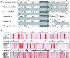
At least three IclR family transcription factors are predicted in the M. tuberculosis genome, namely Rv1719, Rv1773c and Rv2989.16 Phylogenetic analysis showed that Rv2989, instead of Rv1719 and Rv1773c, is the most conserved (Fig. 1A) and present in different clades, suggesting an independent origin of Rv2989 (Fig. S1†). The amino acid identity between Rv2989 and its homologs is greater than 80% in all cases (Table 1); its neighboring genes leuD and leuC are conserved too (Fig. 1A). The secondary structure elements and important motifs of Rv2989 are also conserved (Fig. 1B) even in distant homologs, such as those in Thermotoga maritima and Rhodococcus, identified by a structure-based homologous protein searching algorithm.17
 |
| Fig. 1 Rv2989 is a conserved IclR family transcriptional factor among mycobacteria. (A) Genomic context of Rv2989 among mycobacteria. The grey shading represents regions of conservation between genomes. (B) Structure-based sequence alignment of Rv2989 and other homologue proteins with the IclR transcription factor derived from Thermotoga maritima indicated by the PDB ID of their crystal structure 1MKM. α-Helices and β sheets are indicated above the residues as coils and arrows, respectively. | |
Table 1 Percentage amino acid identity and similarity between IclR orthologs
strains |
Homologous genes (identity with M. tuberculosis H37Rv%) |
M. tuberculosis H37Rv |
Rv1719 |
Rv1773c |
Rv2989 |
M. tuberculosis H37Ra |
|
|
MRA_3018 (99.57) |
M. bovis AF2122/97 |
Mb1748 (100) |
Mb1802c (100) |
Mb3013 (100) |
M. marinum M |
|
|
MMAR_1725 (84.98) |
M. leprae TN |
ML1375 (pseudogene) |
|
ML1686 (pseudogene) |
M. smegmatis mc2155 |
|
MSMEG_2483 (29.57) and MSMEG_6757 (25) |
MSMEG_2386 (81.12) |
Rv2989 homolog MSMEG_2386 is associated with isoniazid susceptibility
To explore the function of Rv2989, we used M. smegmatis mc2155 as a model. A deletion strain of an Rv2989 homolog MSMEG_2386 (ΔMS_2386) was constructed by homologous recombination (Fig. 2A) and confirmed by PCR (Fig. 2B). The complemented strain was constructed by a multiple copy plasmid vector pMV261 and the M. smegmatis mc2155 wild type gene MSMEG_2386 (Fig. 2B). The growth rates of wild-type M. smegmatis mc2155 (WT), ΔMS_2386 and ΔMS_2386-com were monitored by measuring OD600 with the same initial inoculation. As shown in Fig. 3A, there was no growth difference between WT and ΔMs2386, demonstrating that knockout MSMEG_2386 does not affect the growth of M. smegmatis in 7H9 or M9 medium. Upon exposure to INH, initial WT cell growth was not disturbed and entered the logarithmic phase 45 h post inoculation, reaching the stationary phase after 66 h post inoculation (Fig. 3A). In contrast, ΔMS_2386 growth was not obvious until 66 h post inoculation and entered the stationary phase after 92 h post inoculation (Fig. 3A). The 21 h delay suggested that ΔMS_2386 growth was severely compromised upon the stress of INH. ΔMS_2386-com can completely revert the growth defect of ΔMS_2386 in the presence of INH, and sometimes even thrive to a greater extent. A similar phenotype also was found when grown in a minimal M9 medium (Fig. S2†). To confirmed that ΔMS_2386 was relatively more susceptible to INH, the growth rates of wild-type M. smegmatis mc2155 (WT), ΔMS_2386 and ΔMS_2386-com were monitored on 7H10 solid medium containing different concentration INH (Fig. 3B). Consistent with the growth curves in liquid medium, ΔMS_2386 exhibited increased growth defect in the presence of INH. We determined the MICs of INH for each strain using the broth microdilution method previously described. As presented in Table 2, the MIC of ΔMS_2386 was 4 μg ml−1, whereas the MICs for other strains were 2-fold higher. In contrast, M. smegmatis WT, ΔMS_2386 and ΔMS_2386-com were equally susceptible to the other drugs tested, including rifampin, capreomycin and norfloxacin (Table 2). To explore whether Rv2989 and MSMEG_2386 possess the same function, Rv2989 was used to complement ΔMS_2386. As shown in Fig. S3,† Rv2989 can completely revert the growth defect of ΔMS_2386 in the presence of INH on 7H10 plate.
 |
| Fig. 2 Construction of MSMEG_2386 knockout strain. (A) The upper panel shows the genetic organization of the MSMEG_2386 gene locus. Gene location and orientation are indicated by large arrows. Primer location and orientation are shown by small arrows. (B) The lower panel shows the PCR for verification of MSMEG_2386 knockout strain. 1, wild type; 2, ΔMS_2386; 3, ΔMS_2386-com. | |
 |
| Fig. 3 Growth of WT, ΔMS_2386 and ΔMS_2386-com under INH exposure. (A) Growth curves of WT, ΔMS_2386 and ΔMS_2386-com in 7H9 medium (supplemented with 0.05% Tween80 and 0.2% glycerinum) with or without INH (3 μg ml−1). The OD600 were determined at an interval of 3 h. (B) Ten-fold serial dilutions of WT, ΔMS_2386 and ΔMS_2386-com were spotted on Middlebrook 7H10 containing concentration of INH. Then the result was recorded when incubated at 37 °C for 3 days. | |
Table 2 MIC of various antibiotics for WT, ΔMS_2386 and ΔMS_2386-coma
Strains |
MIC/μg ml−1 |
INH |
RIF |
CAP |
NOR |
ETH |
INH, isoniazid; RIF, rifampicin; CAP, capreomycin; NOR, norfloxacin; ETH, ethionamide. |
WT |
8 |
5 |
1.25 |
2 |
10 |
ΔMS_2386 |
4 |
5 |
1.25 |
2 |
10 |
ΔMS_2386-com |
8 |
5 |
1.25 |
2 |
10 |
MSMEG_2386 knockout does not affect the susceptibility of M. smegmatis to ethionamide
InhA is the shared target of activated INH and ethionamide (ETH). INH is activated by KatG, while ETH is also an anti-tuberculosis prodrug but activated by EthA (Fig. 4A). The observed INH susceptibility in the Rv2989 homolog deletion mutant ΔMS_2386 might contribute to the step of prodrug activation or the effect of the activated prodrug. If this is the effect of activated prodrugs, an obvious growth defect should be observed between the wild type and deletion mutant. To discriminate the two possibilities, the growth of WT, ΔMS_2386, and ΔMS_2386-com upon ETH treatment was compared. The same growth ratio was found on the plate containing the indicated concentration of ETH (Fig. 4B), and the MIC of ETH was 10 μg ml−1 among three strains (Table 1). These results suggested that the MSMEG_2386-mediated INH susceptibility change is independent of inhA, but katG may be responsible for the specific activation of the prodrug INH.
 |
| Fig. 4 MSMEG_2386 knockout does not affect the susceptibility of M. smegmatis to ETH. (A) The action mechanism of INH and ETH. (B) Ten-fold serial dilutions of WT, ΔMS_2386 and ΔMS_2386-com were spotted on a Middlebrook 7H10 containing a concentration of ETH. The result was then recorded after incubation at 37 °C for 3 days. | |
The transcription of katG was elevated in ΔMS_2386
INH is a prodrug that is converted into the active form by the mycobacterial catalase-peroxidase, KatG. The above results suggest that MSMEG_2386 knockout induces INH susceptibility via katG in an inhA-independent manner. To investigate the effect of MSMEG_2386 on the expression of katG, total RNA was extracted from M. smegmatis WT, ΔMS_2386 and ΔMS_2386-com cells and used for qRT-PCR quantification of katG expression. The levels of katG mRNA were determined by the comparative threshold cycle method and normalized to sigA level. As expected, the level of katG mRNA in ΔMS_2386 cells was 2.8-fold higher than in the WT cells (Fig. 5A). The transcriptional level of katG is similar between ΔMS_2386-com and the WT strain (Fig. 5A).
 |
| Fig. 5 Analysis of katG and furA expression by qRT-PCR. Transcription of katG (A) and furA (B) were quantified by qRT-PCR. The relative expression levels of katG and furA in ΔMS_2386 and ΔMS_2386-com were compared with that in WT after normalization to the housekeeping gene, sigA. The cDNA level is expressed as the average of three replicates. | |
The catalase activity is increased in ΔMS_2386
The discrepancy between transcription and translation level is common. To test whether the upregulated transcription of katG in ΔMS_2386 can translate into increased enzymatic activity, as functionally evidenced by raised INH resistance,18 we measured the catalase activity of WT, ΔMS_2386 and ΔMS_2386-com. The catalase activity of the WT lysate was 56.11 ± 22.4 units per mg, whereas that of ΔMS_2386 was 166.21 ± 25.8 units per mg and ΔMS_2386-com was 61.06 ± 21.2 units per mg (Fig. 6). The results of the catalase activities of KatG in WT and ΔMS_2386 suggested that the catalase activity of KatG was about 3-fold higher in WT than that in ΔMS_2386 during normal growth, and ΔMS_2386-com has similar catalase activity to WT.
 |
| Fig. 6 Effect of MSMEG_2386 knockout on catalase activity. The whole cell lysates from WT, ΔMS_2386 and ΔMS_2386-com were prepared using sonication. Experiments were performed in triplicate. The standard deviation is indicated by error bars. | |
The growth of the MSMEG_2386 deletion mutant within the macrophage is significantly decreased
No growth defect was found between WT and ΔMS_2386 under the normal culture environment, suggesting that MSMEG_2386 is a non-essential gene in vitro. To determine whether MSMEG_2386 knockout alters M. smegmatis survival within the host, we compared the growth of WT, ΔMS_2386 and ΔMS_2386-com within U937 macrophages. The survival of the MSMEG_2386 deletion mutant within U937 macrophages was markedly reduced and the complementary strain ΔMS_2386-com showed a similar survival with WT (Fig. 7). These results suggest that MSMEG_2386 (Rv2989) may play a role in mycobacterial survival within macrophages.
 |
| Fig. 7 Survival of WT, ΔMS_2386 and ΔMS_2386-com in U937 macrophages. U937 macrophages were infected with WT, ΔMS_2386 and ΔMS_2386-com at an MOI of 10. CFU were determined at 6, 24, 48 and 72 h after infection. | |
Discussion
Rv2989 and its homolog MSMEG_2386 are conserved IclR family transcriptional factors present in mycobacteria including M. tuberculosis, M. bovis, M. marinum, M. leprae, and M. smegmatis. We firstly showed that Rv2989 and its homolog MSMEG_2386 play a role in mycobacterial INH resistance via regulating the expression of the catalase-peroxidase KatG.
The action of INH, a potent prodrug discovered in 1952 (ref. 19) and indispensable for tuberculosis treatment, involves a wide variety of molecules. However, spontaneous INH-resistant M. tuberculosis clinical isolates emerged shortly after the introduction of INH into the clinical environment. Multiple mechanisms of resistance to INH have been discovered,20 including alteration of the INH target InhA,21–23 change of redox potential,24 shift of mycothiol biosynthesis,25,26 and overexpression of efflux pumps.27 Both INH and its structural analog ethionamide (ETH) are pro-drugs. However, their activation is different. INH is activated by the catalase-peroxidase KatG, while ETH is activated by the monooxygenase EthA. The incidence of katG mutations, including complete deletion of katG and point mutations,28 can be found in 30–95% INH-resistant clinical isolates. The high-level resistance to INH associated with KatG (S315T) was reported to be specific to the Ser–Thr amino acid change.29 Alterations of katG expression also associated with INH resistance.18 Mycobacterial furA, cotranscribed with katG, is a negative regulator of catalase-peroxidase gene katG.30 Deletion of furA in M. tuberculosis results in overexpression of katG and hypersusceptibility to INH.31 Overexpression of BCG_2013, an M. tuberculosis Rv1996 homologue, increased KatG activity and susceptibility to INH in M. bovis.32 Expression of MDP1, a negative regulator of katG, was increased in the stationary phase and conferred growth phase-dependent tolerance to INH in M. smegmatis.33 As a successful pathogen, M. tuberculosis can sense subtle environment cues and switch its regulatory system for survival. Rv2989 belongs to the IclR family and is conserved in Mycobacterium (Fig. 1). In this study, we explored the role of the M. smegmatis Rv2989 homologue, MSMEG_2386, in INH resistance. We observed differences in both the MIC of INH and bacterial growth in the presence of INH in WT and ΔMS_2386 (Fig. 3). The similar MIC of ETH and bacterial growth in both WT and ΔMS_2386 indicated that MSMEG_2386 is involved in KatG-dependent INH resistance. Furthermore, RT-PCR and enzymatic activity assays support the conclusion that knockout MSMEG_2386 desuppressed the repression of katG expression and thereby increased the INH susceptibility. The transcription of katG was controlled by two promoters, pfurA, located immediately upstream of the furA gene, and pkatG, located within the terminal coding sequence. The increased level of furA was also observed in ΔMS_2386, indicating that the pfurA was affected by MSMEG_2386, but not pkatG. In the future, it will be intriguing to explore the hierarchy of the multiple factors involved in the regulation of katG.
M. smegmatis, a non-pathogenic Mycobacterium species, cannot multiply within macrophages; it can be readily killed by macrophages and delay phagosomal acidification. M. smegmatis is a suitable model system to study intracellular mycobacterial survival.34,35 The reduced survival of ΔMS_2386 within U937 (Fig. 7) suggested a role of the Rv2989 homolog MSMEG_2386 in mycobacterial survival within the host. These data also supported the predicted functional diversity of IclR family transcriptional factors.
Experimental
Bacterial strains and culture conditions
Liquid cultures of M. smegmatis mc2155 were grown in a 7H9 medium supplemented with 0.2% glycerinum and 0.05% Tween80. Luria–Bertani medium was used to culture the E. coli strains. Antibiotics were added at following concentrations: ampicillin, 100 μg ml−1 for E. coli; kanamycin, 50 μg ml−1 for E. coli and 20 μg ml−1 for M. smegmatis mc2155; hygromycin, 75 μg ml−1 for E. coli or 50 μg ml−1 for M. smegmatis mc2155. All cultures were incubated at 37 °C.
Construction of knockout strains and corresponding complemented
An Xer-cise system was used to introduce a 602 bp deletion into MSMEG_2386 encoding the putative IclR family transcriptional regulator by Xer Site-Specific Recombination according to the method of Alessandro Cascioferro.36 Two DNA fragments, 500 and 600 bp (including the first 50 bp and the last 50 bp of the gene, respectively), were amplified using primers Ms2386LF, Ms2386LR and Ms2386RF, Ms2386RR, respectively, cloned into a pMD19-T simple vector, and cloned at the borders of the Hyg excisable cassette at a unique BglII site. The resulting DNA fragment was purified and introduced by recombineering in an M. smegmatis mc2 155 derivative containing pJV53, a replicative plasmid expressing two phage recombinases and conferring kanamycin (Kan) resistance, and by selection of plates containing Kan and Hyg. Two Hyg-resistant colonies were isolated and verified by PCR for the correct integrating of the excisable cassette into the chromosome. Subsequently, four generations were consecutively grown without Hyg and Kan to allow the excision of the Hyg cassette and loss of pJV53. Hyg and Kan sensitive colonies were recovered at the expected frequency. One of them was analyzed in parallel with a colony of the wild-type (wt) parental strain by PCR with primers flanking the region used for the recombination, which produced mutant ΔMS_2386.
Complementation of M. smegmatis MSMEG_2386 gene in ΔMS_2386
The intact MSMEG_2386 gene of M. smegmatis was PCR amplified using a forward primer MSMEG_2386F and a reverse primer MSMEG_2386R containing a BamH I site and a SalI site. The PCR product was cloned into a pMD 19-T simple vector and digested with BamHI and SalI. The recycled product was cloned into a modified version of the mycobacterial expression vector pMV261,37 pretreated with the same enzymes, to generate pMV261-MS_2386. The recombinant plasmid was electroporated into a M. smegmatis mutant strain ΔMS_2386. The transformants were selected on 7H9 agar containing 25 μg ml−1 kanamycin, which produced the complementation strain ΔMS_2386-com.
Bacterial growth curves
Growth patterns of the wild-type mycobacterial strain, the MS_2386-deleted mutant and the complementary strains were cultured in a 7H9 medium containing 0.5% glycerol, 0.05% Tween 80 until OD600 reached 0.8–1.0. Then, the cultures were reinoculated in fresh 7H9 medium (with or without 3 μg ml−1 INH) at a ratio of 1
:
1000 dilution. Cultures were incubated at 37 °C with shaking through the entire growth phase. Samples were collected at the same growth stage, and the OD600 values were measured every 3 h after growth initiation. Experiments were performed in triplicate, and the average values were used to generate growth curves.
RNA isolation and real-time quantitative RT-PCR (qRT-PCR)
Log-phase cultures (OD600 = 0.8–1.0) of the test strains were harvested by centrifugation. Bacterial pellets were ground in liquid nitrogen and then resuspended in TRIzol (Ambion); RNA was then purified according to the manufacturer’s instructions. cDNA was synthesized using the high-capacity reverse transcription kit (Roche) according to the manufacturer’s instructions. The resulting cDNA was amplified using real-time quantitative RT-PCR (RT-qPCR). RT-qPCR was performed in a CFX Connect™ Real-Time PCR Detection System (Bio-Rad) in 20 μl reaction volumes using Power SYBR® select Master Mix (life technologies) with primers. All samples underwent three experiments run in triplicate. Relative expression levels were estimated using the 2−ΔΔCT method and the 16S rRNA or sigA gene served as a reference for normalization.
Catalase activity assay
Exponentially growing cultures were washed with phosphate-buffered saline (PBS, pH = 7), and resuspended in 1 ml buffer containing 10 mM MgCl2, 1 mM EDTA, 1 mM DTT, and 30% glycerol. Cell lysates were prepared using sonication by a scientz-IID processor with intermittent cooling on ice for 5 min and centrifuged. Protein concentrations were quantified by a Bradford Protein Assay Kit (TIANGEN) as per the instructions. Then, the supernatants were transferred to a sterilized 1.5 ml tube, and the catalase activity was measured with a Catalase Assay Kit (Beyotime, Jiangsu, China) using the method for measuring peroxidase activity. Standard curves are listed in the ESI.†
Bacterial growth within macrophages
U937 macrophages were thoroughly suspended and seeded into 24-well plates 107 cell per well, infected with M. smegmatis WT strains, the mutant strain and the complementary strain for 30 min, respectively, and then added to macrophage cells at an MOI of 10
:
1. Hygromycin was added to kill extracellular bacteria, and the culture was maintained. 0, 6, 24, 48 and 72 h after infection, macrophages were collected and washed with PBS; then lysed in PBS containing 0.05% SDS. Cell lysates were serially diluted and plated on 7H9 agar plates to count the colony forming units (CFU).
Statistical analysis
Data from at least three biological replicates were used to calculate means and standard deviations (SD) for graph purposes. Statistical analysis employed the unpaired student’s t test; asterisks indicate a statistically significant difference (*P < 0.05; **P < 0.01; ***P < 0.001).
Acknowledgements
This work was supported by the National Natural Science Foundation [grant numbers 81371851, 81071316, 812718 82, 81301394, 81172806, 81471563], the New Century Excellent Talents in Universities [grant number NCET-11-0703], the National Megaprojects for Key Infectious Diseases [grant numbers 2008ZX10003-006, 2008ZX10003-001], the Excellent PhD Thesis Fellowship of Southwest University [grant numbers kb2010017, ky2011003], the Fundamental Research Funds for the Central Universities [grant numbers XDJK2013D003, XDJK2014D040, XDJK2016D025], the Graduate research and Innovation Project of Graduates in Chongqing (CYS14044), and the Undergraduates Teaching Reform Program [grant numbers 2013JY201]. This study is also partly inspired by the work of Eugene Dubuna and Jianping Xie during the 2009–2010 visit of the latter sponsored by the China Scholarship Council (No. 2006104680).
Notes and references
- A. J. Molina-Henares, T. Krell, M. Eugenia Guazzaroni, A. Segura and J. L. Ramos, FEMS Microbiol. Rev., 2006, 30, 157–186 CrossRef CAS PubMed.
- T. Ghosh, D. Bose and X. Zhang, FEMS Microbiol. Rev., 2010, 34, 611–627 CrossRef CAS PubMed.
- A. Ishihama, FEMS Microbiol. Rev., 2010, 34, 628–645 CrossRef CAS PubMed.
- S. R. Maloy and W. D. Nunn, J. Bacteriol., 1982, 149, 173–180 CAS.
- A. Sunnarborg, D. Klumpp, T. Chung and D. C. Laporte, J. Bacteriol., 1990, 172, 2642–2649 CAS.
- W. Nasser, S. Reverchon and J. Robert-Baudouy, Mol. Microbiol., 1985, 311, 1488–1492 Search PubMed.
- Z. Hindle and C. P. Smith, Mol. Microbiol., 1994, 12, 737–745 CrossRef CAS PubMed.
- R. G. Kok, D. A. D’Argenio and L. N. Ornston, J. Bacteriol., 1998, 180, 5058–5069 CAS.
- G. P. Van Wezel, J. Van der Meulen, S. Kawamoto, R. G. Luiten and K. Hk, J. Bacteriol., 2000, 182, 5653–5662 CrossRef CAS PubMed.
- M. E. Guazzaroni and X. J. Zhang, J. Bacteriol., 1976, 21, 2921–2927 Search PubMed.
- R. Colangeli, D. Helb, C. Vilchèze, M. H. Hazbón, C.-G. Lee, H. Safi, B. Sayers, I. Sardone, M. B. Jones and R. D. Fleischmann, PLoS Pathog., 2007, 3, e87 Search PubMed.
- Y. Zhang, B. Heym, B. Allen, D. Young and S. Cole, Nature, 1992, 358, 591–593 CrossRef CAS PubMed.
- N. Makoto, N. Mamiko, T. Yoshitaka, O. Yuriko, K. Teruo, L. Astrid, I. Yusuke, M. Makoto, J. L. Dahl and O. Hisashi, J. Biol. Chem., 2012, 287, 27743–27752 CrossRef PubMed.
- H. Xinling, L. Xiaojing, H. Lige, C. John, C. Yuling, D. Haiteng and M. Kaixia, J. Proteome Res., 2014, 14, 1445–1454 Search PubMed.
- H. I. Boshoff, T. G. Myers, B. R. Copp, M. R. McNeil, M. A. Wilson and C. E. Barry, J. Biol. Chem., 2004, 279, 40174–40184 CrossRef CAS PubMed.
- S. Cole, R. Brosch, J. Parkhill, T. Garnier, C. Churcher, D. Harris, S. Gordon, K. Eiglmeier, S. Gas and C. R. Barry, Nature, 1998, 393, 537–544 CrossRef CAS PubMed.
- X. Robert and P. Gouet, Nucleic Acids Res., 2014, 42, W320–W324 CrossRef CAS PubMed.
- A. Hiroki, K. Tomoe, M. A. Tohru, K. Seiya, M. Toru and K. Teruo, Mol. Microbiol., 2011, 79, 1615–1628 CrossRef PubMed.
- H. H. Fox, Science, 1952, 116, 129–134 CAS.
- C. Vilchèze and W. R. Jacobs Jr, Microbiol. Spectrum, 2014, 2(4), MGM2-0014-2013 Search PubMed.
- F. Brossier, N. Veziris, C. Truffot-Pernot, V. Jarlier and W. Sougakoff, J. Clin. Microbiol., 2006, 44, 3659–3664 CrossRef CAS PubMed.
- B. Müller, E. Streicher, K. Hoek, M. Tait, A. Trollip, M. Bosman, G. Coetzee, E. Chabula-Nxiweni, E. Hoosain and N. Gey van Pittius, Int. J. Tuberc. Lung Dis., 2011, 15, 344–351 Search PubMed.
- A. Banerjee, E. Dubnau, A. Quemard, V. Balasubramanian, K. S. Um, T. Wilson, D. Collins, G. Lisle De and W. R. Jacobs, Science, 1994, 263, 227–230 CAS.
- V. Catherine, T. R. Weisbrod, C. Bing, K. Laurent, M. H. Hazbón, W. Feng, A. David, J. C. Sacchettini and W. R. Jacobs, Antimicrob. Agents Chemother., 2005, 49, 708–720 CrossRef PubMed.
- V. Catherine, A. G. Yossef, B. S Whitney, M. H. Larsen, J. R. Walker, R. J. Glynne and W. R. Jacobs, Antimicrob. Agents Chemother., 2011, 55, 4422–4423 CrossRef PubMed.
- X. Xia, V. Catherine, A. G. Yossef, G. V. Anaximandro and W. R. Jacobs, Antimicrob. Agents Chemother., 2011, 55, 3133–3139 CrossRef PubMed.
- M. R. Pasca, P. Guglierame, E. D. Rossi, F. Zara and A. Riccardi, Antimicrob. Agents Chemother., 2005, 49, 4775–4777 CrossRef CAS PubMed.
- M. Igor, N. Olga, O. Tatiana, L. Elena, S. Lidia and V. Boris, Antimicrob. Agents Chemother., 2002, 46, 1417–1424 CrossRef.
- D. Soolingen Van, P. E. Haas De, H. R. Doorn Van, E. Kuijper, H. Rinder and M. W. Borgdorff, J. Infect. Dis., 2000, 182, 1788–1790 CrossRef PubMed.
- T. C. Zahrt, J. Song, J. Siple and V. Deretic, Mol. Microbiol., 2001, 39, 1174–1185 CrossRef CAS PubMed.
- A. S. Pym, P. Domenech, N. Honoré, J. Song, V. Deretic and S. T. Cole, Mol. Microbiol., 2001, 40, 879–889 CrossRef CAS PubMed.
- X. Hu, X. Li, L. Huang, J. Chan, Y. Chen, H. Deng and K. Mi, J. Proteome Res., 2015, 14, 1445–1454 CrossRef CAS PubMed.
- M. Niki, M. Niki, Y. Tateishi, Y. Ozeki, T. Kirikae, A. Lewin, Y. Inoue, M. Matsumoto, J. L. Dahl and H. Ogura, J. Biol. Chem., 2012, 287, 27743–27752 CrossRef CAS PubMed.
- M. P. Kuehnel, R. Goethe, A. Habermann, E. Mueller, M. Rohde, G. Griffiths and P. Valentin-Weigand, Cell. Microbiol., 2001, 3, 551–566 CrossRef CAS PubMed.
- E. Anes, P. Peyron, L. Staali, L. Jordao, M. G. Gutierrez, H. Kress, M. Hagedorn, I. Maridonneau-Parini, M. A. Skinner and A. G. Wildeman, Cell. Microbiol., 2006, 8, 939–960 CrossRef CAS PubMed.
- A. Cascioferro, F. Boldrin, A. Serafini, R. Provvedi, G. Palù and R. Manganelli, Appl. Environ. Microbiol., 2010, 76, 5312–5316 CrossRef CAS PubMed.
- C. Stover, V. De La Cruz, T. Fuerst, J. Burlein, L. Benson, L. Bennett, G. Bansal, J. Young, M. Lee and G. F. Hatfull, Nature, 1991, 351, 456–460 CrossRef CAS PubMed.
Footnotes |
† Electronic supplementary information (ESI) available. See DOI: 10.1039/c6ra07733a |
‡ These authors contributed equally to this work. |
|
This journal is © The Royal Society of Chemistry 2016 |
Click here to see how this site uses Cookies. View our privacy policy here.