Mesoporous nanocrystalline TiO2 loaded with ferulic acid for sunscreen and photo-protection: safety and efficacy assessment

Ortensia Ilaria Parisi a, Daniela Aiellob, Maria Francesca Casulac, Francesco Puoci*a, Rocco Malivindia, Luca Scrivanoa and Flaviano Testab
aDepartment of Pharmacy, Health and Nutritional Sciences, University of Calabria, 87036 Rende (CS), Italy. E-mail: francesco.puoci@unical.it
bDepartment of Environmental and Chemical Engineering, University of Calabria, 87036 Arcavacata di Rende (CS), Italy
cDepartment of Chemical and Geological Sciences, INSTM, University of Cagliari, 09042 Monserrato (Ca), Italy

Received 23rd March 2016 , Accepted 11th August 2016

First published on 15th August 2016


Abstract

In the present study, the use of surfactant-free mesoporous TiO2 combined with an antioxidant and photo-protecting agent, such as ferulic acid (FA), as a sunscreen was investigated for the first time. Ferulic acid is a natural antioxidant characterized by UV absorption capacity and radical scavenging activities and, due to these properties, it has been approved as an active ingredient in several skin lotions and sunscreens. However, despite the double function exerted by FA, the use of this molecule in the cosmetic field is limited by its poor stability. Aiming to overcome this drawback, mesoporous TiO2, prepared by using a sol–gel route assisted by a polyoxyethylene–polyoxypropylene block copolymer template followed by solvothermal treatment, was used as a matrix for the encapsulation of ferulic acid. The stability studies performed confirmed the ability of the prepared material to preserve the active molecule from degradation induced by light and, therefore, its properties. Antioxidant and anti-inflammatory activities of FA-loaded titania (TiO2@FA) and titania matrix (TiO2) were evaluated and high scavenging activity towards DPPH, ABTS and NO radicals were recorded. The in vitro assessment of the spectrophotometric Sun Protection Factor (SPF) was also performed and a value of 14.7 was observed for TiO2@FA while mesoporous TiO2 showed a lower SPF value equal to 2.6. These results suggested the potential application of the titania-doped FA as a “booster of SPF” that is able to enhance the SPF of a sunscreen. Furthermore, in vitro safety studies confirmed the biocompatibility of the prepared material and the absence of skin irritation.

Introduction

In recent years, mesoporous materials prepared by supramolecular templating methods that have provided high specific surface area, well ordered mesopores and large pore volumes, have attracted much attention for a wide range of applications as adsorbents and carrier matrices for various drugs.1,2 Indeed, their interesting textural properties indicated their potential as matrices to host and release a large variety of guest species for biotechnological applications.3,4

Mesoporous TiO2 materials with unique properties, such as high porosity and surface area, chemical durability as well as good stability and biocompatibility of the drug encapsulated within, have been reported.5,6 For instance, the controlled release of phenytoin for epilepsy treatment was investigated using nanostructured titania and silica tubes,7 while mesostructured titania films have been used as matrices in order to bind linear polypeptides for the development of a biosensor.8

Furthermore, a constant drug release rate for antiepileptic drugs directly into the central nervous system was obtained by using nanostructured TiO2.9

Titanium dioxide also shows a high refractive index and is able to adsorb, reflect and disperse sunlight.10 For this reason, TiO2 powder is widely used as an inorganic UV filter, with particle morphology, within sunscreen formulations often in association with zinc oxide. TiO2 is extensively used in sunscreen lotions as an active broadband sunscreen that screens both UVB (290–320 nm) and UVA (320–400 nm) sunlight radiation; and as a protective compound because it does not penetrate the skin, it is inert and non-irritating to sensitive skin. Furthermore it appears to be safe as well as effective.11 In sunscreens, its use is limited to a concentration of 25%.

The Sun Protection Factor (SPF) measures the relative time length a product protects against skin reddening from UVB compared to how long the skin takes to redden without protection.12 In particular, SPF values in the range of 15 to 30 are an indication of effective shielding against most UVA and UVB rays.

Several research works have investigated the effect of TiO2 particle size on UVB protection efficiency as determined by SPF values. In particular, for TiO2 nanoparticles used in aqueous media, relative agglomeration into larger particles resulted in higher SPF.13 Sunscreens with enhanced SPF values were also obtained by the use of TiO2 particles with sizes less than 50 nm.14 Silica coating has been also proposed as a means to provide a larger SPF and a two-fold larger UVA protection in sunscreens based on micron0sized TiO2 particles was acheived.15

In this research work, we address the issue of obtaining cosmetics able to reduce the detrimental effects of ultraviolet radiation by the combined use of an inorganic filter, such as mesoporous TiO2, and an organic filter such as ferulic acid (FA).

FA, namely (E)-3-(4-hydroxy-3-methoxy-phenyl)prop-2-enoic acid (Fig. 1), is a natural antioxidant compound widely found in plants, and characterized by significant UV absorption capacity and radical scavenging activities. Due to these important properties, this compound is able to prevent damage induced by UV radiation (including pre-cancerous and cancerous skin injuries and the effects of sun exposure on skin ageing) and by free radicals. FA, indeed, offers protection against the action of these reactive species and it is able to reduce UVB-induced erythema due to its nitric oxide scavenging properties. Based on these considerations, FA has been approved as an active ingredient in many skin lotions and sunscreens16–19 and, therefore, it was chosen as a suitable organic UV absorber to be encapsulated in the prepared mesoporous TiO2. However, despite the double function exerted by FA due to its ability to act as both UV filter and radical scavenger, the use of this molecule in the cosmetic field is limited by its poor stability when exposed to UV light.


image file: c6ra07653j-f1.tif
Fig. 1 Chemical structure of ferulic acid (FA).

Although several research works investigated the employment of antioxidants for mitigating UV radiation skin damage in sunscreen formulations within different carrier matrices, such as with polymeric backbones, Fe oxides and crystalline powders of ZnO or TiO2, so far the use of surfactant-free mesoporous TiO2 as a sunscreen has not been investigated.

In the present study, mesoporous TiO2 was used as inorganic matrix for the encapsulation of FA, by a simple impregnation method, to overcome the drawbacks associated to the poor stability of this compound. Loading of FA into the mesoporous titania matrix, indeed, allows for preservation of the radical scavenging abilities, thus obtaining a long-lasting effect. This was quantitatively assessed by thermogravimetric analysis. The structure, morphology and texture of the resulting material were studied by X-ray diffraction (XRD), transmission electron microscopy (TEM) and nitrogen physisorption measurements, respectively. The shielding activity of FA against UV radiation after its insertion onto the mesoporous TiO2 matrix was investigated by determining the SPF value for the new TiO2@FA sunscreen material. Moreover, photo-stability and safety studies were also performed in order to investigate the stability after light exposure of the prepared material, the skin irritation potential and the biocompatibility.

Experimental

Chemicals

Titanium(IV) tetraisopropoxide (TTIP, reagent grade >99%), poly(ethylene glycol)-block-poly(propylene glycol)-block-poly(ethylene glycol) (Pluronic P123, MW = 5800), acetylacetone (Hacac, reagent grade >99.8%), anhydrous ethanol (EtOH, reagent grade >99.98%), 2,2′-diphenyl-1-picrylhydrazyl radical (DPPH), sodium nitroprusside (SNP), disodium hydrogen phosphate, sodium dihydrogen phosphate, sodium chloride, Griess reagent, 2,2′-azinobis-(3-ethylbenzothiazoline-6-sulfonic acid) (ABTS), sodium dodecyl sulfate (SDS), neutral red (NR), 3-[4,5-di-methyl-thiazol-2-yl]-2,5-diphenyl tetrazolium bromide (MTT) and potassium persulfate were used as supplied by Sigma-Aldrich.

Deionized water was produced from a Milli-Q system.

Ferulic acid was purchased from Fluka.

All commercially available solvents and reagents were used without further purification.

Cell culture

Balb/c 3T3 mouse fibroblast, clone A31 cells were obtained from the ATCC (ATCC® CCL-163™); 3T3 cells were cultured in Dulbecco's modified Eagle's medium (DMEM; Sigma, Milan, Italy) supplemented with 10% fetal bovine serum (FBS; Sigma, Milan, Italy) and 1% penicillin–streptomycin (10[thin space (1/6-em)]000 unit per mL) at 37 °C in a 5% CO2 atmosphere.

Mesoporous TiO2 synthesis and loading procedure

A mesoporous TiO2 sample was prepared using a procedure similar to the one described in the literature.20

In a typical preparation, 2.84 g of TTIP was dissolved in a solution of EtOH (19.81 g), Pluronic P123 (2.90 g) and Hacac (0.50 g) under continuous stirring. Deionized water (0.27 g) was then slowly added to this solution and was left to react overnight at room temperature in a closed vessel under magnetic stirring. The molar ratios in the solution were TTIP[thin space (1/6-em)]:[thin space (1/6-em)]P123[thin space (1/6-em)]:[thin space (1/6-em)]Hacac[thin space (1/6-em)]:[thin space (1/6-em)]H2O[thin space (1/6-em)]:[thin space (1/6-em)]EtOH = 1[thin space (1/6-em)]:[thin space (1/6-em)]0.005[thin space (1/6-em)]:[thin space (1/6-em)]0.5[thin space (1/6-em)]:[thin space (1/6-em)]1.5[thin space (1/6-em)]:[thin space (1/6-em)]43. The resulting mixtures were sealed within a Teflon-lined autoclave (100 mL) for a solvothermal treatment at 80 °C for 24 h and then 150 °C for 24 h. The white sol was centrifuged for 30 min at 5000 rpm, then filtered and washed with ethanol and water. The solid was dried in air at 100 °C overnight. The mesoporous product was labeled as TiO2.

TiO2 mesoporous powder was loaded with a solution of FA in ethanol through a soaking procedure.21 In particular, 200 mg of TiO2 powder was mixed with 80 mg of FA dissolved in 4 mL of EtOH at room temperature and stirred for 24 h. After this adsorption time, the drug-loaded product, labeled as TiO2@FA, was recovered by filtration under vacuum and washed with ethanol to remove excess unbound FA from the sample. The amount of FA loaded was determined by the weight loss using a thermogravimetric analyzer (TGA) with a heating rate of 10 °C min−1 until 600 °C (Netzsch STA409 apparatus).

Characterization

Nitrogen adsorption–desorption measurements were carried out at 77 K using a Micromeritics ASAP 2010 apparatus over a relative pressure range 0.01–0.99. Before the adsorption experiment, TiO2 and TiO2@FA samples were outgassed under vacuum by heating at 378 K for 12 h. The specific surface areas were estimated by using the Brunauer–Emmett–Teller (BET) and pore sizes by the Barrett–Joyner–Halenda (BJH) methods.22,23

The XRD patterns were obtained at room temperature with CuKα radiation (λ = 1.5416 Å) at 40 kV and 20 mA using a Philips 1730/10 diffractometer. Diffraction intensities were measured in the 2θ range between 20° and 70° with step of 0.02° and 1 s per point.

TEM micrographs of mesostructured substrates were obtained in the bright field mode on a JEOL 200CX microscope equipped with a tungsten cathode operating at 200 kV. Samples for observation were prepared by dispersing finely grounded mesoporous titania powders in alcohol and by depositing a drop of the suspension on a carbon coated copper grid. Selected area electron diffraction patterns have been obtained by using a camera length of 82 cm.

Scanning electron microscopy (SEM) experiments were performed on a dual beam FEI Nova Nanolab 600 high resolution SEM at the Polaris Facility (Pula, CA). The titania powders were deposited on conductive tape for observation.

UV-Vis absorption spectra were obtained with a Jasco V-530 UV/Vis spectrometer.

Evaluation of stability after light exposure

Aiming to evaluate the stability of FA in its free form or after incorporation into the prepared mesoporous matrix, FA was mixed with sodium chloride and an amount of the obtained homogeneous mixture, containing 2 × 10⁻⁶ mol of the active compound, was used to prepare a sample (thickness of ∼3 mm). Then, the sample was exposed to light radiation (4500 ± 500 Lx) that was comparable to natural conditions at room temperature.24

An amount of TiO2@FA, containing the same quantity of FA and characterized by a similar thickness was exposed to the same irradiation conditions.

After one day, 2 mL of ethanol was added to each sample and the obtained solutions were analyzed by UV/VIS spectroscopy in order to determine FA concentration, which was calculated using the equation achieved from the calibration curve of five different standard solutions.

Determination of scavenging effect on the DPPH radical

In order to evaluate the free radical scavenging properties of TiO2@FA, its reactivity towards a stable free radical, 2,2′-diphenyl-1-picrylhydrazyl radical (DPPH), was evaluated.25

With this goal, 20 mg of TiO2@FA in a test tube was dispersed in 12.5 mL of EtOH and then 12.5 mL of an ethanol solution of DPPH (200 μM) was added to obtain a solution of DPPH with a final concentration of 100 μM.

The sample was incubated in a water bath at 25 °C and after 30 min the absorbance of the remaining DPPH was determined through a colorimetric method at 517 nm.

The same reaction conditions have been applied to the blank TiO2 powder in order to evaluate the interference of the material on the DPPH assay.

The scavenging activity of the tested materials has been measured as the decrease in the absorbance of the DPPH, and it has been expressed as percent inhibition of the DPPH radical calculated according to the following eqn (1):

 
image file: c6ra07653j-t1.tif(1)
where A0 is the absorbance of a standard prepared in the same conditions without any material, and A1 is the absorbance of the samples.

All samples have been assayed in triplicate and all data are expressed as mean values (±SD).

Determination of scavenging effect on the ABTS radical cation

The scavenging activity towards the hydrophilic ABTS radical cation has been assessed according to a literature method with minor modifications.26,27

ABTS was dissolved in water at a 7 mM concentration. ABTS radical cation (ABTS˙+) was produced by reacting ABTS stock solution with 2.45 mM potassium persulfate (final concentration) and allowing the mixture to stand in the dark at room temperature for 12–16 h before use. Because ABTS and potassium persulfate react at a ratio of 1[thin space (1/6-em)]:[thin space (1/6-em)]0.5, this leads to an incomplete oxidation of the ABTS. Oxidation of the ABTS started immediately, but the absorbance was not maximal and stable until more than 6 hours. Finally, the concentration of the resulting ABTS˙+ solution was adjusted to an absorbance of 0.970 ± 0.020 at 734 nm. The radical was stable in this form for more than two days when stored in the dark at room temperature.

In order to evaluate the scavenging effect of TiO2@FA on ABTS, 20 mg of the sample was mixed with 25 mL of the ABTS radical solution. The obtained mixture was incubated in a water bath at 37 °C and protected from light for 5 min. The decrease of absorbance at 734 nm was measured at the endpoint of 5 min. The same reaction conditions were applied to blank TiO2 powder with the aim to evaluate the interference of the material on the ABTS assay.

The antioxidant activity was expressed as a percentage of scavenging activity on the ABTS radical according to eqn (1).

All samples were assayed in triplicate and data expressed as means (±SD).

Anti-inflammatory effect

NO˙ radical generated from sodium nitroprusside (SNP) was measured according to the method reported in the literature.28

Briefly, the reaction mixture (5.0 mL) containing SNP (5 mM) in phosphate-buffered saline (pH 7.3), with or without 20 mg of TiO2@FA, was incubated at 25 °C for 180 min in front of a visible polychromatic light source (25 W tungsten lamp). The NO˙ radical thus generated interacted with oxygen to produce the nitrite ion (NO2), which was assayed at 30 min intervals by mixing 1.0 mL of incubation mixture with an equal amount of Griess reagent (1% sulfanilamide in 5% phosphoric acid and 0.1% naphthylethylenediamine dihydrochloride). The absorbance of the chromophore (purple azo dye) formed during the diazotization of nitrite ions with sulfanilamide and subsequent coupling with naphthylethylenediamine dihydrochloride was measured at 546 nm.

The antioxidant activity was expressed as a percentage of scavenging activity according to eqn (1).

The same reaction condition was applied to blank TiO2 powder in order to evaluate the interference of the material on the assay.

In vitro determination of sun protection factor (SPF)

The SPF value is defined as the ratio between UV energy required to produce a minimum erythemal dose (MED), or redness, on protected skin and the UV energy required to produce MED on unprotected skin.29

In the literature, spectrophotometric methodologies are used for the in vitro determination of chemical barrier behavior in sunscreen. Sayre et al. consider that SPF is an average of the inverse transmission (1/T) of the respective sunscreen in the spectral range between 290 and 400 nm, including weighing with erythemal efficiency spectrum multiplied by the solar simulator spectrum.30 The same procedure, with the same sunscreen concentration, has been also carried out on human volunteers for in vivo tests and the results have been compared with the measurement of light transmission through an alcoholic solution and with the scattering of light on the epidermis of a hairless mouse.

Of the various in vitro techniques that have been developed for the evaluation of SPF, the SPF model used in the present study was based on the methodology proposed by Mansur, which is a simplification of Sayre, Lowe and Breeding equations.31,32 Mansur considered the absorbance directly proportional to the SPF and multiplied them by the Sayre pondered values.

The Mansur eqn (2) is:31

 
image file: c6ra07653j-t2.tif(2)
where CF is a correction factor, equal to 10, determined by two sunscreens with known SPF, EE(λ) the erythemal efficiency spectrum, I(λ) the solar simulator spectrum as measured with a calibrated spectroradiometer and Abs(λ) the absorbance of the sunscreen product.

The preparation procedure for UV-Vis analysis involves sample dilution with ethanol to a final concentration of 0.2 mg mL−1. The absorption spectra of samples were obtained in the range of 290 to 320 nm using a 1 cm quartz cell, and ethanol as a blank. The absorption data was obtained in the range of 290 to 320, every 5 nm; 3 determinations were made at each point using the Mansur equation (eqn (2)).

Evaluation of cytotoxicity by the NRU assay

The 3T3 NRU test was performed as described in the guidelines of the Organization for Economic Co-operation and Development (OECD) 432 (OECD, 2004) and ISO 10993-5 Part 5: tests for in vitro cytotoxicity.

3T3 mouse fibroblast cells were seeded at 50 μL per well from a 2.5 × 10⁴ cells per well suspension into the central 60 wells of a 96 well-plate and 100 μL of culture medium only were suspended into the peripheral wells of a 96-well plate. Two plates were prepared, one for TiO2 and one for TiO2@FA. Plates were then incubated at 37 °C in a humidified atmosphere of 5% CO2 in air for 24 h until half-confluent. Cells were exposed to 10, 20 and 30 μg mL−1 dilutions of TiO2; and 10, 20 and 30 μg mL−1 dilutions of TiO2@FA in DMEM for 24 h. The medium was removed and replaced by 50 μg mL−1 NR medium. Cells were then incubated at 37 °C in a humidified atmosphere of 5% CO2 for 3 h. NR medium was removed and gently washed with DMEM. Uptake of NR medium was extracted with a NR extraction solution (150 μL) of acetic acid, ethanol and water (1[thin space (1/6-em)]:[thin space (1/6-em)]50[thin space (1/6-em)]:[thin space (1/6-em)]49). Plates were shaken for 10 min prior to plate reading. The optical density (OD) was then calculated after a 540 nm spectrophotometric reading with a microplate reader (Bio-Tek).

Cell viability was expressed as the percentage of the OD of the studied condition with respect to the OD of cells incubated with the culture medium only according to the following eqn (3):

 
image file: c6ra07653j-t3.tif(3)
where ODtest item is the optical density of cells incubated with the sample, ODblank is the optical density of cells incubated with the culture medium and without any material, and ODnegative control is the optical density of cells incubated with a 5% (w/v) SDS solution.

Results were reported as the average of three independent biological replicates.

A cellular vitality reduction more than 30% was considered a cytotoxic effect.

EPISKIN™ test for the assessment of skin irritation

The EPISKIN™ RHE/L/13 human skin equivalent kit was purchased from SkinEthic Laboratories (Lyon, France).

The RHE/L/13 tissue constructs are 1.07 cm2 tissues shipped on the 13th day of culture required for acceptable tissue differentiation (http://www.episkin.com). The kit includes maintenance medium (MM), which is a proprietary DMEM-based medium that allows acceptable differentiated morphology of the tissue for ∼5 days upon receipt by end users. Upon receipt, the EPISKIN™ tissues were equilibrated overnight with their MM at 5% CO2 and 37 °C before use in the experiment.

In order to evaluate skin irritation of the prepared materials, the EPISKIN™ RHE/L/13 human skin was treated with 20 μg mL−1 of TiO2, 20 μg mL−1 of TiO2@FA, PBS without Ca++ and Mg++ as a negative control and 5% (w/v) SDS as a positive control. Each sample was applied on triplicate tissues.

After a 42 min exposure at room temperature, RHE tissue was taken with forceps and rinsed with sterile PBS. After the last rinsing, the RHE insert was emptied as much as possible and the bottom of the insert was dried on a sterile absorbent paper or gauze. The surface of the stratum corneum was swept gently with both ends of a cotton-tip. RHE tissue was transferred in 2 mL growth medium into a 6-well plate.

After 42 h post-incubation at 37 °C and 5% CO2, tissue viability was assessed by MTT reduction.

The negative control was validated if the mean OD was >1.2 and mean standard deviation (SD) ≤18%. The positive control was validated when the mean percentage of viability was <40% and mean SD ≤ 18%.

Cell viability by MTT assay

The RHE tissues were incubated in 300 μL of MTT solution (1 mg mL−1) for 3 h at 37 °C and 5% CO2 in the dark.

Formazan crystal inserts were dissolved in 1.5 mL of isopropanol in 24-well plates and, after 2 h extraction at room temperature, the OD was measured at 570 nm against a blank consisting of isopropanol.

The quantification of cell viability was obtained by comparing the OD of the extracts in percentage to the negative PBS treated controls.

Results and discussion

Mesoporous TiO2 synthesis

Preparation of the titania matrix was performed according to an approach already reported as reliable in the preparation of mesoporous nanocrystalline TiO2 by combining the advantages of sol–gel and solvothermal approaches.16 The role of the sol–gel is to provide chemical homogeneity and mild reaction conditions in order to assemble the mesostructure through controlled hydrolysis and condensation of TTIP in the presence of the surfactant and the chelating agent, which drives the self-assembly process. The solvothermal process (first at 80 °C and then at 150 °C) allows one to remove the surfactant and to promote the formation of anatase particles under mild temperatures.

In order to validate the adopted protocol and to monitor the textural and structural features of the TiO2 and TiO2@FA samples, XRD, TEM, TGA and N2 physisorption investigations were performed.

XRD analysis

The XRD patterns of the TiO2 and TiO2@FA samples, shown in Fig. 2a, were very similar and indicated the occurrence of a nanocrystalline phase. In particular, the XRD patterns of both samples were consistent with the occurrence of the anatase phase with the most intense peak at 2θ = 25.3°, and other distinctive TiO2 peaks at 37.9°, 48°, 54.1°, 55°, 62.8° and 68.8° that corresponded to reflections in the (101), (004), (200), (105), (211), (204) and (116) crystal planes, respectively. Loading of FA into the TiO2 matrix does not have any effect on the peak positions or broadening, ruling out any significant loss of sample ordering upon FA loading into the TiO2 sample.
image file: c6ra07653j-f2.tif
Fig. 2 (a) Wide angle XRD patterns of TiO2 and TiO2@FA samples. (b) Low-angle XRD pattern of TiO2 and TiO2@FA samples.

However, a reduction of the relative intensity of the (101) diffraction peak was observed, which was ascribed to the presence of FA molecules introduced into the mesopores of the TiO2 host.

The partial loss of long-range order of the mesostructure of pores upon FA loading was confirmed by low-angle XRD results (Fig. 2b) that show only a wide peak at 2θ = 2.35° in both samples. In particular, the TiO2@FA had a broader peak with respect to the peak of the as-made sample.

Textural characterization

In order to investigate the porous structure of TiO2 and TiO2@FA powders, characterization by nitrogen adsorption–desorption isotherms was carried out.

The corresponding isotherms, displayed in Fig. 3, are very similar and can be defined as type-IV curves according to the IUPAC classification.


image file: c6ra07653j-f3.tif
Fig. 3 N2 adsorption/desorption isotherms at 77 K of the TiO2 and TiO2@FA samples. In the inset: BJH pore size distribution of the TiO2 and TiO2@FA samples.

A well-defined step at relative pressure (P/P0) ≈ 0.5–0.7, and the related hysteresis, indicates the mesoporous structure of the synthesized TiO2 samples. Both materials have a narrow pore size distribution in the mesoporous size range and high special surface area (inset Fig. 3). The pore diameter, specific surface area and pore volume of the samples were calculated by the BJH method, and the results are summarized in Table 1. FA loading on the TiO2 sample causes a decrease in surface area (25%), pore volume (20%) and diameter (6%). After the loading procedure, indeed, FA is dispersed into the pores of the prepared titania matrix and the established interactions involve OH groups of the titania surface and OH moieties of the antioxidant compound.

Table 1 BET surface area, pore volume, and pore diameters of TiO2 and TiO2@FA
Sample BET surface area (m2 g−1) Total pore volume (cm3 g−1) Pore diameter (nm)
Blank TiO2 190 0.20 3.6
TiO2@FA 142 0.16 3.4


Thermogravimetric analysis

The weight losses of TiO2 and TiO2@FA samples subjected to a reactive thermal treatment were studied using TGA measurements. Both mesostructured samples showed the absence of the surfactant (which is expected to contribute with a weight loss at relative low temperatures) due to the effective solvothermal treatment. The TiO2@FA exhibits a larger weight loss than the blank TiO2 sample. The difference of weight loss (4.3%) can be ascribed to the presence of FA in the TiO2 mesostructure. These results are consistent with XRD measurements and nitrogen sorption analysis, indicating that the presence of FA induces some variations in the surface parameters.

Transmission electron microscopy

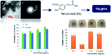
TEM investigation of the TiO2 sample by bright field imaging (Fig. 4a) indicates the presence of nanoparticle agglomerates that give rise to a mesoporous non-periodic texture. Selected area electron diffraction (Fig. 4b) indicates that the sample is made out of nanocrystalline TiO2 particles in the anatase polymorph. The most relevant observed diffraction rings can be indexed as related to the (101), (112), (200) and (211) family of planes of the anatase structure. SEM investigation (Fig. 4c) fully supports TEM observations, pointing out that the material was made out of primary nanoparticles with relatively uniform size that were connected to form the titania network and features mesoporosity. In particular, extended porosity was observed throughout the material, which could be ascribed to mesopores with a size around 5 nm (see Fig. 4c, inset).
image file: c6ra07653j-f4.tif
Fig. 4 TEM bright field image (a) and corresponding selected area diffraction (b). SEM image (c) of the TiO2 sample.

Evaluation of stability after light exposure

The photo-stability of free FA and FA contained in the TiO2@FA sample was investigated adopting experimental irradiation conditions comparable to the natural ones.

After one day of light exposure, 87% of FA was preserved by the developed mesoporous matrix; on the contrary, only 14% was recovered from the free sample.

The obtained results confirmed the ability of the prepared material to preserve the active molecule from degradation induced by light and, therefore, its functional properties.

Determination of scavenging effect on DPPH and ABTS radicals

The antioxidant properties of TiO2@FA were evaluated by rapid and reliable methods measuring the disappearance of colored stable free radicals such as the 2,2′-diphenyl-1-picryl-hydrazyl radical (DPPH˙) and the 2,2′-azino-bis(3-ethylbenzothiazoline-6-sulphonic acid) radical cation (ABTS˙+).

DPPH˙ is one of the few stable and commercially available organic nitrogen radicals characterized by an absorption maximum band around 515–528 nm and, therefore, it represents a useful reagent for evaluating the antioxidant activity of compounds.33 This radical accepts an electron or hydrogen radical to become a stable molecule and, thus, the antioxidant effect is proportional to the disappearance of DPPH˙ in tested samples.

ABTS is a preformed stable organic radical with an absorption maximum band at around 734 nm and, also in this case, the reaction depends on the hydrogen donating ability of the antioxidant compound.

The TiO2@FA scavenger ability was evaluated in terms of DPPH and ABTS reduction, and all data were expressed as percent inhibition. The FA-loaded material showed high scavenging activity towards the selected radicals while blank TiO2 did not interfere with the scavenger process (Table 2).

Table 2 Inhibition percentages of DPPH, ABTS and NO radicals and in vitro spectrophotometric SPF
Sample Inhibition (%) SPF values
DPPH ABTS NO
Blank TiO2 0 ± 0.1 0 ± 0.2 0 ± 0.2 2.6 ± 0.2
TiO2@FA 87 ± 0.3 75 ± 0.2 79 ± 0.4 14.7 ± 0.1


Anti-inflammatory effect

NO is one of the reactive oxygen species (ROS) that plays a key role in several physiological processes including vasodilatation, neurotransmission and immune responses.34

Despite its possible beneficial effects, NO is also implicated in a variety of pathological conditions and diseases, such as cardiovascular and inflammatory diseases (including sepsis, arthritis, multiple sclerosis and systemic lupus erythematosus), stroke, diabetes and cancer.35,36 Furthermore, NO is a pivotal pro-inflammatory mediator and its uncontrolled production by the inducible NO synthase (iNOS) leads to the generation of reactive nitrogen species, which induce biomolecular and cellular damage.37 NO, indeed, may contribute to oxidative damage due to its reaction with superoxide to form the peroxy nitrite anion, a potentially strong oxidant that can decompose to produce ˙OH and NO2.

In the performed assay, SNP was used as a NO radical donor to evaluate the NO˙ scavenging activity of TiO2@FA.

The obtained data (Table 2) was reported as percent of inhibition and shows the high scavenging activity of the FA-loaded material while blank TiO2 powder did not interfere with the test.

In vitro determination of SPF

Titanium dioxide is widely employed in sunscreen formulations as a UV filter in combination with other substances due to its capability to reflect and scatter UV light and, in the present study, it was combined with FA, a natural antioxidant compound able to protect against the damages due to oxidative stress induced by UV radiation.

Aiming to test the UVB shielding ability of the TiO2 and TiO2@FA samples, their in vitro SPFs were determined.

The in vitro SPF provides a useful parameter to assess the potential of a material in view of the development of sunscreen formulations for in vivo use. Table 2 summarizes the SPF values obtained for TiO2 and TiO2@FA by applying the above reported mathematical (eqn (2)) to the spectrophotometry data.

The in vitro SPF analysis of TiO2@FA indicates that FA has a synergistic effect on boosting the SPF of blank titania. For TiO2@FA, indeed, high absorbance values have been observed in the 290–320 nm wavelength range while, at the same concentration, TiO2 showed a significantly lower SPF value.

Evaluation of cytotoxicity by the NRU assay

The 3T3 NR uptake test was designed to detect toxicity induced by TiO2 and TiO2@FA using an in vitro cytotoxicity assay in the Balb/c 3T3 mouse fibroblast cell line.

This test was based on the ability of viable cells to incorporate and bind the NR dye in the lysosomes; therefore, the quantity of incorporated dye is directly proportional to the number of cells with an intact membrane.

In the present study, cells were treated with medium containing different dilutions of the tested item (10, 20 and 30 μg mL−1) and cell viability was evaluated at 24 h.

No differences in viability were observed for blank TiO2 and TiO2@FA treated cells (Fig. 5). Therefore, both the prepared materials were found to be non-cytotoxic at the tested concentrations.


image file: c6ra07653j-f5.tif
Fig. 5 Effect of blank TiO2 and TiO2@FA on cell viability.

Similar results, which confirm the absence of cellular toxicity for TiO2 nanoparticles, are also reported in the literature.38–40

EPISKIN™ test for the assessment of skin irritation

EPISKIN™ is an in vitro reconstructed human epidermis from normal human keratinocytes cultured on a collagen matrix at the air–liquid interface, and this model is histologically similar to the in vivo human epidermis.

Aiming to evaluate the skin irritation potential of TiO2 and TiO2@FA, EPISKIN™ RHE/L/13 human skin was treated with 20 μg mL−1 of TiO2 or TiO2@FA according to the “42 bis” protocol. This protocol consists of a 42 min exposure time followed by a rinsing step and a 42 h post-incubation period before quantitative measurement of cell viability by MTT reduction.

The obtained results are presented in Fig. 6 and, as it is possible to observe, no significant differences in viability were observed for the two tested materials. Therefore, irritation for TiO2 and TiO2@FA were not exhibited in EPISKIN™ RHE/L/13 human skin equivalent. However, 5% SDS provided as a positive control, induced a significant reduction in viability.


image file: c6ra07653j-f6.tif
Fig. 6 Effect of blank TiO2 and TiO2@FA on EPISKIN™ RHE/L/13 human skin.

Conclusions

In this study, the applicability of a new material obtained by combining titania and FA as a photo-protecting agent to be used in cosmetic formulations was demonstrated.

The prepared material was made of mesoporous TiO2, a common physical UV filter for sunscreen formulations, as an inorganic matrix for the encapsulation of FA by a simple impregnation method. This composite material was also used in many skin lotions due to its antioxidant properties, which prevents UV radiation damage.

The matrix was made out of nanocrystalline anatase TiO2 and featured a mesoporous ordered porous structure with relatively high surface area (190 m2 g−1). The impregnation with FA results in loading of more than 4 wt% of FA into the titania matrix. The loading process was accompanied by significant changes of the textural properties with respect to the pure matrix, such as the decrease of the specific area and pore diameter. Nevertheless, mesostructure and hexagonal symmetry were unaffected by the presence of the FA into internal and onto the external TiO2 surfaces, respectively.

Due to its nature, FA exerts a double role by the ability to act as both UV filter and radical scavenger at the same time. The proposed mechanism involves the release of loaded FA from the prepared mesoporous matrix. It is reasonable to assume that an amount of released FA is retained on the skin surface, absorbing UV radiations, while another fraction penetrates into deeper skin layers to act as a radical scavenger.

Antioxidant and anti-inflammatory properties of the synthesized TiO2@FA were evaluated by performing different assays and high scavenging activity towards DPPH, ABTS and NO radicals were recorded in the tested conditions.

The in vitro determination of the spectrophotometric SPF was also performed according to the method proposed by Mansur. A value of 14.7 was observed for TiO2@FA while, at the same concentration, TiO2 showed a lower SPF value equal to 2.6, confirming the potential application of the titania-doped FA as a “booster of SPF” that is able to enhance the SPF of a sunscreen.

Moreover, in vitro safety studies were also performed confirming the biocompatibility of the prepared material and the absence of skin irritation.

Conflict of interests

The authors declare that there is no conflict of interests regarding the publication of this paper.

Acknowledgements

The authors thank the Italian Minister of University and Research and University of Calabria for the financial support of the work.

Notes and references

  1. L. Pasqua, F. Testa, R. Aiello, S. Cundari and J. B. Nagy, Microporous Mesoporous Mater., 2007, 103, 166 CrossRef CAS.
  2. M. Manzano, M. Colilla and M. Vallet-Regí, Stud. Surf. Sci. Catal., 2008, 174, 13 CrossRef.
  3. J. M. Rosenholm, J. Zhang, W. Sun and H. Gu, Microporous Mesoporous Mater., 2011, 145, 14 CrossRef CAS.
  4. S. Giri, B. G. Trewyn and V. SY Lin, Nanomedicine, 2007, 2, 99 CrossRef CAS PubMed.
  5. J. D. Bass, D. Grosso, C. Boissiere, E. Belamie, T. Coradin and C. Sanchez, Chem. Mater., 2007, 19, 4349 CrossRef CAS.
  6. T. Lopez, E. Ortiz-Islas, E. Vinogradova, J. Manjarrez, J. A. Azamar, J. J. Alvarado-Gil and P. Quintana, Opt. Mater., 2006, 29, 82 CrossRef CAS.
  7. T. Lopez, E. Ortiz, D. Meza, E. Basaldella, X. Bokhimi, C. Magana, A. Sepulveda, F. Rodriguez and J. Ruiz, Mater. Chem. Phys., 2011, 126, 922 CrossRef CAS.
  8. S. Mura, G. Greppi, A. M. Roggio, L. Malfatti and P. Innocenzi, Microporous Mesoporous Mater., 2011, 142, 1 CrossRef CAS.
  9. T. Lopez, E. Ortis, P. Quintana and R. D. Gonzalez, Colloids Surf., A, 2007, 300, 3 CrossRef CAS.
  10. K. Awazu, M. Fujimaki, C. Rockstuhl, J. Tominaga, H. Murakami, Y. Ohki, N. Yoshida and T. Watanabe, J. Am. Chem. Soc., 2008, 130, 1676 CrossRef CAS PubMed.
  11. N. Serpone, A. Salinaro, A. V. Emeline, S. Horikoshi, H. Hidaka and J. Zhao, Photochem. Photobiol. Sci., 2002, 1, 970 CAS.
  12. COLIPA, 2006, COLIPA Guidelines, International Sun Protection Factor (SPF) Test Method.
  13. J. R. Villalobos-Hernández and C. C. Müller-Goymann, Eur. J. Pharm. Biopharm., 2005, 60, 113 CrossRef PubMed.
  14. C.-C. LIN and W.-J. LIn, J. Food Drug Anal., 2011, 19, 1 CAS.
  15. M. F. Sojka, P. G. Cummins, L. A. G. Declercq, C. G. Fthenakis, M. C. Ionita-Manzatu, W. A. Lee, D. H. Maes, M. A. McKeever-Alfieri, L. J. Najdek, N. Pernodet, I. M. E. Sente, L. P. Teta, K. Van Rillaer, D. B. Yarosh and P. U. Giacomoni, Photochem. Photobiol. Sci., 2011, 10, 1146 CAS.
  16. F. Iemma, F. Puoci, M. Curcio, O. I. Parisi, G. Cirillo, U. G. Spizzirri and N. Picci, J. Appl. Polym. Sci., 2010, 115, 784 CrossRef CAS.
  17. MHW Ministry of health and welfare, Notification No. 331/2000, Standards for cosmetics, 2000.
  18. E. Graf, Free Radical Biol. Med., 1992, 13, 435 CrossRef CAS PubMed.
  19. N. Kumar and V. Pruthi, Biotechnology Reports, 2014, 4, 86 CrossRef.
  20. L. Wu, J. C. Yu, X. Wang, L. Zhang and J. Yu, J. Solid State Chem., 2005, 178, 321 CrossRef CAS.
  21. O. I. Parisi, M. Fiorillo, L. Scrivano, M. S. Sinicropi, V. Dolce, D. Iacopetta, F. Puoci and A. R. Cappello, Biomacromolecules, 2015, 16, 3126 CrossRef CAS PubMed.
  22. S. Brunauer, P. H. Emmett and E. J. Teller, J. Am. Chem. Soc., 1938, 62, 309 CrossRef.
  23. E. P. Barrett, L. G. Joyner and P. P. Halenda, J. Am. Chem. Soc., 1951, 73, 373 CrossRef CAS.
  24. O. I. Parisi, F. Puoci, F. Iemma, M. Curcio, G. Cirillo, U. G. Spizzirri and N. Picci, Pharm. Dev. Technol., 2013, 18, 1058 CrossRef CAS PubMed.
  25. F. Puoci, C. Morelli, G. Cirillo, M. Curcio, O. I. Parisi, P. Maris, D. Sisci and N. Picci, Anticancer Res., 2012,(7), 2843 CAS.
  26. R. Re, N. Pellegrini, A. Proteggente, A. Pannala, M. Yang and C. Rice-Evans, Free Radical Biol. Med., 1999, 26, 1231 CrossRef CAS PubMed.
  27. O. Vittorio, G. Cirillo, F. Iemma, G. Di Turi, E. Jacchetti, M. Curcio, S. Barbuti, N. Funel, O. I. Parisi, F. Puoci and N. Picci, Pharm. Res., 2012,(9), 260 Search PubMed.
  28. F. M. Awah, P. N. Uzoegwu, J. O. Oyugi, J. Rutherford, P. Ifeonu, X. J. Yao, K. R. Fowke and M. O. Eze, Food Chem., 2010, 119, 1409 CrossRef CAS.
  29. F. L. Melquiades, D. D. Ferreira, C. R. Appoloni, F. Lopes, A. G. Lonni, F. M. Oliveira and J. C. Duarte, Anal. Chim. Acta, 2008, 613, 135 CrossRef CAS PubMed.
  30. R. M. Sayre, P. P. Agin, G. J. LeVee and E. Marlowe, Photochem. Photobiol., 1979, 29, 559 CrossRef CAS PubMed.
  31. J. S. Mansur, M. N. R. Breder, M. C. A. Mansur and R. D. Azulay, An. Bras. Dermatol., 1986, 61, 121 Search PubMed.
  32. N. J. Lowe and J. Breeding, Cosmet. Toiletries, 1983, 98, 65 CAS.
  33. A. Ardestani and R. Yazdanparast, Food Chem., 2007, 104, 21 CrossRef CAS.
  34. M. S. Sinicropi, A. Caruso, F. Conforti, M. Marrelli, E. Kashef, J. C. Lancelot, S. Rault, G. A. Statti and F. Menichini, J. Enzyme Inhib. Med. Chem., 2009, 24(5), 1148–1153 CrossRef CAS PubMed.
  35. B. G. Lee, S. H. Kim, O. P. Zee, K. R. Lee, H. Y. Lee and J. W. Han, Eur. J. Pharmacol., 2000, 406, 301 CrossRef CAS PubMed.
  36. K. D. Kroncke, K. Fehsel and V. Kolb-Bachofen, Clin. Exp. Immunol., 1998, 113, 147 CrossRef CAS PubMed.
  37. Y. S. Huang and S. C. Ho, Food Chem., 2010, 119, 868 CrossRef CAS.
  38. Y. H. Park, S. H. Jeong, S. M. Yi, B. H. Choi, Y. R. Kim, I. K. Kim, M. K. Kim and S. W. Son, Toxicol. In Vitro, 2011, 25, 1863 CrossRef CAS PubMed.
  39. T. Xia, M. Kovochich, J. Brant, M. Hotze, J. Sempf, T. Oberley, C. Sioutas, J. I. Yeh, M. R. Wiesner and A. E. Nel, Nano Lett., 2006, 6, 1794 CrossRef CAS PubMed.
  40. S. Hackenberg, G. Friehs, M. Kessler, K. Froelich, C. Ginzkey, C. Koehler, A. Scherzed, M. Burghartz and N. Kleinsasser, Environ. Mol. Mutagen., 2010, 52, 264 CrossRef PubMed.

Footnote

These authors contributed equally to the manuscript (O. I. P. and D. A.).

This journal is © The Royal Society of Chemistry 2016
Click here to see how this site uses Cookies. View our privacy policy here.