DOI:
10.1039/C6RA07236D
(Paper)
RSC Adv., 2016,
6, 59677-59683
A novel fluorescein-based colorimetric probe for Cu2+ detection†
Received
19th March 2016
, Accepted 13th June 2016
First published on 13th June 2016
Abstract
A new colorimetric probe, 5-chlorosalicylaldehyde fluorescein hydrazone (CSFH), has been synthesized and characterized by infrared spectroscopy, nuclear magnetic resonance spectroscopy, mass spectrometry, elemental analysis and X-ray single crystal diffraction. CSFH had nearly no absorption in visible region. In the presence of copper(II) ion a rapid color changes from colorless to yellow along with an obvious new absorption band appeared at 495 nm. CSFH was highly selective to Cu2+ only in the aqueous solution, instead of common metal ions. The absorbance intensity and the color of CSFH solution increased gradually with the increase of Cu2+ concentration. Common metal ions did not show any interference on the determination of Cu2+. The chemical stoichiometric ratio between the CSFH and Cu2+ was 1![[thin space (1/6-em)]](https://www.rsc.org/images/entities/char_2009.gif) :1 and corresponding association constant was 6.85 × 10⁵ L mol−1, and the linear range for Cu2+ detection was 0.25–14 μmol L−1. The detection limits were 0.25 μmol L−1 and 1.0 μmol L−1 of Cu2+ using the UV-Vis changes and the visual color changes by the naked eye respectively. The proposed method was applied to the determination of Cu2+ in drinking water samples and the recoveries were 96–106%. The preparation of CSFH exhibited the quick, simple and facile advantages. The results showed that CSFH can be a good candidate for simple, rapid and sensitive colorimetric detection of Cu2+ in aqueous solution.
:1 and corresponding association constant was 6.85 × 10⁵ L mol−1, and the linear range for Cu2+ detection was 0.25–14 μmol L−1. The detection limits were 0.25 μmol L−1 and 1.0 μmol L−1 of Cu2+ using the UV-Vis changes and the visual color changes by the naked eye respectively. The proposed method was applied to the determination of Cu2+ in drinking water samples and the recoveries were 96–106%. The preparation of CSFH exhibited the quick, simple and facile advantages. The results showed that CSFH can be a good candidate for simple, rapid and sensitive colorimetric detection of Cu2+ in aqueous solution.
1. Introduction
Copper (Cu2+) is the most essential transition metal in the human body.1,2 It plays a vital role in various physiological processes, such as cellular respiration, hematopoietic function, bone formation, prevention of cardiovascular diseases and connective tissue development.3,4 It also serves as a significant catalytic co-factor for several metalloenzymes like cytochrome c oxidase, superoxide dismutase and tyrosinase.5 The recommended daily uptake of copper is 0.4–0.6 mg for infants, 1.5–2.5 mg for children, 1.5–3.0 mg for adults.6 Disruption of copper homeostasis results in a number of neurodegenerative diseases, such as Alzheimer's disease (AD), prion diseases, Parkinson's disease (PD), amyotrophic lateral sclerosis (ALS), Menkes disease, and Wilson's disease.7,8 To date, many methods for detecting Cu2+ involved atomic absorption spectroscopy,9,10 inductively coupled plasma mass spectroscopy,11 the colorimetry,12–17 fluorimetry18–22 and electrochemical method.23–25 Colorimetric method still remains the attraction due to convenience and the simplicity, easy observation by the naked eye, and no requirement for sophisticated instruments than closely related methods.26,27
Recently, numerous colorimetric probes for Cu2+ have been developed based on quinolone,17,28 polythiophene,29 rhodamine,30,31 fluorescein32 etc. These probes possess large rigid π-conjugated system and the unique optical properties. Despite these discoveries, there is still some room to develop aqueous based, facile preparation, cost-effective probes that target Cu2+ with high affinity and specificity.
The fluorescein and its derivatives, excellent chromophores, have received much attention due to their high water solubility, high fluorescence quantum yield, or high molar extinction coefficient as well as good photo-stability and short synthetic routes.33 The fluorescein family dyes have two distinctive structures: ring-opening and spirocycle. The spirocycle structure is colorless and non-fluorescence, some metal ions can make spirocycle opened to become ring-opening form accompanying the change of spectroscopic properties. In recent years, the fluorescein and its derivatives have been used as chemosensors for Cu2+.34,35 Ma and co-worker36 reported a fluorescein hydrazide as the fluorescent probe for Cu2+ and expended to the detection of trace Cu2+ in real biological fluids. However, most of these fluorescein derivatives were used as fluorescent probes and only few works focused on their colorimetric properties toward Cu2+. Li et al.37 synthesized a fluorescein-based colorimetric probe, 1-phenyl-3-methyl-5-hydroxypyrazole-4-benzoyl (fluorescein) hydrazine, for rapid, visual and spectroscopic detection of Cu2+. Qu et al.38 developed a pyridoxal-based fluorescein probe for real-time, simple-to-use and naked-eye detection of Cu2+. Nevertheless, current colorimetric systems usually require organic solvent as a cosolvent which greatly limits their analytical application. Therefore, it is strongly desirable to develop novel fluorescein-based colorimetric probe, via facile preparation, with high selectivity and sensitivity for the rapid detection of Cu2+ in aqueous media.
Salicylaldehyde consists of reactive aldehyde groups and phenolic hydroxyl groups. Active aldehyde group can easily react with amino group to form Schiff base with a suitable coordination oxygen atom. With this in mind, we designed and synthesized a new fluorescein-based probe, 5-chlorosalicylaldehyde fluorescein hydrazone (CSFH). The CSFH might bind Cu2+ via the carbonyl O, amido N and hydroxyl O as donors and exhibits a rapid and selective response to Cu2+ only in aqueous solution by color change from colorless to yellow along with an obvious new band appeared at 495 nm in UV-Vis absorption spectra. No any changes of the colour or absorption spectra were observed to other common metal ions. CSFH can be used to detect Cu2+ as a naked-eye colorimetric probe with simplicity, rapidity and sensitivity. The CSFH was also successfully applied to the determination of Cu2+ in water samples.
2. Materials and methods
2.1 Chemicals and instruments
Fluorescein and 5-chlorosalicylaldehyde were purchased from Aladdin Chemical Co., Ltd. (Shanghai, China). Cationic salts were purchased from Shanghai Experiment Reagent Co., Ltd. (Shanghai, China). All chemicals and reagents were analytical grade and used as received without further purification. Deionized (DI) water was used throughout the work and obtained from a Millipore Milli-QRO4 water purification system with a resistivity higher than 18.25 MΩ cm (Bedford, MA, USA).
Absorption and fluorescence spectra were recorded in PBS buffer solutions (0.01 mol L−1) using a Lambda 950 UV/VIS/NIR Spectrometer (PerkinElmer, USA) and Varian Cary Eclipse spectrometer (Palo Alto, CA, USA), respectively. PBS buffer solutions were obtained by 0.01 mol L−1 Na2HPO4 solution and 0.01 mol L−1 NaH2PO4 solution. The pH value was measured by a pH meter (Mettler-Toledo International Inc, Switzerland). FT-IR spectra were recorded with KBr pellet in the range of 400–4000 cm−1 on Bruker FT-IR Spectrometer (Bruker Optik GmbH, Ettlingen, Germany). 1H NMR and 13C NMR spectra were recorded on a Bruker Avance-300 MHz NMR Spectrometer (Bruker Cryomagnet, Switzerland). Mass Spectroscopy was performed on MALDI-TOF/MS with DHB as matrix. Elemental analyses were performed on an Elementar Analysen-systeme vario EL cube elemental analyzer (Hanau, Germany). The single-crystal structure of CSFH was determined by Bruker SMART APEX diffractometer with CCD as a detector. Data were collected at 296 K using Mo-Kα radiation (λ = 0.710713 Å) and the ω-scan technique, and the absorption correction was applied to the collected reflections with SADABS. The structures were solved by direct methods using the SHELXL97 program, and subsequent difference Fourier maps were inspected and then refined in F2 using a full-matrix least-squares procedure and anisotropic displacement parameters.
2.2 Synthesis and characterisation of CSFH
The synthesis of CSFH is illustrated in Scheme 1, which started from fluorescein and hydrazine to give fluorescein hydrazide (FH) with reference to a reported method with slight modifications.39 CSFH was synthesized with Schiff base formation by the reaction of 5-chlorosalicylaldehyde (0.32 g, 2.0 mmol) with fluorescein hydrazide (0.69 g, 2.0 mmol) in 25 mL ethanol, and then the reaction mixture was refluxed for 8 h under constant stirring with the reaction progress monitored by TLC. After the reaction completed, the reaction mixture was cooled with some off-white precipitate formed. The mixture was filtered and the crude product was recrystallized using ethanol to afford a pale yellow powder of CSFH 0.64 g with 69% yield. The single-crystal for X-ray diffraction was formed by slowly evaporating the resulting solution at room temperature. 1H NMR (300 MHz, 25 °C, DMSO-d6): δ 10.43 (s, 1H), 9.98 (s, 2H), 9.08 (s, 1H), 7.94 (d, J = 7.5 Hz, 1H), 7.63 (d, J = 9.1 Hz, 1H), 7.59 (s, 1H), 7.39 (s, 1H), 7.22 (d, J = 8.8 Hz, 1H), 7.14 (d, J = 7.5 Hz, 1H), 6.81 (d, J = 8.8 Hz, 1H), 6.65 (s, 2H), 6.52 (q, J = 9.1 Hz, 4H). 13C NMR (75 MHz, 25 °C, DMSO-d6): δ 18.24, 64.92 (Cspiro), 102.23, 109.15, 112.20, 117.86, 120.80, 122.72, 122.99, 123.519, 126.19, 127.68, 128.21, 128.89, 130.79, 133.92, 146.22, 150.19, 151.83, 155.40, 158.39, 163.32. FT-IR (KBr, cm−1): 3358 (–OH), 1704 (C![[double bond, length as m-dash]](https://www.rsc.org/images/entities/char_e001.gif) O), 1612 (CN). MS, m/z: 485.5 [CSFH + H]+, 507.5 [CSFH + Na]+ (Fig. S1–S4†). Elemental analysis (calcd%) for C27H17N2O5Cl: C, 66.87; N, 5.78; H, 3.51; O, 16.51; found: C, 65.86; N, 5.41; H, 3.91; O, 16.99. Melting point: >270 °C. Crystal data for C27H17N2O5Cl: crystal size: 0.35 × 0.33 × 0.32, monoclinic, space group C2/c. a = 30.28(4) Å, b = 10.223(14) Å, c = 17.42(2) Å, α = 90(3)°, β = 98.71(3)°, γ = 90(3)°, V = 5330(12) Å3, Z = 8, T = 296 K, θmax = 25.05°, 13448 reflections measured, 4640 unique (Rint = 0.0519). Final residual for 349 parameters and 4640 reflections with I > 2σ(I): R1 = 0.0854, wR2 = 0.2191 and GOF = 1.009.
O), 1612 (CN). MS, m/z: 485.5 [CSFH + H]+, 507.5 [CSFH + Na]+ (Fig. S1–S4†). Elemental analysis (calcd%) for C27H17N2O5Cl: C, 66.87; N, 5.78; H, 3.51; O, 16.51; found: C, 65.86; N, 5.41; H, 3.91; O, 16.99. Melting point: >270 °C. Crystal data for C27H17N2O5Cl: crystal size: 0.35 × 0.33 × 0.32, monoclinic, space group C2/c. a = 30.28(4) Å, b = 10.223(14) Å, c = 17.42(2) Å, α = 90(3)°, β = 98.71(3)°, γ = 90(3)°, V = 5330(12) Å3, Z = 8, T = 296 K, θmax = 25.05°, 13448 reflections measured, 4640 unique (Rint = 0.0519). Final residual for 349 parameters and 4640 reflections with I > 2σ(I): R1 = 0.0854, wR2 = 0.2191 and GOF = 1.009.
 |
| | Scheme 1 Synthetic route for 5-chlorosalicylaldehyde fluorescein hydrazone (CSFH). | |
2.3 UV-Vis and fluorescence spectra measurement
The stock solution of CSFH (1.0 mmol L−1) was prepared in ethanol. A standard Cu2+ solution (1.0 mmol L−1) was prepared in deionized water. The solutions of Ca2+, Al3+, Zn2+, Mg2+, Ba2+, Ni2+, Co2+, Cr3+, Fe3+, Hg2+, Fe2+, Pb2+, Mn2+ and Cu2+ were prepared from their chloride salts, and the solutions of Ag+ was prepared from its nitrate salt, the solutions of Cd2+ was prepared from its sulfate salt, and the solution of Cr6+ was prepared from its potassium salt. In detection, CSFH was diluted to 15 μmol L−1 by PBS buffer (pH 7.4). Each time 3.0 mL of 15 μmol L−1 CSFH was placed into a 1.0 cm pathlength silica cuvette, and the required quantity of Cu2+ solution was added by a micro-pipette. 10 s later the UV-Vis and fluorescence spectra were measured. All the measurements were taken at room temperature.
2.4 Job's plot analyses for determination of stoichiometry
A series of mixed solutions of CSFH and Cu2+ was prepared, with the total concentration of added CFSH and Cu2+ maintained at constant (15 μmol L−1), and varying the relative proportions of CFSH and Cu2+ from 9:1, 8:2, …, until 1:9. The solution (1.0 mmol L−1) of CSFH in ethanol and the Cu2+ standard solution (1.0 mmol L−1) in DI water were mixed based on the above relative proportions, and then diluted to 10 mL with PBS buffer (pH 7.4). The absorbance intensities at 495 nm of these test solutions were recorded for the Job's plot.
3. Results and discussions
3.1 Synthesis and characterization
Schiff base CSFH could be facilely prepared by condensation of FH and 5-chlorosalicylaldehyde in refluxing absolute ethanol and characterized by IR, 1HNMR, 13CNMR, MS, elemental analysis, and single crystal X-ray structural analysis (Fig. 1). The X-ray crystallographic structure indicates that the O1, N2 atoms in FH moiety and phenol oxygen (O5) of 5-chlorosalicylaldehyde moiety can provide suitable coordination sites to a certain metal ion. Moreover, the O1 atom in FH moiety and phenol oxygen (O5) of 5-chlorosalicylaldehyde moiety exist in trans form and there is an intramolecular hydrogen bonding between the phenolic (O5) proton and imino nitrogen atom (N2). However, the trans form and the hydrogen bonding may be changed due to the coordination of metal ion.
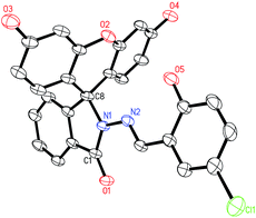 |
| | Fig. 1 Crystal structure of CSFH (hydrogen atoms are omitted for clarity). | |
3.2 Selectivity of CSFH over metal ions
The binding activity of CSFH with different metal ions, such as Ag+, Ca2+, Al3+, Zn2+, Mg2+, Ba2+, Fe2+, Co2+, Fe3+, Ni2+, Hg2+, Mn2+, Pb2+, Cd2+, Cr3+, Cr6+, Cu2+, were investigated respectively by absorption spectral changes as shown in Fig. 2A. It could be observed that CSFH had almost no absorption in the visible region, which attributed to the closed spirocyclic form of CSFH, and there was no obvious absorption spectral change of CSFH with addition of other metal ions except Cu2+. However, with the addition of Cu2+, the CSFH solution immediately changed from colorless to yellow with a new absorption band centred at 495 nm appeared. It suggested there could be a complex formation between CSFH and Cu2+. The results showed that CSFH could be exploited as a colorimetric probe for the recognition of Cu2+.
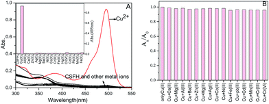 |
| | Fig. 2 (A) UV-Vis absorption spectra of CSFH (15 μmol L−1, pH 7.4, PBS) with addition of different metal ions (15 μmol L−1). Inset: Absorbance intensity of CSFH (15 μmol L−1) in the presence of different metal ions at 495 nm. (B) Absorption intensity change of CSFH (15 μmol L−1, pH 7.4, PBS) in the presence of Cu2+ (15 μmol L−1) with 45 μmol L−1 of various other metal ion, where Ax/A0 are the absorption intensities of CSFH and Cu2+ in the presence and absence of various other metal ion, respectively. | |
To confirm the selective binding of CSFH towards Cu2+, the competition experiment was conducted by adding 15 μmol L−1 Cu2+ solution to CSFH solution (15 μmol L−1) in the presence of other metal ions, such as Ag+, Ca2+, Al3+, Zn2+, Mg2+, Ba2+, Fe2+, Co2+, Fe3+, Ni2+, Hg2+, Mn2+, Pb2+, Cd2+, Cr3+, Cr6+ at high concentration of 45 μmol L−1. Fig. 2B showed relative absorbance changes of CSFH with addition of Cu2+ in the presence of other different metal ions. There was almost no change for the relative absorbance of CSFH and Cu2+ in the presence of other metal ions as compared with the absorbance only from CSFH and Cu2+. As a result, the selective binding of CSFH toward Cu2+ over other metal ions was remarkably high, and these coexisting metal ions had no interference on the detection of Cu2+.
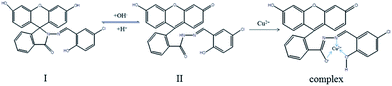
3.3 Effect of pH
As shown in Fig. 3, CSFH equilibrated with two types in the solution. The spirocycle type I dominated in acidic solution, while in alkaline solution, the spirocycle ring was opened to become the ring-opening type II. Fig. 4 demonstrated that the solution of free CSFH had nearly no absorbance relatively at 495 nm within pH 4.0–13.0. The strong absorbance at 495 nm was observed upon the addition of Cu2+ at pH great than 7.0. It revealed that the complex of CSFH with Cu2+ was formed suitably for alkaline condition. It is attributed to CSFH was deprotonated and spirocycle ring was opened in alkaline solution and then further coordinate with Cu2+ to form CSFH–Cu complex. However, the high concentration of OH− would induce precipitation of Cu(OH)2, which might result in the decrease in the absorbance of CSFH–Cu complex, especially at pH 13. The absorbance of CSFH–Cu complex remained constant in the pH range 7.0–12.0 and pH 7.4 was chosen for the further studies.
 |
| | Fig. 3 Structure change of CSFH. | |
 |
| | Fig. 4 (A) Absorption spectra of CSFH + Cu2+ under different pH condition. Inset: Absorption spectra of CSFH (15 μmol L−1) under different pH condition. (B) Absorbance intensities of free CSFH (15 μmol L−1) and in the presence of Cu2+ (15 μmol L−1) under different pH condition. | |
3.4 UV-Vis spectra for Cu2+ titration
The absorption spectra and the color changes of CSFH (15 μmol L−1) with addition of Cu2+ in pH 7.4 PBS buffer solution were shown in Fig. 5. CSFH was colorless and had almost no absorption after 450 nm, which mainly existed in the spirocyclic form. When Cu2+ was added into the CSFH solution, yellow could be observed clearly by naked-eye, with a new peak centred at 495 nm appeared in the absorption spectra. With further addition of the Cu2+, the absorption intensity at 350 nm decreased and intensities of absorbance at 495 nm increased dramatically, along with two isosbestic points at 330 nm and 372 nm. The colour change and the appearance of new absorption band were obvious signatures for the molecular binding between CSFH and Cu2+.
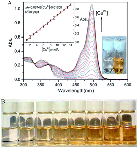 |
| | Fig. 5 (A) Absorption spectral changes of CSFH (15 μmol L−1, PBS buffer, pH 7.4) with addition of Cu2+ at 0, 1.0, 2.0, 3.0, 4.0, 5.0, 6.0, 7.0, 8.0, 9.0, 10.0, 11.0, 12.0, 13.0, 14.0, 15.0, 16.0, 17.0, 18.0 and 19.0 μmol L−1, respectively. Inset: Linear range of Cu2+ concentration (0–14.0 μmol L−1). Photograph showing the color change of free CSFH (15 μmol L−1) (left) and in the presence of Cu2+ (15 μmol L−1) (right). (B) The color changes observed upon the addition of the different concentration of Cu2+ (from left to right: 0, 1, 3, 5, 7, 9, 11, 14 μmol L−1) to the solution of CSFH (15 μmol L−1, PBS buffer, pH 7.4). | |
CSFH (15 μmol L−1) was titrated with various concentrations of Cu2+ (0–19 μmol L−1) and relative absorbance intensity at 495 nm was plotted as a function of Cu2+ concentration as shown in Fig. 5A. There was a good linear relationship between the change of absorbance value (ΔA) and the Cu2+ concentrations (0–14 μmol L−1) was found based on the regression equation: ΔA = 0.06048[Cu2+] − 0.01208 (R2 = 0.9991). By the definition of IUPAC (DL = 3 Sb m−1),40 the limit of detection with 0.25 μmol L−1 from 11 blank solutions was obtained. The CSFH showed a high sensitivity toward Cu2+.
The color change of CSFH with the different concentration of Cu2+ was shown in Fig. 5B. The color change could be clearly observed by the naked eye. The colorless solution of CSFH turned to a light yellow upon addition of Cu2+ at 1.0 μmol L−1 and the color of CSFH solution increased gradually with the increase of Cu2+ concentration, which meant that the detection limit of Cu2+ by the naked eye could be as low as 1.0 μmol L−1.
According to linear Benesi–Hildebrand expression, the measured absorbance [1/(A − A0)] at 495 nm varied as a function of 1/[Cu2+] in a good linear relationship (R2 = 0.9996) (Fig. 6). Which indicates the stoichiometry of 1:1 between Cu2+ and CSFH. The association constant of CSFH with Cu2+ was calculated to be 6.85 × 10⁵ L mol−1, it revealed that CSFH formed stable complex with Cu2+.
 |
| | Fig. 6 The measured intensity [1/(A − A0)] at 495 nm varied as a function of 1/[Cu2+](106) in a linear relationship (R2 = 0.9996). CSFH (15 μmol L−1) was treated with various concentrations of Cu2+ (0–14 μmol L−1). | |
3.5 Binding kinetics
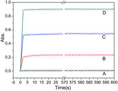
Binding kinetics were showed in Fig. 7. After addition different concentration of Cu2+ into CSFH solution, all the absorbance at 495 nm quickly reached the maximum within 10 s and then remained the same intensity, indicating the molecular binding between CSFH and Cu2+ extreme rapidly reached equilibrium under the experimental conditions. CSFH could be used as an effective probe for the real-time and quantitative detection of Cu2+ without any pretreatment of samples.
 |
| | Fig. 7 The response time of CSFH (15 μmol L−1) to Cu2+ in PBS buffer (pH 7.4) at different concentrations, (A) 0 μmol L−1, (B) 3 μmol L−1, (C) 8 μmol L−1, (D) 14 μmol L−1. | |
3.6 Job's plot analyses
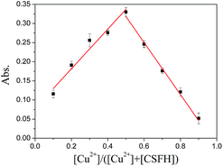
All Job's analyses were conducted with [CSFH] + [Cu2+] = 15 μmol L−1. A plot of absorbance intensities at 495 nm versus the molecular fraction of [Cu2+]/([CSFH] + [Cu2+]) was shown in Fig. 8. The absorbance maximum was obtained when the molar fraction of Cu2+ was close to 0.5, and then the absorbance decreased again with the further increasing the molar fraction of Cu2+ above 0.5. This indicated a 1:1 stoichiometry between CSFH and Cu2+. This result is consistent with the result of Benesi–Hildebrand.
 |
| | Fig. 8 Job's plot of absorption at 495 nm from CSFH and Cu2+ in aqueous solution at various molar fractions. | |
3.7 Fluorescence spectra of CSFH in the presence of Cu2+
Fig. 9 showed the fluorescence spectra of the CSFH in the presence of Cu2+. The fluorescence intensity of CSFH in the aqueous solution was extremely weak at 516 nm. In the presence of equiv. of Cu2+, CSFH exhibited quenched fluorescence from binding with Cu2+. The coordination interaction induced the spirocycle ring of CSFH open and led to fluorescence quenching, whereas the solution did not show the strong fluorescence emission of fluorescein moiety. It could be attributed to paramagnetic effect from spin–orbit coupling of the Cu2+.6 Similar phenomena had also been observed in other studies.5,41,42 Considering the fluorescence of CSFH was relatively weak in the solution, in this situation, it would be hard to establish sensitive quenching fluorescence detection for Cu2+. So, this work studied mainly the absorbance and color changes.
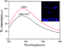 |
| | Fig. 9 The fluorescence spectra of CSFH (15 μmol L−1, PBS buffer, pH 7.4) in the absence and presence of Cu2+ (15 μmol L−1). Inset: The corresponding colors in UV light in the absence (left) and presence (right) of Cu2+. The excitation (λex)/emission (λem) are 495/516 nm. | |
3.8 Interaction mechanism
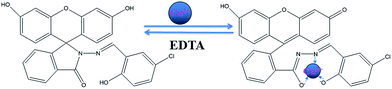
The proposed binding motif of CSFH with Cu2+ was shown in Fig. 10. The CSFH had spirocyclic and ring-opening form, the spirocyclic form was colorless and non-fluorescent possibly due to all π–π* and n–π* transitions in ultra-visible region. The addition of Cu2+ into CSFH made the spirocycle open via coordination or irreversible chemical reaction to form a complex resulting in color change, which has a metal centered d–d transition. Therefore, the new absorbance band centered at 495 nm could arise from this metal centered d–d transition, and the solution appeared as yellow. The isosbestic points at 330 nm and 372 nm indicated an equilibrium between CSFH and CSFH–Cu2+. On addition of EDTA to the solution of CSFH–Cu2+, the solution color changed from yellow to colorless, and the absorption at 495 nm disappeared (Fig. 11). Probably EDTA has a much larger binding constant with Cu2+ than CSFH with Cu2+. With addition of EDTA, the equilibrium shifted towards EDTA–Cu2+, with CSFH released from complexation and color changed from yellow to colourless. These results indicated that a reversible coordinated between CSFH and Cu2+.
 |
| | Fig. 10 The proposed binding motif of CSFH with Cu2+. | |
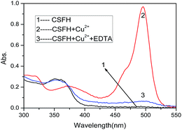 |
| | Fig. 11 Absorption spectra of CSFH (15 μmol L−1), CSFH + Cu2+ (15 μmol L−1) and CSFH + Cu2+ upon addition of EDTA disodium (15 μmol L−1) in PBS buffer solution. | |
3.9 Assay of Cu2+ in water samples
To investigate whether the proposed method was feasible to real samples, recovery experiments were performed in Kangshifu drinking mineralized water samples obtained from the local supermarket (Table 1). The found content of Cu2+ were analysed by the calibration equation of ΔA = 0.06048[Cu2+] − 0.01208 and the recoveries of Cu2+ were in range of 96–106%. The results revealed the designed method was reliable and CSFH could be applied for the detection of Cu2+ in real samples.
Table 1 Determination of Cu2+ in drinking water samples
| Sample |
Cu2+ added (μmol L−1) |
Cu2+ found (μmol L−1) |
Recovery (%) |
RSD (n = 3) (%) |
| 1 |
0.5 |
0.53 |
106 |
3.2 |
| 2 |
2.0 |
1.92 |
96 |
2.1 |
| 3 |
8.0 |
8.2 |
102.5 |
1.7 |
3.10 Comparison with other colorimetric probes
As shown in Table 2, the comparison of some different fluorescein-based colorimetric probes for the determination of Cu2+ was summarized. Compared with these reported methods, our method had higher sensitivity in aqueous solution and fast reaction time. Although a lower LOD was presented in one reported method, the organic solvent was required as a co-solvent. And other reported methods had a wide linear range of Cu2+, however, the sensitivity would remain to be further improved. On the whole, our proposed method possessed a lower detection limit without any organic reagent and fast reaction time, which would expand its analytical application.
Table 2 Comparison of colorimetric probe for the detection of Cu2+
| Probe |
Linear range/μmol L−1 |
Detection limit/μmol L−1 |
Reaction media |
Reaction time |
Reference |
| 1-Phenyl-3-methyl-5-hydroxypyrazole-4-benzoyl(fluorescein)hydrazone |
2–40 μmol L−1 |
2 μmol L−1 |
DMSO/H2O = 4:6, v/v |
2 min |
37 |
| Pyridoxal-based fluorescein derivative |
0.14–15 μmol L−1 |
0.14 μmol L−1 |
CH3OH/HEPES (pH 7.4) = 1:1, v/v |
30 s |
38 |
| Furfuraldehyde fluorescein hydrazone |
6.6–330 μmol L−1 |
6.6 μmol L−1 |
B–R buffer solution (pH 9.0) |
2 min |
5 |
| 3-Bromo-5-methylsalicylal-dehyde fluorescein hydrazone |
3.0–330 μmol L−1 |
3.0 μmol L−1 |
B–R buffer solution (pH 7.4) |
2 min |
32 |
| 5-Nitrosalicylaldehyde fluorescein hydrazone |
1.89–25 μmol L−1 |
1.89 μmol L−1 |
HEPES buffer solution (pH 7.0) |
15 s |
6 |
| 2-Hydroxynaphthaldehyde fluorescein hydrazone |
1.5–21 μmol L−1 |
1.5 μmol L−1 |
2% DMSO/Tris–HCl (pH 7.0) |
— |
26 |
| 2-Pyridylaldehyde fluorescein hydrazone |
1.5–120 μmol L−1 |
1.5 μmol L−1 |
HEPES buffer solution (pH 7.0) |
10 s |
35 |
| 5-Chlorosalicylaldehydefluorescein hydrazone |
0.25–14 μmol L−1 |
0.25 μmol L−1 |
PBS buffer solution (pH 7.4) |
10 s |
This work |
4. Conclusions
A new fluorescein derivative, 5-chlorosalicylaldehyde fluorescein hydrazone (CSFH), was facile fabricated based on the reaction between 5-chlorosalicylaldehyde and fluorescein hydrazine in ethanol, and characterized by FTIR, NMR, mass and X-ray spectrometry. CSFH showed a remarkable colorimetric selectivity to Cu2+ over common metal ions, and could form stable 1:1 complex with Cu2+ and generated color change from colorless to bright yellow in aqueous media. It could be serve as an effective probe for colorimetric detection of Cu2+ with a detection limit as low as 0.25 μmol L−1 and 1.0 μmol L−1 using the UV-Vis spectra and the visual color changes by the naked eye respectively. Combining its simple and fast synthetic method and high sensitivity and selectivity to Cu2+, especially, CSFH could be utilized as a colorimetric probe in aqueous solution and no need for organic co-solvent. It would be a good colorimetric candidate for Cu2+ detection.
Acknowledgements
This work was supported by the National Science Foundation of China (21575084 and 21405099), the Hundred Talent Programme of Shanxi Province, and Shanxi Province “Sanjin scholars” Project of China.
Notes and references
- W. Zhang, J. Wei, H. J. Zhu, K. Zhang, F. Ma, Q. S. Mei, Z. P. Zhang and S. H. Wang, J. Mater. Chem., 2012, 22, 22631–22636 RSC.
- W. Gao, Y. T. Yang, F. J. Huo, C. X. Yin, M. Xu, Y. B. Zhang, J. B. Chao, S. Jin and S. P. Zhang, Sens. Actuators, B, 2014, 193, 294–300 CrossRef CAS.
- Y. H. Chan, J. X. Chen, Q. S. Liu, S. E. Wark, D. H. Son and J. D. Batteas, Anal. Chem., 2010, 82, 3671–3678 CrossRef CAS PubMed.
- J. Zhang, L. Zhang, Y. Wei, J. Chao, S. Shuang, Z. Cai and C. Dong, Spectrochim. Acta, Part A, 2014, 132, 191–197 CrossRef CAS PubMed.
- J. Zhang, L. Zhang, Y. Wei, J. Ma, S. Shuang, Z. Cai and C. Dong, Spectrochim. Acta, Part A, 2014, 122, 731–736 CrossRef CAS PubMed.
- Y. Yang, F. Huo, C. Yin, Y. Chu, J. Chao, Y. Zhang, J. Zhang, S. Li, H. Lv, A. Zheng and D. Liu, Sens. Actuators, B, 2013, 177, 1189–1197 CrossRef CAS.
- E. Que, D. Domaille and C. Chang, Chem. Rev., 2008, 108, 1517–1549 CrossRef CAS PubMed.
- M. Taki, S. Iyoshi, A. Ojida, I. Hamachi and Y. Yamamoto, J. Am. Chem. Soc., 2010, 132, 5938–5939 CrossRef CAS PubMed.
- M. Chan and S. Huang, Talanta, 2000, 51, 373–380 CrossRef CAS PubMed.
- M. Tuzen, Microchem. J., 2003, 74, 289–297 CrossRef CAS.
- J. Wu and E. Boyle, Anal. Chem., 1997, 69, 2464–2470 CrossRef CAS PubMed.
- S. Banthia and A. Samanta, New J. Chem., 2005, 29, 1007–1010 RSC.
- L. Tang, F. Li, M. Liu and R. Nandhakumar, Spectrochim. Acta, Part A, 2011, 78, 1168–1172 CrossRef PubMed.
- N. Kaur and S. Kumar, Tetrahedron, 2011, 67, 9233–9264 CrossRef CAS.
- Q. Lin, P. Chen, J. Liu, Y. Fu, Y. Zhang and T. Wei, Dyes Pigm., 2013, 98, 100–105 CrossRef CAS.
- J. Tan and X. Yan, Talanta, 2008, 76, 9–14 CrossRef CAS PubMed.
- L. Tang, P. Zhou, Q. Zhang, Z. Huang, J. Zhao and M. Cai, Inorg. Chem. Commun., 2013, 36, 100–104 CrossRef CAS.
- M. Kim, H. Jang, S. Yi, S. Chang and M. Han, Chem. Commun., 2009, 32, 4838–4840 RSC.
- L. Qu, C. Yin, F. Huo, Y. Zhang and Y. Li, Sens. Actuators, B, 2013, 183, 636–640 CrossRef CAS.
- R. Tang, K. Lei, K. Chen, H. Zhao and J. Chen, J. Fluoresc., 2011, 11, 141–148 CrossRef PubMed.
- B. Chen and P. Zhong, Anal. Bioanal. Chem., 2005, 381, 986–992 CrossRef PubMed.
- L. Zeng, E. Miller, A. Pralle, E. Isacoff and C. Chang, J. Am. Chem. Soc., 2006, 128, 10–11 CrossRef CAS PubMed.
- M. Kopylovich, K. Mahmudov and A. Pombeiro, J. Hazard. Mater., 2011, 186, 1154–1162 CrossRef CAS PubMed.
- L. Singh and J. Bhatnagar, Talanta, 2004, 64, 313–319 CrossRef CAS PubMed.
- M. Ghanei-Motlagh, M. Taher, V. Saheb, M. Fayazi and I. Sheikhshoaie, Electrochim. Acta, 2011, 56, 5376–5385 CrossRef CAS.
- F. Abebe and E. Sinn, Tetrahedron Lett., 2011, 52, 5234–5237 CrossRef CAS.
- B. Zhu, Y. Xu, W. Liu, C. Shao, H. Wu, H. Jiang, B. Du and X. Zhang, Sens. Actuators, B, 2014, 191, 473–478 CrossRef CAS.
- X. Li, Z. Niu, L. Chang, M. Chen and E. Wang, Chin. Chem. Lett., 2014, 5, 80–82 CrossRef.
- Z. Yao, B. Huang, X. Hu, L. Zhang, D. Li, M. Guo, X. Zhang, H. Yuan and H. Wu, Analyst, 2013, 138, 1649–1652 RSC.
- X. Chen, M. Jou, H. Lee, S. Kou, J. Lim, S. Nam, S. Park, K. Kim and J. Yoon, Sens. Actuators, B, 2009, 137, 597–602 CrossRef CAS.
- Z. Xu, L. Zhang, R. Guo, T. Xiang, C. Wu, Z. Zheng and F. Yang, Sens. Actuators, B, 2011, 156, 546–552 CrossRef CAS.
- L. Zhang and X. Zhang, Spectrochim. Acta, Part A, 2014, 133, 54–59 CrossRef CAS PubMed.
- T. Egawa, Y. Koide, K. Hanaoka, T. Komatsu, T. Terai and T. Nagano, Chem. Commun., 2011, 47, 4162–4164 RSC.
- B. Muthuraj, R. Deshmukh, V. Trivedi and P. Iyer, ACS Appl. Mater. Interfaces, 2014, 6, 6562–6569 Search PubMed.
- F. Huo, C. Yin, Y. Yang, J. Su, J. Chao and D. Liu, Anal. Chem., 2012, 84, 2219–2223 CrossRef CAS PubMed.
- X. Chen and H. Ma, Anal. Chim. Acta, 2006, 575, 217–222 CrossRef CAS PubMed.
- T. Li, Z. Yang, Y. Li, Z. Liu, G. Qi and B. Wang, Dyes Pigm., 2011, 88, 103–108 CrossRef CAS.
- L. Qu, C. Yin, F. Huo, J. Chao, Y. Zhang and F. Cheng, Sens. Actuators, B, 2014, 191, 158–164 CrossRef CAS.
- X. Yang, D. Wu and H. Li, Microchim. Acta, 2004, 149, 123–129 CrossRef.
- B. Joshi, J. Park, W. Lee and K. Lee, Talanta, 2009, 78, 903–909 CrossRef CAS PubMed.
- L. M. Hyman, C. J. Stephenson, M. G. Dickens, K. D. Shimizu and K. J. Franz, Dalton Trans., 2010, 39, 568–576 RSC.
- G. Li, F. R. Tao, H. Wang, Y. C. Li and L. P. Wang, Sens. Actuators, B, 2015, 211, 325–331 CrossRef CAS.
Footnote |
| † Electronic supplementary information (ESI) available: FT-IR spectra of CSFH, 1H NMR of CSFH, 13C NMR of CSFH, MADLI-MS of CSFH. See DOI: 10.1039/c6ra07236d |
|
| This journal is © The Royal Society of Chemistry 2016 |
Click here to see how this site uses Cookies. View our privacy policy here. 


