Minmin Donga,
Liyan Zhanga,
Rui Lia,
Shuying Lia,
Yao Jianga,
Yuchun Qiaoa,
Zhiqiang Duana,
Ru Lia,
Quanfu Wangb and
Hua Wang*a
aShandong Province Key Laboratory of Life-Organic Analysis, College of Chemistry and Chemical Engineering, Qufu Normal University, Qufu City, Shandong Province 273165, P. R. China. E-mail: huawangqfnu@126.com; Web: http://wang.qfnu.edu.cn Fax: +86 5374456306; Tel: +86 5374456306
bSchool of Marine and Technology, Harbin Institute of Technology, Weihai, Shandong Province, P. R. China
First published on 4th May 2016
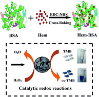
An enzyme mimic synthesis protocol has been proposed by simply cross-linking the redox active center of peroxidase onto a protein scaffold. Colorimetric assays and kinetic studies indicate that the developed peroxidase mimic can present much stronger catalysis and better aqueous stability than native hemin.
Hem is an iron–porphine complex occurring naturally from heme, which is the active center of heme enzymes embedded deeply in the protein pockets of the catalytic hemeprotein family, which includes cytochromes, peroxidases, myoglobins, and hemoglobins.20–23 As a commercially available and inexpensive catalyst, Hem derivatized with carboxyl groups was utilized for a variety of catalytic oxidation reactions,24,25 e.g., it was used as a substitute for HRP in the catalytic reactions of hydrogen peroxide.22,24 Nevertheless, Hem may be trapped by its low catalytic activity, poor aqueous solubility, oxidative self-degradation, and especially molecular aggregation to yield inactive dimers in aqueous solution.26 Moreover, this iron–porphine complex can present considerably high affinity towards plasma proteins like hemopexin and albumins for the study of different biological transfers and toxic effects of extracellular heme.16,27–29 For example, the coupling of heme with hemopexin was studied to induce the inhibition of lipid peroxidation in artificial liposomes30 and rat liver microsomes.31 To the best of our knowledge, however, the interaction of Hem with albumins for applications in enzymatic catalysis has rarely been investigated to date.
It is well recognized that bovine serum albumin (BSA), as a commercially available and inexpensive protein, contains 583 amino acid residues. It is often utilized in enzymatic media to protect and stabilize protein enzymes. Considering that BSA can combine with many small molecules like heme and drugs,29,32,33 we initially tried to directly mix the native Hem derivatized with carboxyl groups (Fig. S1, ESI†) with BSA to form a Hem@BSA composite to mimic the natural enzyme. Surprisingly, the yielded composite was discovered to present much higher catalytic activity than native Hem. Nevertheless, the resulting Hem@BSA might not be stable enough in water so as to readily aggregate over time. Alternatively, in the present work, the native Hem derivatized with carboxyl groups was covalently bound onto the protein scaffold of BSA to produce a Hem–BSA composite. The main synthetic and catalytic application procedures of the Hem–BSA composite are schematically illustrated in Scheme 1. In addition to the pretty high aqueous stability, the obtained enzyme mimic could display much stronger catalytic activity than Hem@BSA in the typical redox reactions of 3,3′,5,5′-tetramethylbenzidine (TMB) and H2O2.
The topological structure of the developed Hem–BSA composite was characterized by transmission electron microscope (TEM) imaging, taking BSA as a control (Fig. 1). In contrast to native BSA blocks (Fig. 1A), the Hem–BSA composites could be uniformly dispersed with flake-like shapes, showing a average size of about 50 nm in diameter (Fig. 1B). Also, the Hem–BSA composites were examined by dynamic light scattering (DLS), showing an average hydrodynamic diameter of about 95 nm (Fig. S2A, ESI†). Noteworthily, the Hem–BSA composites could be stored in water with high aqueous stability, as evidenced by their hydrodynamic diameters that could be sustained over seven months (Fig. S2B, ESI†). Moreover, the composition of the Hem–BSA composites was explored by Fourier transform infrared (FTIR) spectroscopy, taking Hem and BSA as the controls (Fig. S3, ESI†). As expected, the spectra of the obtained Hem–BSA composite included the typical characteristic peaks of native Hem and BSA, i.e., the peaks at about 2990 cm−1 and 3410 cm−1 that are assigned to the stretching vibrations of –C–H and –NH2,34 respectively. In particular, the peak at about 1700 cm−1 that is ascribed to the bending vibration of –CO–OH of Hem disappeared in the spectra of the yielded Hem–BSA. Instead, typical peaks at 1575 cm−1 and 1425 cm−1 of the bending vibrations of –C
O–NH35 were observed, thus confirming the formation of Hem–BSA composites through the covalent conjugation of Hem and BSA. Moreover, UV-vis spectra of the Hem–BSA composite were recorded in comparison with BSA, native Hem, and Hem@BSA (Fig. 2A). One can note from Fig. 2A that the enzyme mimic of Hem–BSA could apparently manifest two absorption peaks at 288 nm and 405 nm, corresponding to those of BSA (280 nm) and Hem (385 nm), respectively, and so could the Hem@BSA. By comparing to native Hem, both of the peroxidase mimics showed a red shift in the absorption peak of Hem, implying that a change in the conformational composition of Hem might occur after the interaction with BSA. Such an optical characterization might be one of the possible reasons responsible for enhancing the catalytic activity of the resulting Hem@BSA composite, in addition to the high aqueous solubility and well defined structure.
Colorimetric investigations were carried out to probe the catalytic performances of the prepared Hem–BSA composite by comparing with Hem@BSA and native Hem, taking BSA as the control (Fig. 2B). The results of the catalytic TMB–H2O2 reactions indicate that the Hem–BSA composite could exhibit over 4-fold and 2-fold stronger catalytic activities than native Hem and the Hem@BSA, respectively, as clearly witnessed in the photographs of the solutions of the reaction products (inset). Accordingly, the covalent coupling of BSA with Hem could dramatically improve the intrinsic catalysis of Hem by forming the Hem–BSA composite. Despite the detailed mechanism remaining unclear for the interaction between BSA and Hem,29 some important factors regarding the unique functions of BSA scaffolds could be taken into account for clarifying the increased catalysis of the Hem–BSA composite. On the one hand, the cross-linking of Hem onto the BSA scaffold to form the Hem–BSA composite could help to enhance the aqueous stability of catalytic Hem so as to prevent it from the formation of molecular aggregations to yield inactive dimers. On the other hand, BSA might improve the solubility of Hem in water and provide a hydrophilic micro-environment to achieve a higher substrate affinity for pre-organizing more redox substrates to the catalytically active sites of Hem,36 thus leading to improved catalytic activity of the Hem–BSA composite. More importantly, the environmental stability of the developed artificial peroxidase was explored by monitoring the catalysis for the TMB–H2O2 reactions (Fig. S4, ESI†). No significant change was observed in the catalytic performances of the Hem–BSA composites even though they were stored in water for up to seven months, thus confirming the considerably high environmental stability.
The main synthesis conditions of the Hem–BSA composites were optimized, with the results shown in Fig. S5 (ESI†). Apparently, the catalytic performances of the Hem–BSA composite could depend on the Hem concentrations used in the synthetic reactions, showing maximum catalysis at 1.0 mg mL−1 Hem (Fig. S5A, ESI†). Meanwhile, the effects of the amount of BSA on the catalytic activities of the Hem–BSA composites were investigated, showing that 40 mg mL−1 BSA was the most suitable concentration (Fig. S5B, ESI†). Also, NaOH was found to play a vital role in fabricating the artificial enzyme. Fig. S5C (ESI†) illustrates that 0.050 M NaOH should be selected as the optimal dosage to create a moderate alkaline environment for the controlled formation of the desired enzyme mimic.
The best conditions, in terms of pH values and temperature, for maximum catalytic activity of the Hem–BSA composites were studied (Fig. 3). It was found that the developed peroxidase mimic could catalyze the TMB–H2O2 reactions ideally at pH 6.0–7.0 (Fig. 3A), as clearly illustrated by the photographs of the reactant products (inset). Also, the catalytic redox reactions could proceed at the optimal temperature of 37 °C (Fig. 3B), which is in good consistency with the catalysis temperature of most protein enzymes in biological systems. Moreover, colorimetric evaluation of the enzyme dosage-dependent catalysis behaviours was conducted for the Hem–BSA composite in comparison with native Hem, each containing Hem of different concentrations (Fig. S6A, ESI†). Once again, the results stress that the catalytic activity of the Hem–BSA composite was much higher than that of native Hem. Furthermore, the catalytic reaction time of the developed artificial enzyme was probed by monitoring the absorbance changes of the TMB–H2O2 reaction products over different time periods (Fig. S6B, ESI†). Accordingly, the Hem–BSA composite could display a much faster catalysis rate than native Hem, showing the greatly improved catalytic efficiency.
Steady-state catalysis kinetic studies were conducted for the Hem–BSA composite, taking native Hem as a comparison. Herein, the apparent Michaelis constants (Km) were calculated by using Lineweaver–Burk plots of the double reciprocal of the Michaelis–Menten equation.37 The double reciprocal curves involved are comparably shown for the varying substrates of TMB and H2O2 (Fig. S7A and B, ESI†). The obtained kinetic parameters are shown comparably in Table S1 (ESI†). It can be noted that the apparent Km value of Hem–BSA for the TMB substrate (2.97) was much lower than that for native Hem (4.81), indicating that the Hem–BSA may possess a higher affinity for TMB than native Hem. Notably, the higher substrate affinity of Hem–BSA would be expected to pre-organize more TMB substrates close to the catalytically active sites of the enzymes for more efficient substrate transformation.36 Moreover, the Km value of the Hem–BSA composite for the H2O2 substrate (2.46) was slightly lower than that of native Hem (2.85), implying that both might present approximately equal affinity to H2O2. Additionally, as can be found from Table S1 (ESI†), by comparing to HRP,9 both Hem–BSA and native Hem display higher Km values for the TMB substrate, and inversely, lower Km values for the H2O2 substrate. This suggests that there still is a difference in the redox substrate affinities between the tested enzyme mimics and natural peroxidases. Furthermore, the possible catalytic mechanism of the developed Hem–BSA composite was thereby investigated in the TMB–H2O2 reaction. The double reciprocal plots of the initial velocities against three fixed concentrations of one substrate over a range of concentrations of another substrate were obtained separately for TMB and H2O2 (Fig. S7C and D, ESI†). It is found that parallel plotting lines could be achieved separately at three concentrations of H2O2 (Fig. S7C, ESI†) and TMB (Fig. S7D, ESI†). Therefore, the characteristic catalysis of Hem–BSA was considered to feature a ping–pong mechanism, as confirmed elsewhere for HRP,38 indicating that the Hem–BSA composite may work as a peroxidase mimic for a broad range of catalytic applications.
Preliminary catalytic applications of the developed Hem–BSA composite were comparably examined by colorimetric assays for H2O2 with different concentrations (Fig. 4). Fig. 4A shows a comparison of colorimetric H2O2 results between the Hem–BSA composite and native Hem. One can find that the developed composite could provide much stronger colorimetric performances than native Hem in terms of the response sensitivity and linear range in targeting H2O2. Noteworthily, the developed composite could tolerate toxic H2O2 with concentrations up to 20 mM without a significant change in catalysis activity. Fig. 4B manifests the calibration curve of the Hem–BSA-based colorimetric assays for H2O2. Accordingly, H2O2 can be detected with concentrations ranging linearly from 0.015 mM to 2.5 mM, with a high correlation coefficient (R2 = 0.9980). A detection limit of about 6.0 μM was obtained as estimated by the 3σ rule, which is much lower than that of Hem (about 25 μM) obtained here. Therefore, the fabricated Hem–BSA composite can allow for colorimetric H2O2 analysis a with much higher sensitivity detection and wider analysis range than native Hem.
To summarize, a simple and highly efficient fabrication method has been successfully developed by mimicking natural peroxidases to produce artificial enzymes with powerful catalysis. Herein, native Hem derivatized with carboxyl groups, that was commercially obtained from heme of the peroxidase-active center, was skilfully cross-linked onto the protein scaffold of BSA, yielding a Hem–BSA composite. The BSA scaffold could not only help in enhancing the aqueous stability of catalytic Hem, so as to avoid the formation of molecular aggregations to yield inactive dimers, but also improve the aqueous solubility of Hem with a hydrophilic micro-environment. More importantly, the so fabricated peroxidase mimic can display high environmental stability and especially powerful intrinsic catalysis, which was over 4-fold stronger than that of native Hem. Kinetic studies indicate that the Hem–BSA composite could display much higher substrate affinity than native Hem, which is comparable to natural peroxidases, like HRP. Subsequently, their outstanding catalytic performances were confirmed in preliminary catalytic applications for probing H2O2, with the detection limit down to about 6.0 μM. This facile and efficient fabrication protocol may open a new door toward the preparation of diverse enzyme mimics with high catalysis and aqueous stability, with promise for wide applications in chemical, environmental, and biomedical fields.
Footnote |
† Electronic supplementary information (ESI) available. See DOI: 10.1039/c6ra07139b |
This journal is © The Royal Society of Chemistry 2016 |