Mehdi Jaymanda,
Meherdad Lotfi*ab and
Rana Lotfic
aResearch Center for Pharmaceutical Nanotechnology, Tabriz University of Medical Sciences, P.O. Box: 51656-65811, Tabriz, Iran. E-mail: mehrlotfi@yahoo.com; Fax: +98-41-33367929; Tel: +98-41-33367914
bDepartment of Endodontics, Dental Faculty, Tabriz University of Medical Sciences, P.O. Box: 51666-14711, Tabriz, Iran
cFaculty of Health, York University, Toronto, ON, Canada
First published on 26th April 2016
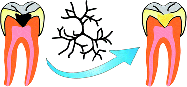
Dental caries and dentin hypersensitivity are the most common clinical diseases in oral health worldwide. Depending on the extent of tooth damage, various approaches can be used to repair teeth. When caries break in more than half of the tooth enamel, a dentist removes the decayed materials and replaces it with appropriate materials. The most commonly used restorative materials are amalgam, dental porcelain (dental ceramic), gold, glass ionomer, and resin-based composites. It is a decisive fact that a great deal of research effort has focused on the design of new photoinitiators, monomers, and telechelic oligomers in the field of resin-based dental composites in order to achieve interesting materials with improved physicochemical properties. In this respect, a novel achievement with multifunctional dendrimers is their dental restorative materials performance, because they exhibit suitable polymer mechanical properties, and reduce shrinkage or shrinkage stress when compared with other similar molecular weight molecules. They possess a high number of functional end groups, and a high degree of conversion. Furthermore, in dendrimer-based dental restorative materials the high cross-link density, as a result of the large number of reactive functional end groups provide some advantages such as a three-dimensional network, decreases solubility and water sorption, improve the mechanical and thermal properties of the resin. Moreover, multifunctional dendrimers can be used in regeneration of human tooth enamel. From the practical point of view, approximately all conducted research in this area has been focused on regeneration of human tooth enamel by a poly(amido amine) (PAMAM) dendrimer, in part due to the capability of this dendrimer in the crystallization process of hydroxyapatite (HA). This review provides a snapshot of recent progress in the synthesis and application of dendrimers in dental restorative materials, and in situ re-mineralization of human tooth enamel.
The common type of restorative dental materials are amalgam, dental porcelain (dental ceramic), gold, glass ionomer, and resin-based composites (RBCs). Among them amalgam and resin-based composites are of particular interest. Amalgam is a self-hardening material made from silver, mercury, copper or other metals. Amalgam has many advantages such as low cost, excellent polishability, good resistance to compression and abrasion, self-sealing, versatility and eases of use, minimal-to-no shrinkage and resists leakage, and completed in one visit. However, some drawbacks of amalgam are the lack of adhesion to the residual tooth structure, requires removal of some healthy tooth, poor resistance to torsion, possible oxidation, corrosion, and tattoos, mercuroscopic expansion, and gray color.8–12
It is an unquestionable fact that the resin-based composite fillings have stimulated great interest as materials of choice for replacing amalgam as a restorative material for posterior restorations. Resin-based dental composite generally made from synthetic functional photoactive resin (e.g., dimethacrylate), high percentage (up to 60 vol%) of inorganic filler (e.g., silica), and a photoinitiator system.13–15 However, resin based restorative materials have not yet achieved the level of mechanical properties found in dental amalgam. Moreover, the cost of resin-based composite fillings more than amalgam, and generally are not covered by some insurance plans. Thus, from the practical point of view, some progress will be made in matrix resins, reinforcing fillers, and curing agents towards the very desirable attainments in this area.16–22
There are numerous reviews are available around the synthesis, and properties of resin based restorative materials. However, to the best of our knowledge, the presented review is the first comprehensive article around the synthesis and application of dendritic dental restorative materials. These type of components were neglected in the field of dental restorative materials and there were only a few researches were conducted in this area. Moreover, recent progresses in the application of dendrimers for in situ re-mineralization of human tooth enamel are also highlighted.
In comparison with dental amalgams and dental porcelain, resin-based dental composites possess several advantages, such as tooth colored, maximum amount of tooth preserved, and the ability to bond to enamel surface. However, these materials possess several disadvantages like costs more than dental amalgam, low degree of conversion, undesirable consequences of residual monomer into the human body (e.g., allergic reactions and sensitization in patients), oxygen inhibition, water sorption, shrinkage stress and strain during their photopolymerization.28–36 As mentioned low degree of conversion in these materials has led to monomer release in vivo, and this can cause irritation, inflammation, and an allergic reaction of the oral mucosa in patients. Thus, the biocompatibility of the resin-based dental composites and their biodegradation by-products are important factors that require consideration beside mechanical and other physicochemical properties of these materials.37,38 The potential cytotoxic effect of an implantable biomaterial can be examined using either a monolith of the material, a particulate form, or extracted solutions, which may contain several types of toxic compounds.39,40 The potential cytotoxic effect of the resin-based dental composites can be evaluated by all above mentioned approaches. The potential in vitro cytotoxic, genotoxic, mutagenic, estrogenic effects of some dental monomers such as Bis-GMA, UDMA, Bis-EMA, and TEGDMA (the most commonly used monomers in dental composites) have been proved.22,41,42 Considering these facts, the find innovative strategies to design and development of new or more efficient resin-based dental composites with controlled duration and total amount of monomer(s) release or polymerization condition which may have fewer negative biological consequences are necessary in the future.
Among mentioned disadvantages polymerization shrinkage is the main drawback of resin-based dental restorative composites which may bring on marginal gaps between the tooth and the material leading to secondary caries. This problem originated from the conversion of intermolecular van der Waals distances of the resin monomers to the covalent bond lengths during photopolymerization.43–47 Thus, many research efforts have been focused to reduce polymerization shrinkage of resin-based dental composites. In this respect, the use of monomers can be polymerized by ring-opening polymerization (e.g., siloranes),48–50 incorporation of bulky cross-linking agents such as (polyhedral oligomeric silsesquioxanes (POSS)),51–53 and thiol–ene or methacrylate–thiol–ene systems15,54–56 could be reduce the polymerization shrinkage and the associated stress.
It is well established that, the thiol–ene or methacrylate–thiol–ene systems can be reduce polymerization shrinkage of resin-based dental composites. During the last decade, a considerable amount of academic researches has been devoted to the thiol–ene or methacrylate–thiol–ene systems including design of new photoinitiators, monomers, and telechelic oligomers.63,64
It is demonstrated that the conventional methacrylate systems polymerize via a radical chain growth polymerization mechanism. In contrast, the thiol–ene reactions proceed via a radical step growth addition mechanism that comprises the addition of a thiyl radical to an ene functional group, followed by propagation and chain transfer to a thiol, thus regenerating the thiyl radical. Step growth addition mechanism has some advantages such as rapid loss of monomer early in the reaction, ends remain active, little sensitivity to oxygen inhibition, terminal functionality tenability, improved cure depths, delayed gel point, and enhanced control of the polymerization in comparison with conventional radical chain growth polymerization mechanism.65–68 Moreover, in step growth addition mechanism monomeric thiol–ene system exhibit 12–15 mL shrinkage per mol CC polymerized, which represents a notable reduction in polymerization shrinkage when compared with the 22.5 mL shrinkage per mol C
C polymerized for methacrylate systems. Since the efficiency of this method has been extensively discussed elsewhere.69
There are various types of dendrimers including tecto, liquid crystalline, chiral, triazine, peptide, polyether, glyco, poly(amido amine-organosilicon) (PAMAM-OS), poly(amido amine) (PAMAM), poly(propylene imine) (PPI), polyester, hybrid, multilingual, and micellar dendrimers have been synthesized.75,76,83–91 Dendrimers have many different commercial and technological applications such as electrochemical nanobiosensing, diagnostic applications, biomedical and pharmaceutical sciences, and nanoscale catalysts.92–101
It is well established that, one successful approach for circumvention of these problems is increase the molecular weight or molar volume of the monomers in order to decrease the concentration of the reactive double bonds. In this respect, a novel achievement with dendrimers is their dental restorative materials performance, because they exhibit acceptable polymer mechanical properties, and reduce shrinkage or shrinkage stress when compared with other similar molecular weight molecules. They have high number of functional end groups, and high degree of conversion. Moreover, in dendrimer-based dental composites the higher cross-link density of dendrimer provide some benefits such as three-dimensional network, decreases water sorption and solubility, improve the mechanical properties and melting temperature of the resin. In addition, multifunctional dendrimers can be used in regeneration of human tooth enamel. From the practical point of view, approximately all conducted researches in this area have been focused on regeneration of human tooth enamel by poly(amido amine) (PAMAM) dendrimer, in part due to the capability of this dendrimer in the crystallization process of hydroxyapatite (HA).
In 2005 Matinlinna et al.104 described the bond strengths of Bis-GMA, and two novel experimental methacrylated polyester dendrimer with 12 methacrylate groups (DR1 and DR2) to grit-blasted titanium substrate with three silanes (3-methacryloxypropyltrimethoxysilane, ESPE Sil™, and Monobond-S™). The chemical structure of DR1 dendrimer is shown in Scheme 3. The shear bond test results and storage conditions for applied silanes are summarized in Table 1. As seen in this table the highest shear bond for thermocycled samples was obtained for Bis-GMA with Monobond-S™ (19.4 ± 7.1 MPa), and after water storage with a laboratory-made silane the shear bond was obtained as 26.4 ± 8.1 MPa. They reported that statistical analysis by ANOVA test showed that independent factors (storage conditions, resins, and silanes) differed significantly (p < 0.001).
![]() | ||
Scheme 3 The chemical structure of DR1 dendrimer.104 |
Silane | Bis-GMA | DR1 | DR2 | |||
---|---|---|---|---|---|---|
WS | TC | WS | TC | WS | TC | |
a Groups with * and ** superscripts do not differ statistically according to the t-test (WS, water storage, TC, thermocycled). | ||||||
0.5% MPS | 26.4 (8.1) | 13.6 (3.2) | 17.9 (9.3) | 11.0 (3.8) | 18.4 (6.1)** | 16.6 (4.3)** |
Monobond-S™ | 19.1 (6.5)* | 19.4 (7.1)* | 19.4 (7.3) | 5.7 (3.8) | 19.8 (5.2) | 9.0 (5.0) |
ESPE Sil™ | 26.2 (7.7) | 10.8 (6.2) | 15.7 (5.4) | 5.3 (2.3) | 13.9 (7.3) | 6.3 (2.1) |
The degree of conversion (DC) was calculated by means of Fourier transform infrared (FTIR) spectroscopy. The values for the Bis-GMA after 40 s light-curing with a hand-unit and after light-curing in an oven were obtained 47.4 ± 1.6 and 53.9 ± 2, respectively. Moreover, for DR1 the DC-values were 44.3 ± 1.4 and 61.0 ± 3.2, and finally for DR2 resin 45.8 ± 0.7 and 60.4 ± 5.7 after 40 s light-curing with a hand-unit and after light-curing in an oven, respectively. Based on these findings, the degree of conversion results for DR1 and DR2 were higher in comparison with the Bis-GMA resin, when the post-cure in the light oven had taken place. They concluded that after thermocycling, the combinations of laboratory-made 0.5% MPS with DR2 resin, and Monobond-S™ with Bis-GMA resin yielded the highest shear bond values. DR2 and the Bis-GMA resin systems conferred statistically equivalent bonding properties to grit-blasted Ti after thermocycling.
The effect of a photopolymerizable dendritic compound with 12 methacrylate groups (D12) on the degree of conversion and mechanical properties of the resultant dental composites has been extensively studied by the Viljanen group. The chemical structure of dendrimer is shown in Scheme 3. In their older study105 the experimental resin system consisted of dendrimer D12, and methyl methacrylate (MMA) in a mass ratio of 80:
20. The initiator and activator used were camphorquinone (CQ), and 2-(N,N-dimethylamino)ethyl methacrylate (DMAEMA), whose concentrations varied individually from 1 to 4 wt%. The experimental design for screening the influence of CQ and DMAEMA concentrations in the D12/MMA copolymer is shown in Fig. 1.
![]() | ||
Fig. 1 The experimental design for screening the influence of CQ and DMAEMA concentrations in the D12/MMA copolymer. Each point represents a resin mixture prepared with the composition indicated by the variable values. In addition to the nine single measurements, three repetitions were made at the center (red spot) (this figure has been adapted/reproduced from ref. 105 with permission from Elsevier). |
The degree of conversion was determined using FTIR spectroscopy, according to the following equation:
In this equation A(CC) is the absorbance intensity of the methacrylate peak and A(C
O) is the absorbance intensity of the internal standard peak. Moreover, the flexural strength, and flexural modulus were studied by the three-point bending tested, and results obtained are summarized in Table 2. As seen in this table the degree of conversion values ranged from 56.2 to 65.5% and increased with increasing initiator and activator concentrations. The mechanical properties (flexural strength and flexural modulus) demonstrated an opposite trend.
Specimen no. | CQ (wt%) | DMAEMA (wt%) | DC (%) | FS (MPa) | FM (MPa) |
---|---|---|---|---|---|
1 | 1.0 | 1.0 | 56.2 | 46.7 | 720 |
2 | 2.5 | 1.0 | 60.1 | 50.6 | 642 |
3 | 4.0 | 1.0 | 61.2 | 35.1 | 329 |
4 | 1.0 | 2.5 | 59.6 | 35.6 | 712 |
5 | 2.5 | 2.5 | 64.7 | 31.7 | 505 |
6 | 4.0 | 2.5 | 64.0 | 28.7 | 382 |
7 | 1.0 | 4.0 | 62.4 | 35.9 | 605 |
8 | 2.5 | 4.0 | 64.3 | 21.6 | 427 |
9 | 4.0 | 4.0 | 65.5 | 18.1 | 189 |
10 | 2.5 | 2.5 | 62.5 | 34.9 | 546 |
11 | 2.5 | 2.5 | 64.3 | 39.2 | 550 |
12 | 2.5 | 2.5 | 65.3 | 39.3 | 574 |
As they concluded, the high concentrations of initiator probably restrained the transmittance of the active wavelengths to the depths of the samples (the inner filter effect), resulting in inhomogeneous conversion and thus decreased mechanical properties. In addition, the high concentrations of radicals that would be present with the high initiator concentrations could also result in reduced polymer chain lengths that could potentially affect polymer properties, although the inner filter effect is likely to be much more significant.105 Based on these findings, this study was demonstrated the hypothesis that both mechanical properties and degree of conversion were increased by increasing the initiator and activator concentrations was not true.
Finally, it should be emphasized that double bond conversion and monomer conversion is not completely the same, and in some cases it is complex. For example, in the case of conducted research by Viljanen et al.22 they mentioned that the measured DC is an overall value of the copolymer (composed of an experimental monomer with four methacrylate groups (D4), and methyl methacrylate (MMA) monomer), and the relative amounts of D4 and MMA reacted in polymerization cannot be determined on the basis of FTIR alone. Therefore, high-performance liquid chromatographic (HPLC) measurements of remaining unreacted monomers are necessary.
In another study, Viljanen and co-workers analyzed the residual monomer content of photopolymerized resin mixtures consisted of dendritic methacrylate monomer, methyl methacrylate (MMA), acetoacetoxyethyl methacrylate (AAEM), and 1,4-butanediol dimethacrylate (BDDMA) (Schemes 3 and 4) in varied proportions by means of headspace-gas chromatography/mass spectrometry (HS-GC/MS), and HPLC. Camphorquinone (CQ), and 2-(N,N-dimethylamino)ethyl methacrylate were used as the light-activated initiator system.106
![]() | ||
Scheme 4 The chemical structures of methyl methacrylate (MMA) (a), acetoacetoxyethyl methacrylate (AAEM) (b), and 1,4-butanediol dimethacrylate (BDDMA) (c).106 |
The compositions of the uncured samples are summarized in Table 3. The content of residual monomers was analyzed after 40 s photopolymerization (visible light-curing). For example the residual MMA and AAEM monomers were extracted with tetrahydrofuran (THF) inhibited with 20 ppm of hydroquinone. The polymer specimens were placed in the solution and stirred with a magnetic stirrer for 72 ± 2 hours at room temperature. An aliquot of the supernatant was diluted with methanol inhibited with 20 ppm of hydroquinone. The sample solution was filtered to remove precipitated polymers, and analyzed with HPLC and HS-GC/MS equipments. Fig. 2 is shown the residual monomer content in dendrimer D12/methyl methacrylate/acetoacetoxyethyl methacrylate copolymers and composites analyzed with HPLC (N = 6), and HS-GC/MS (N = 2).
D12 | MMA | AAEM | BDDMA | Initiator system | Particulate fillera | |
---|---|---|---|---|---|---|
a The composition of the particulate filler composites was similar to dental restorative materials.b The monomers in PFC1 consisted of 90 wt% of the basic mixture D12![]() ![]() ![]() ![]() |
||||||
AAEM0 | 78.4 | 19.6 | — | — | 2.0 | — |
AAEM4 | 75.3 | 18.8 | 3.9 | — | 2.0 | — |
AAEM8 | 72.1 | 18.0 | 7.8 | — | 2.0 | — |
AAEM12 | 69.0 | 17.2 | 11.8 | — | 2.0 | — |
AAEM16 | 65.9 | 16.5 | 15.7 | — | 2.0 | — |
PFC1 | 20.7 | 5.2 | 2.9 | — | × | 71.3 |
PFC2 | 15.6 | 3.9 | 2.8 | 5.6 | × | 71.2 |
![]() | ||
Fig. 2 The residual monomer content in dendrimer D12/methyl methacrylate/acetoacetoxyethyl methacrylate copolymers and composites analyzed with (a) HPLC (N = 6) and (b) HS-GC/MS (N = 2). The error bars represent standard deviation (this figure has been adapted/reproduced from ref. 106 with permission from Elsevier). |
As shown in this figure the addition of AAEM was found to promote the photopolymerization of MMA (p = 0.014). In the other hand, the amount of residual MMA decreased from 2.7 to 1.6% of specimen weight as the initial concentration of AAEM increased from 0 to 16 wt%, but at the same time the amount of residual AAEM increased from 0.3 to 1.7%. In addition, the residual amounts of methyl methacrylate in the PFC1 and PFC2 specimens were 0.10 and 0.08% of resin weight and the amounts of AAEM 0.65 and 0.77%, respectively. Bases on these finding, they suggested that the small amount of residual methyl methacrylate monomer in PFC specimens compared to unfilled resins was probably mainly due to earlier evaporation of MMA at the manufacturing stage. They concluded that the addition of acetoacetoxyethyl methacrylate enhanced the copolymerization of methyl methacrylate and the dendritic monomer, however, the final total residual monomer content did not decrease. The results obtained from HS-GC/MS and HPLC analysis are similar in the analysis of low-boiling residuals in dental polymers. HS-GC/MS has the advantages of being less laborious and also its ability to identify other volatile compounds.106
In another study, Viljanen group investigated the degree of conversion and thermal properties of the above mentioned dental composites.107 The compositions of the uncured samples (wt%) in this study is summarized in Table 4. In this study camphorquinone and 2-(N,N-dimethylamino)ethyl methacrylate were used as the light-activated initiation system. The degree of conversion and thermal properties of the samples were investigated by means of FTIR and differential scanning calorimetry (DSC), respectively. The obtained results are summarized in Tables 5 and 6. As seen in Table 5 the degree of conversion was improved by increasing the concentration of acetoacetoxyethyl methacrylate (AAEM), due to the less viscosity and more flexibility of the resultant resin. The relatively same result was observed in the dilution of viscose Bis-GMA with TEGDMA. It is important to note that, the initiator system concentration in the control material was lower, which may have resulted in a, respectively, lower DC. In addition, PFC2 has higher degree of conversion in comparison with PFC1, due to the higher mobility of the resin system in PFC2 which contained more mono- and dimethacrylates and less dendritic monomer.
D12 | MMA | AAEM | BDDMA | Bis-GMA | TEGDMA | Initiator system | Particulate filler | |
---|---|---|---|---|---|---|---|---|
a A commercially available restorative dental composite (3M Dental Products, USA). The Z100 made from Bis-GMA, TEGDMA, initiator, activator, inhibitor, zirconia/silica particulate filler. The concentrations of the composites are proprietary information of the manufacturer (*). | ||||||||
AAEM0 | 78.4 | 19.6 | — | — | — | — | 2.0 | — |
AAEM4 | 75.3 | 18.8 | 3.9 | — | — | — | 2.0 | — |
AAEM8 | 72.1 | 18.0 | 7.8 | — | — | — | 2.0 | — |
AAEM12 | 69.0 | 17.2 | 11.8 | — | — | — | 2.0 | — |
AAEM16 | 65.9 | 16.5 | 15.7 | — | — | — | 2.0 | — |
Bis-GMA![]() ![]() |
— | — | — | — | 49.3 | 49.3 | 1.4 | — |
PFC1 | 20.7 | 5.2 | 2.9 | — | — | — | * | 71.3 |
PFC2 | 15.6 | 3.9 | 2.8 | 5.6 | — | — | * | 72.2 |
Z100a | — | — | — | — | * | * | * | 66 (vol%) |
DC% | Residual reactivity | ||
---|---|---|---|
ΔHa (J g−1) | Tmax (°C) | ||
a ΔH: reaction exotherms (determined by DSC). | |||
AAEM0 | 52.1 (0.3) | 0.25 | 138 |
AAEM4 | 52.1 (0.2) | 5.22 | 127 |
AAEM8 | 53.1 (0.8) | 8.54 | 123 |
AAEM12 | 57.7 (0.2) | 9.56 | 126 |
AAEM16 | 59.6 (0.6) | 8.46 | 124 |
Bis-GMA![]() ![]() |
51.7 (0.2) | — | — |
PFC1 | 32 (2) | 4.38 | 122 |
PFC2 | 44 (2) | 2.99 | 129 |
Z100 | 38.4 (0.1) | — | — |
Tg | ||||
---|---|---|---|---|
Photo-polymerized (°C) | Post-polymerized (°C) | |||
Ramp 1 | Ramp 2 | Ramp 1 | Ramp 2 | |
a Determined by DSC. | ||||
AAEM0 | 56 | 83 | 51 | 119 |
AAEM4 | 48 | 116 | — | — |
AAEM8 | 48 | 113 | — | — |
AAEM12 | 50 | 112 | 46 | 115 |
AAEM16 | 48 | 116 | 46 | 112 |
Bis-GMA![]() ![]() |
54 | 83 | — | — |
PFC1 | 46 | 84 | — | — |
PFC2 | 45 | 87 | — | — |
Z100 | 46 | 68 | — | — |
Additional evidence for the improving the degree of conversion by increasing the concentration of the acetoacetoxyethyl methacrylate (AAEM) was also obtained from DSC results and summarized in Table 6. It is a decisive fact that, the degree of conversion increased by decreasing of residual reactivity. The low transition temperature determined from the first heating ramp probably results from a relaxation by the thermal movement of short pendant chains or local chain segments in the cross-linked polymers (Table 6). Based on these finding, they concluded that the addition of AAEM enhanced the degree of conversion of the dendritic resin system. Exchanging methyl methacrylate to another diluent comonomer might further improve the polymerization.
Paul et al.108 investigated the influence of globular and highly branching 2,3-dihydroxybenzyl motif on material properties and degree of polymerization of formulated dental composites made from bisphenol A dimethacrylate (Bis-EMA):
hexane-1,6-diol dimethacrylate (HDMA)
:
dimethylaminoethyl methacrylate (DMAEMA)
:
camphorquinone (CQ) (70
:
30
:
1
:
0.5), and varying percentages of multi-methacrylated dendritic additives. The chemical structure of the dendritic 2,3-dihydroxybenzyl motifs synthesized by Paul et al. are shown in Schemes 5–7. The dental resin mixture was formulated as follows: Bis-EMA
:
HDMA
:
DMAEMA
:
CQ (70
:
30
:
1
:
0.5) was added to a solution of purified dendritic additive (1, 2, 3, 4, or 5) dissolved in dichloromethane (0.5 to 5 mL) in ratios of 98
:
2, 90
:
10, 70
:
30, and 50
:
50 (resin mixture
:
dendritic additive) by weight. The solvent was evaporated using a stream of nitrogen followed by high vacuum. This modified resin was then blended with 3 parts by weight of silanated filler (Schott Glass GM27884, UF1.0, 3.2% silane) to produce samples containing 0.5%, 2.5%, 7.5%, and 12.5% dendritic 2,3-dihydroxybenzyl motif by weight.
![]() | ||
Scheme 5 The chemical structures of the two type (compounds 1 and 2) of dendritic 2,3-dihydroxybenzyl motifs synthesized by Paul et al.108 |
![]() | ||
Scheme 6 The chemical structures of the two type (compounds 3 and 4) of dendritic 2,3-dihydroxybenzyl motifs synthesized by Paul et al.108 |
![]() | ||
Scheme 7 The chemical structure of the highly branched dendritic 2,3-dihydroxybenzyl motif (compound 5) synthesized by Paul et al.108 |
The degree of polymerization (DP) was investigated for prepared composite samples using TA Instruments DSC. The DP was calculated using the equation DP = j/(n × 54.8 J mmol−1) × 100, where DP is the percentage of the theoretical maximum conversion, j is the measured energy released upon photopolymerization (in J g−1), n is the mmol of methacrylate group per g of a given sample, and 54.8 J mmol−1 is the theoretical energy value of methacrylate group polymerization. The results obtained are summarized in Table 7. As seen in this table the photopolymerization studies are not significantly different than the controls at low concentrations of additive, and degree of polymerization decreases with higher concentration as expected.
Sample name | 0% | 0.5% | 2.5% | 7.5% | 12.5% |
---|---|---|---|---|---|
1 | 49.7 (1.7) | 44.6 (2.5) | 40.0 (3.1) | — | — |
2 | 49.7 (1.7) | 51.9 (0.7) | 50.1 (0.7) | 39.5 (0.3) | — |
3 | 49.7 (1.7) | 45.6 (1.7) | 44.7 (1.3) | 37.6 (1.5) | 34.5 (0.6) |
4 | 49.7 (1.7) | 49.9 (2.5) | 36.6 (2.8) | 41.5 (1.1) | 35.2 (1.2) |
5 | 49.7 (1.7) | 42.4 (0.9) | 36.5 (1.6) | 31.9 (1.3) | 23.5 (0.4) |
Moreover, they studied acetone extraction of the samples as follow: bar samples (2 mm square and 25 mm in length) were weighed (Wi) into vials, then steeped in acetone at room temperature for 96 hours. The samples were dried under a flow of nitrogen until a constant weight (Wf) was reached. The percentage of organic material extracted was calculated by (Wi − Wf)/Wi × 100. The results obtained are presented in Table 8. As seen in this table an increase in the amount of extractables at higher concentrations of additives are observed. Although there was no apparent trends that correlated with molecular weight, 4 afforded the least amount of extractables.
Sample name | 0% | 0.5% | 2.5% | 7.5% | 12.5% |
---|---|---|---|---|---|
1 | 0.46 (0.10) | 0.33 (0.08) | 0.42 (0.07) | — | — |
2 | 0.46 (0.10) | 0.20 (0.05) | 0.36 (0.04) | 0.77 (0.11) | — |
3 | 0.46 (0.10) | 0.21 (0.07) | 0.20 (0.01) | 0.31 (0.01) | 0.70 (0.04) |
4 | 0.46 (0.10) | 0.12 (0.03) | 0.07 (0.03) | 0.09 (0.04) | 0.57 (0.10) |
5 | 0.46 (0.10) | 0.52 (0.07) | 0.39 (0.02) | 0.91 (0.23) | 1.60 (0.20) |
Table 9 is shown the flexural strength tests results. As seen in this table, improvements in flexural strength were uniformly observed at low concentrations of dendritic additive, particularly with compounds 3, 4, and 5. The largest improvements in flexural strength were observed at the lowest dendritic additive concentrations employed; whereas, formulations using 7.5% and 12.5% of dendritic additive showed significant decreases in flexural strength, in comparison with control, in all samples except dendrimer 4. Based on these finding, it was demonstrated that at a given concentration, the improvement in mechanical properties positively correlated with the generation level of the dendritic additive. The lower flexural strengths observed at higher concentrations with dendrimer 5, in part due to the lower degree of polymerization (DP) that affords a weaker, less cross-linked composite.108 They concluded that highly globular and highly cross-linking additives could be improved composite properties even at very low concentrations.
Sample name | 0% | 0.5% | 2.5% | 7.5% | 12.5% |
---|---|---|---|---|---|
1 | 60.1 (6.7) | 60.9 (7.7) | 62.2 (12.1) | — | — |
2 | 60.1 (6.7) | 62.5 (5.2) | 65.4 (7.0) | 56.3 (8.2) | — |
3 | 60.1 (6.7) | 72.9 (12.0) | 72.6 (12.4) | 56.1 (8.4) | 50.4 (11.0) |
4 | 60.1 (6.7) | 73.9 (9.9) | 68.3 (9.6) | 63.0 (10.5) | 75.6 (7.2) |
5 | 60.1 (6.7) | 81.3 (12.3) | 81.0 (19.9) | 69.4 (11.3) | 44.8 (8.7) |
Kawaguchi et al.109 investigated the mechanical properties of denture base resin cross-linked with methacrylated dendrimer (D12; Scheme 3). The monomer liquid of resin was a mixture of methyl methacrylate (MMA), and cross-linker dendrimer (D12) or cross-linker ethyleneglycol dimethacrylate (EGDMA) with five different volume percentages. Due to the high viscosity of the dendrimer (D12), a 20 wt% amount of MMA was added to the dendrimer liquid. The amount of cross-linking agent used was at 1.1, 2.3, 4.6, 6.9, 9.1 vol% relative to MMA. Powder were made by adding 2% benzoyl peroxide by weight as radical initiator. The specimens were polymerized in distilled water maintained at 55 °C under pressure of 0.4 MPa for 20 minutes. Mechanical property study has revealed that the highest flexural strength was achieved by the addition of 2.3% D12, and followed by the addition of 1.1% EGDMA. Addition of 9.1% showed the lowest value with both cross-linking agents.
As can be seen in Fig. 3 the surface hardness test photographs showed that there was a semi-IPN intermediate layer between the matrix and polymer PMMA beads with the specimens containing 4.6, 6.9, and 9.1% of D12. Based on these finding, they concluded that the statistical analysis by ANOVA test showed that the addition of cross-linker dendrimer (D12) had a significantly higher effect (p < 0.05) on flexural modulus and hardness of matrix area than EGDMA but on flexural strength (p > 0.05). The effect of quantity differences of cross-linker was statistically significant only on flexural strength (p < 0.05).
![]() | ||
Fig. 3 Typical photographs of the semi-IPN layer between PMMA beads and polymer matrix containing of (a) 1.1%, (b) 2.3%, (c) 4.6%, (d) 6.9%, and (e) 9.1% D12-cross-linked polymer. Arrows indicate the semi-IPN layer (original magnification 400×; bar = 30 μm) (this figure has been adapted/reproduced from ref. 109 with permission from Elsevier). |
In another study, Kawaguchi et al.110 described the influence of molecular weight of poly(methyl methacrylate) (PMMA) beads on the mechanical properties of cross-linked denture base polymers. The materials tested in Kawaguchi et al. study are summarized in Table 10. The monomer liquid of the resin was a mixture of MMA containing 4.6 vol% dendrimer (D12; Scheme 3), as the cross-linker. The three PMMA powders (MW 120000, 350
000, and 996
000) were used in groups 1, 3, and 4 were made by adding 2 wt% benzoyl peroxide (B.P.O) by weight as the initiator. Group 2 tested a commercial autopolymerizable denture base resin powder (MW 220
000) containing benzoyl peroxide, which was added as an initiator to start the autopolymerization process. The mechanical properties (flexural strength, and flexural modulus), microhardness of PMMA beads (MHN), and thickness of a swollen layer of the prepared dental composites were tested and summarized in Table 11. As can see groups 2 and 3 showed significantly higher values of flexural strength in comparison with groups 1 and 4; the values for groups 1 and 4 ranged from 49 to 100 MPa. The flexural modulus results indicated that group 1 had the lowest values, and there were no significant differences among the other groups. The micrographs of prepared dental composites groups are shown in Fig. 4. The micrographs showed that in all groups, the semi-IPN layer was located around the PMMA beads, and the swollen layer was found parallel to the interface line. Moreover, the statistical analysis by ANOVA test revealed that the molecular weight of the powder significantly affected the thickness of the swollen semi-IPN layer, while did not significantly affect the surface microhardness. They concluded that the molecular weight of the PMMA beads of multiphase denture base polymers considerably influences their flexural properties and formation of semi-IPN layer between the matrix polymer and the PMMA beads.110
Component | Function | Manufacturer | Lot | |
---|---|---|---|---|
Liquid | ||||
MMA (methyl methacrylate) | Monomer | Aldrich | S76535-139 | |
DMPT (N,N-dimethyl-p-toluidine) | Activator | Aldrich | 03007KH-108 | |
D12 | Gross-liker | VTT processes | PRO6/259/02 | |
![]() |
||||
Powder | ||||
Poly(methyl methacrylate) | MW 120![]() |
Aldrich | MKBC2531 | |
MW 220![]() |
Heraeus | Kulzer 012 527 | ||
MW 350![]() |
Polymer | Aldrich | 15713KC | |
MW 996![]() |
Aldrich | 03722TC | ||
BPO (benzoyl peroxide) | Initiator | Fluka | MKAA0075 |
Molecular weight | 120![]() |
220![]() |
350![]() |
996![]() |
||||
---|---|---|---|---|---|---|---|---|
a Identical letters indicate that the values are not statistically different (p > 0.05). | ||||||||
Flexural strength (MPa) | 48.7 | (14.2)a | 100.1 | (5.7)b | 83.3 | (11.4)b | 63.9 | (15.8)a |
Flexural modulus (GPa) | 2.37 | (0.08) | 2.72 | (0.08)a | 2.74 | (0.07)a | 2.68 | (0.05)a |
Microhardness of PMMA beads (MHN) | 24.9 | (3.8)a | 22.2 | (3.2)a | 24.0 | (5.2)a | 22.5 | (3.2)a |
Thickness of a swollen layer (μm) | 27.9 | (11.0) | 12.2 | (4.2)a | 16.6 | (2.2)a | 13.1 | (1.1)a |
![]() | ||
Fig. 4 Micrographs of the swollen layer in each group. Arrows indicate the thickness of the swollen layer. Upper side indicates new resin; lower side indicates old resin (original magnification ×400; bar = 30 μm) (this figure has been adapted/reproduced from ref. 110 with permission from Elsevier). |
Based on findings, enamel regeneration cannot occur in vivo following failure. Regeneration of lost dental tissue can be achieved by means of stem cell biology or gene therapy technology. However, the complexity and high cost of these approaches seriously limit them wide applications. This problem can be circumvented through in situ re-mineralization of dentine by biomaterials. In this respect, the key point of biomimetic mineralization is to seek the proteins which will be the regulating template for the mineralization of inorganic crystals. Due to difficulty of extraction or purification the natural proteins, design of a biomacromolecular structure that can mimic the self-assembly behavior and the morphology of the natural proteins may be the best and first choice.115–117 From the practical point of view, biodegradable poly(amido amine) (PAMAM) dendrimer has been proposed as the first choice for biomimetic mineralization of human tooth enamel.118–122 It is important to note that, approximately all conducted researches for biomimetic mineralization of human tooth enamel have been focused on the PAMAM dendrimers, in part due to the capability of this dendrimer in the crystallization process of hydroxyapatite. The selected examples for biomimetic mineralization of human tooth enamel are further discussed in the following context.
Chen et al. described the synthesis and application of carboxy-modified G7 PAMAM dendrimers as nanoprobes of the biological hydroxyapatite nanorod surfaces.123 For this purpose, approximately 5 μL of methanol containing the enamel crystals was pipetted onto the mica, the methanol was evaporated rapidly, and the PAMAM dendrimer solution (285 nM in water, pH 7.4) was then flowed over the crystals. They were used a 120 seconds exposure period for the interaction of the PAMAM dendrimer solution with the enamel crystals.
The tapping-mode AFM images of the enamel crystals on a mica surface before, and after binding PAMAM dendrimers (G7) with carboxylic acid end groups are shown in Fig. 5 and 6, respectively. As shown in AFM images the PAMAM dendrimers on the mica surface were 2.3 ± 1.0 nm in height and 15 ± 3.3 nm in width; however, those on the hydroxyapatite crystal surface were of 6.9 ± 1.4 nm in height and 19 ± 2.4 nm in width. Based on these findings, the binding energy between the dendrimer and crystal surface, 2A, equals 460 ± 100 eV. Each G7 carboxylic acid-capped PAMAM dendrimer contains 512 carboxylic acid end groups. Therefore, by using Monte Carlo method the binding energy of each –COOH group exerted upon the crystal surface is 0.9 ± 0.2 eV or 90 ± 20 kJ mol−1. They concluded that carboxylic acid-capped PAMAM dendrimer could be used as nanoprobes to mimic the pattern and determine the strength of binding of proteins to biological nanorod surfaces.
![]() | ||
Fig. 5 Tapping-mode AFM images of enamel crystals on a mica surface [all image sizes 1 × 1 μm; (A) height image, z-range, 200 nm; (B) phase image, z-range, 30°] (this figure has been adapted/reproduced from ref. 123 with permission from American Chemical Society). |
![]() | ||
Fig. 6 Tapping-mode AFM images of enamel nanorods after binding generation 7 PAMAM dendrimers with carboxylic acid end groups [all image sizes 1 × 1 μm; (A) height image, z-range, 200 nm; (B) phase image, z-range, 30°] (this figure has been adapted/reproduced from ref. 123 with permission from American Chemical Society). |
Wu et al.124 described the synthesis, characterization, and application of carboxyl-terminated poly(amido amine) (PAMAM-COOH)-alendronate (ALN) conjugate (ALN-PAMAM-COOH) for in situ re-mineralization of human tooth enamel. It is well accepted that PAMAM-COOH has a highly ordered architecture and is capable of promoting the hydroxyapatite crystallization process. On the other hand, it is well demonstrated that the alendronate (ALN) could easily adsorb on the HA crystals.125 A schematic of the specific adsorption of ALN-PAMAM-COOH on the surface of tooth enamel and the subsequent in situ re-mineralization of HA is shown in Fig. 7. Alendronate (ALN) (usually alendronate sodium) is a hydrophilic bisphosphonate drug that acts as a specific inhibitor of osteoclast mediated bone resorption. Bisphosphonates are synthetic analogs of pyrophosphate that bind to the hydroxyapatite. It is well established that ALN increases bone formation and enhances osteoblast proliferation and maturation and leads to inhibition of osteoblast apoptosis.126,127
![]() | ||
Fig. 7 Specific adsorption of ALN-PAMAM-COOH on the surface of tooth enamel and the subsequent in situ re-mineralization of HA (this figure has been adapted/reproduced from ref. 124 with permission from Elsevier). |
The binding capability of ALN-PAMAM-COOH on human tooth enamel was investigated by means of attenuated total reflection-infrared (ATR-IR), and confocal laser scanning microscopy (CLSM) after labeling with fluorescein isothiocyanate (FITC). ATR-IR spectroscopy results demonstrated that ALN-PAMAM-COOH can bind tightly on tooth enamel surface due to the HA-anchored capability of its ALN functional unit. The binding capability between ALN-PAMAM-COOH and enamel surface is so strong that phosphate buffered saline (PBS) cannot wash it off in a large quantity, while the PAMAM-COOH dendrimer without ALN unit can be washed off from the tooth enamel surface by PBS. Moreover, as seen in CLSM and the surface morphology images (Fig. 8) after labeling with FITC the yellow-green fluorescence can be seen and is sporadically dispersed on the surface. In addition, they find that the surface roughness of the sample obviously decreases after the coating of dendrimer (Fig. 8a2 and b2). When the tooth enamel was etched by phosphoric acid, its surface roughness greatly increases, resulting in a significant increase of total surface area (Fig. 8c2). The enlarged enamel surface provided more adsorption sites, thus there were much more FITC labeled ALN-PAMAM-COOH adsorbed on the acid-etched tooth enamel surface (Fig. 8c1).124
![]() | ||
Fig. 8 CLSM and the surface morphology images of normal tooth enamel surface (a1 and a2), FITC-ALN-PAMAM-COOH treated normal tooth enamel surface (b1 and b2), and FITC-ALN-PAMAM-COOH treated acid-etched tooth enamel surface (c1 and c2) (this figure has been adapted/reproduced from ref. 124 with permission from Elsevier). |
In order to observe the cross section of the regenerated minerals, the tooth enamel samples which have been incubated in artificial saliva for 4 weeks were cut and the long axes of the enamel prisms were exposed. Thereafter, the samples were studied by scanning electron microscopy (SEM). As seen in Fig. 9, acid-etched tooth enamel without treatment (Fig. 9; group a) was loosen and accumulated with many big sheet of calcium phosphate crystal on the coarse surface. The tooth enamel samples treated with PAMAM-COOH (Fig. 9; group b) exhibited a certain extent of orientation. However, the morphology of the new crystals in this group was heterogeneous. Also, the arrangement of new crystals became looser from the biomineralization interface to the top area. There was a clear line of interface between original tooth enamel, and the new crystals indicated that the new formed calcium phosphate crystal layer induced by PAMAM-COOH might not bind tightly. However, ALN-PAMAM-COOH treated tooth enamel samples (Fig. 9; group c), exhibited a dense layer of biomineralized crystals on the acid-etched enamel surface. The new generated crystals are parallel bundles of nanorod-like HA with almost uniform size and shape, and mostly oriented perpendicular to the original tooth enamel surface. The morphology of the new regenerated minerals (thicker than 10 mm) was very close to that of normal tissue and should be desirable for in situ re-mineralization of human tooth enamel.124
![]() | ||
Fig. 9 Scanning electron microscope (SEM) micrographs of the cross section of acid-etched tooth enamel without treatment (a group), treated with PAMAM-COOH (b group) and ALN-PAMAM-COOH (c group) (this figure has been adapted/reproduced from ref. 124 with permission from Elsevier). |
They concluded that, the hardness of acid-etched enamel samples treated by ALN-PAMAM-COOH was similar to that of natural tooth enamel and could recovered up to 95.5% of the original value with strong adhesion force. In vivo experiment also demonstrated that ALN-PAMAM-COOH was effective in repairing acid-etched enamel in the oral cavity. Based on these finding, they suggested that ALN-PAMAM-COOH could be a potential restorative nano-material for in situ re-mineralization of human tooth enamel.
Recently, Chen et al.128 reported modulated regeneration of acid-etched human tooth enamel by a phosphate-functionalized PAMAM dendrimer that is an analog of amelogenin. It is well established that the major enamel protein is amelogenin secreted by ameloblasts, and constitutes of approximately 90% of all organic matrix material in developing human tooth enamel. To aim this purpose, PAMAM dendrimer (G4) was modified by dimethyl phosphate to obtain phosphate-end(s) caped dendrimer (PAMAM-PO3H2) since it has a similar dimensional scale and peripheral functionalities to that of amelogenin. The phosphate group has stronger affinity for calcium ion than carboxyl group and can simultaneously provide strong hydroxyapatite-binding capability. A schematic of the adsorption of PAMAM-PO3H2 on the surface of tooth enamel and the subsequent in situ re-mineralization of HA is shown in Fig. 10. The cytotoxicity of the PAMAM-PO3H2 at various concentrations from 15.625 to 1000 μg mL−1 was evaluated by an MTT assay using the L929 cell line and results obtained were summarized in Fig. 11. As seen in this figure the PAMAM-PO3H2 dendrimer has good cell viability, in the range of 0.75–0.99, within 24 hours. That is, PAMAM-PO3H2 has very low cytotoxicity within a wide range of concentrations that are appropriate for further biomedical applications. Adsorption capability of PAMAM-PO3H2 dendrimer on human tooth enamel was evaluated by means of ATR-IR spectroscopy. It is demonstrated that in comparison with PAMAM-COOH the PAMAM-PO3H2 dendrimer has the higher adsorption capability on tooth enamel, in part due to the stronger binding capability of the phosphate group than the carboxyl group. Because it is able form coordination bonds with the calcium ions of HA, thus providing stronger interlinking. The re-mineralization process of tooth enamel was studied by SEM, and X-ray diffraction (XRD). After being incubated in artificial saliva for three weeks, there was a newly generated HA layer of 11.23 μm thickness on the acid-etched tooth enamel treated by PAMAM-PO3H2, while the thickness for the carboxyl-terminated one (PAMAM-COOH) was only 6.02 μm. In addition, XRD results showed that more crystalline HA is newly generated on the PAMAM-PO3H2-treated sample than on the PAMAM-COOH-treated one. Meanwhile, according to the strength of the [002] and [004] peaks in XRD patterns, the newly formed crystals in PAMAM-PO3H2 are oriented almost along the Z-axis, and almost all of the crystals are aligned parallel in the newly generated layer, whereas the interprism crystals are not parallel in natural enamel. Based on these finding, they concluded that the good performance of phosphate-end caped PAMAM dendrimer has a stronger affinity for calcium ions than the PAMAM-COOH and could provide strong HA-binding capability at the same time. Thus, PAMAM-PO3H2 has showed the potential of acting as a biomimetic re-mineralization material for human tooth enamel.
![]() | ||
Fig. 10 Adsorption of PAMAM-PO3H2 on the surface of tooth enamel and the subsequent in situ re-mineralization of HA (this figure has been adapted/reproduced from ref. 128 with permission from Elsevier). |
![]() | ||
Fig. 11 L929 cell viability of PAMAM-PO3H2 at different concentrations, as shown by MTT assay (this figure has been adapted/reproduced from ref. 128 with permission from Elsevier). |
In this respect, a novel achievement with dendrimers is their dental restorative materials performance, because they exhibit acceptable polymer mechanical properties, and reduce shrinkage or shrinkage stress when compared with other similar molecular weight molecules. They have high number of functional end groups, and high degree of conversion. Moreover, in dendrimer-based dental composites the higher cross-link density of dendrimer provides a sufficient number of bridges between linear macromolecules that resulted to some benefits such as three-dimensional network, decreases water sorption and solubility, improve the mechanical properties (e.g., flexural strength and flexural modulus), and melting temperature of the resin. However, the flexural strength and flexural modulus of the dendrimer-based methacrylate was somewhat lower compared to dimethacrylate-based dental resins. The mechanical properties of these composites are strongly depended on initiator and activator concentrations, as well as the structure and concentration of dendritic additive. On the other hand, high degree of polymerization, and highly cross-linking additives could be improved the composite properties even at very low concentrations of initiator, activator, and dendritic additive. It would be expected that further researches will focused on the concentrations optimization of initiator, activator, and dendritic additive to achieve more effective systems.
It is well accepted that in resin-based dental composites the water sorption lowers their mechanical properties and resistance to wear values. These reductions have been are originated from the hydrolytic degradation of the polymer matrix and filler as well as water-induced filler–matrix bond failure.109,129,130 As mentioned, the high cross-link density, as a result of the large number of reactive functional end groups in the dendrimer-based dental composites has a significant effect in the water sorption characterize of the final composite.109 On the other hand, as the dental composites are placed in the oral environment absorption of water takes place, which leads mainly to degradation of the reinforcing filler. However, to the best of our knowledge, no experimental data exist in literature around the water sorption of the dendrimer-based dental composites. Thus, it would be expected further studies will focused on design more efficient systems. In this context, the used of hydrophobic acrylate or thiol-end caped silane and titanate coupling agents for surface modification of inorganic filler can be considered as an efficient approach, mainly due to resistance water diffusion. In addition, these regents provides a crucial link between the matrix and the filler that can have a significant effect on the overall performance of composites as well as improves the resistance of composite to hydrolytic degradation.
It has been well documented that the residual monomers (due to low degree of monomer conversion), and biodegradation by-products of resin-based dental composites can resulted to irritation, inflammation, and an allergic reaction of the oral mucosa in patients. Thus, the creative strategies toward the more efficient resin-based dental composites will be seems necessary to decrease or eliminate their toxicities.
In addition, multifunctional dendrimers can be used in regeneration of human tooth enamel. From the practical point of view, approximately all conducted researches in this area have been focused on regeneration of human tooth enamel by PAMAM dendrimer, in part due to the capability of this dendrimer in the crystallization process of hydroxyapatite.
Considerable advances and promising results such as enhanced the degree of conversion, and mechanical properties have been obtained in the application of dendrimers as dental restorative materials. However, from the practical point of view, some progress will be made towards the very desirable attainments in this area. Thus, it would be expected further studies will focused on the creative design and developmental strategies of new dendrimer-based dental composites including monomers, reinforcing fillers, and curing agents to achieve interesting new materials with improved physicochemical properties. In addition, substantial results in the application of dendrimers for in situ re-mineralization of tooth enamel have been obtained. The literature generally reports basic physicochemical, morphological, and mechanical properties of the dendrimer-based dental composites without an exhaustive characterization in conditions reproducing the real application. Thus, translate into clinical therapies is a demand in the coming decade.
For biomedical applications many companies have continued to develop products based on dendrimers. Despite the commercial availability of some dendrimer (especially PAMAM dendrimers) for large-scale applications the more efficient synthesis strategies will be developed in such a way that the manufacturing costs were kept down.131,132
This journal is © The Royal Society of Chemistry 2016 |