Study of mesoporous magnesium carbonate in contact with whole human blood

S. Frykstranda, J. Forsgrena, O. Cheunga, P. Zhanga, J. Hongb, M. Strømmea and N. Ferraz*a
aDivision for Nanotechnology and Functional Materials, Department of Engineering Sciences, The Ångström Laboratory, Uppsala University, Box 534, SE-751 21 Uppsala, Sweden. E-mail: Natalia.ferraz@angstrom.uu.se
bDepartment of Immunology, Genetics and Pathology, Rudbeck Laboratory, Uppsala University, SE-751 85 Uppsala, Sweden

Received 3rd March 2016 , Accepted 23rd May 2016

First published on 25th May 2016


Abstract

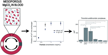
The interaction of mesoporours magnesium carbonate (Upsalite) particles (50–100 μm) with human whole blood was investigated using an in vitro loop model and the effect on the complement system, blood coagulation and red blood cell lysis was assessed. The removal of Ca2+ by Upsalite and the possible exchange with and/or release of Mg2+ were explored as well. Upsalite was found to present anticoagulant properties, most probably due to the uptake of Ca2+ by the particles. No hemolytic activity was detected at Upsalite concentrations up to 1 mg ml−1. Moderate to high levels of C3a and sC5b-9 were observed for Upsalite, however such levels were statistically different from the negative control only when the particle concentrations were 0.25 mg ml−1 and 1.0 mg ml−1, respectively. The presented findings are promising for the future development of mesoporous magnesium carbonate-based materials for biomedical applications.


1. Introduction

Inorganic mesoporous materials are currently being studied worldwide due to their large internal surfaces areas and high pore volumes. These properties open up a number of potential applications in the biomedical field, including their use as drug delivery vehicles,1–6 in regeneration of bone tissue7,8 as vaccine adjuvants9,10 as well as in topical cosmetics applications.11 The materials can be used either per se1,2,5,6,12 or as surface coatings on other functional materials.13

We have recently developed a mesoporous magnesium carbonate commercialized as Upsalite®. The novel mesoporous magnesium carbonate is synthesized without the aid of surfactants and has a narrow pore size distribution centered between 5 and 10 nm.14,15 Furthermore, it was found to have large moisture sorption capacity.14 Upsalite has been shown to exhibit promising properties as amorphous phase stabilizer in drug delivery2,3 and a number of other biomedical applications are also being explored for this material. To ensure a safe implementation of Upsalite within the biomedical field the biocompatibility profile of the material must be known. In this sense, we recently reported the first toxicity screening of Upsalite,16 wherein the material was analyzed for in vitro cytotoxicity using human dermal fibroblasts, for in vivo skin irritation in rabbits and for in vivo acute systemic toxicity in mice. It was concluded that Upsalite is non-toxic for human dermal fibroblast cells at concentrations as high as 1000 μg ml−1 and 48 h exposure, and that topical application of Upsalite powder resulted in negligible cutaneous reaction. It was further found that there was no evidence of significant acute systemic toxicity when saline extracts of Upsalite were injected in mice, but injection of sesame oil extract resulted in transient weight loss in mice. The latter was however attributed to the likely injection of particles rather than a consequence of toxic leachables.

In the present work we further investigate the biocompatibility of Upsalite, focusing on the interactions with blood. If to be used in biomedical applications such as drug delivery or in composites for implant materials, Upsalite will encounter blood. The interaction between blood and artificial materials may lead to the activation of the blood cascades, i.e. coagulation pathways and complement system, together with the activation of platelets, neutrophils and monocytes/macrophages.17 Thus, to continue exploring the use of Upsalite in the biomedical field, it is mandatory to study its blood compatibility.17

Recently B. Ekstrand-Hammarström et al. described an in vitro model to study the interaction between inorganic particles and whole human blood.18 The model uses non-anticoagulated blood from healthy donors to study the possible activation of the coagulation cascade and complement system induced by the particles. Herein a slightly modified version of this model was used to study the interaction of Upsalite particles (50–100 μm) with whole blood. Furthermore, hemolysis induced by the Upsalite particles was analyzed.

2. Materials and methods

2.1 Chemicals and reagents

Magnesium oxide (MgO, >99%), methanol (>99.8%) and CaCl2 (anhydrous mesh, ≥99.99%) were purchased from Sigma-Aldrich. Carbon dioxide (CO2, N48) was purchased from Air Liquide. All reagents were used without further purification.

2.2 Synthesis

The synthesis of Upsalite was carried out as described previously14,15 but on a larger scale. In short, 2.5 l methanol was mixed with 170 g MgO using 500 rpm rotation speed in a 5 l Ecoclave pressure reactor (Büchi). The reaction was carried out at 55 °C under 3 bar CO2 pressure. After four days of reaction time, the temperature was decreased to room temperature and the reactor was depressurized to atmospheric pressure. The product was dried at 75 °C using a rotary evaporator and then heated to 250 °C for 12 h.

After heat treatment, the material was ground in a Planetary Ball Mill (Reestch PM 100, Germany) to reduce the particle size. Thereafter two different sieves, 50 μm and 100 μm, were used to sieve the grinded material in order to obtain samples with a controlled particle size distribution, between 50 and 100 μm. The material was characterized after heat treatment and grinding.

2.3 Material characterization

2.3.1 Nitrogen sorption measurements. Nitrogen sorption measurements were carried out at 77 K using a Micromeritics ASAP 2020 volumetric surface area analyzer. The samples were degassed at 95 °C under dynamic vacuum (1.34 × 10⁻⁴ Pa) for 10 h prior to analysis. The specific surface area (SSA) was determined by applying the Brunauer–Emmet–Teller (BET) equation19 to the relative pressure range 0.05–0.30 of the adsorption branch of the isotherm. The pore size distribution was determined using the Density Functional Theory (DFT) model for slit-shaped pores. The standard deviation of the DFT fit was 4.09 cm3 g−1, Standard Temperature and Pressure (STP).
2.3.2 Helium pyconometry. He pyconometry was performed in order to determine the true density of the sample. Five measurements were performed (n = 5) using an instrument from Micromeritics, USA, model AccuPy 1340.

2.4 Heparinization

The Corline method (Corline Biomedical AB, Sweden) was used to heparin coat the blood collecting tubes, pipette tips and the connectors for the loops. The Corline heparin-coating process includes a layer of a polymeric amine (PAV, proprietary agent Corline, Sweden) onto which a macromolecular heparin conjugate is attached by multiple ionic interactions. The protocol used to heparin-coat the surfaces consisted of an incubation step with PAV, followed by the application of heparin conjugate further followed by an additional incubation step with PAV and a second layer of heparin conjugate.

2.5 Blood sampling

Fresh human blood samples were obtained from seven healthy volunteers. Blood samples were collected in an open system with no soluble anticoagulant in heparinized falcon tubes. In this system, any material that came into contact with blood was coated with heparin to prevent material-induced contact activation.

Ethical approval for the blood experiments was obtained from the regional ethics committee (reference number 2008/264). Informed consent from the blood donors was obtained for the experiments.

2.6 In vitro whole blood model

A modified version of the whole model recently described by B. Ekstrand-Hammarström et al.18 was used to investigate the interaction of Upsalite particles (50–100 μm) with whole blood (Fig. 1). Heparin-coated loops with an internal diameter of 4 mm and a length of 20 cm were used. Various amounts of Upsalite particles, giving final concentrations of 0.05 mg ml−1, 0.25 mg ml−1, 1 mg ml−1 and 10 mg ml−1, were added to the loops. In the next step the loops were filled with 2.0 ml of freshly drawn blood (2.0 ml of blood were also collected in Eppendorf tubes containing ethylenediaminetetraacetic acid, EDTA, or citrate and served as 0 min controls and referred as initial samples). A negative control, i.e. loop filled with blood without Upsalite particles was also included. The loops were then closed using connectors of stainless steel and rotated vertically for 60 min in a 37 °C incubator. Each sample was run in duplicate. After each experiment, the blood was carefully collected from the loops and mixed with EDTA, or citrate, giving final concentrations of 4 mM and 13 mM, respectively. The EDTA-treated blood was then centrifuged at 4500g for 10 min at 4 °C, while the citrate-treated blood was centrifuged twice, first at 1000g and then at 4500g, both for 10 min and at 4 °C. The plasma samples were collected and stored at −70 °C for further analysis.
image file: c6ra05679b-f1.tif
Fig. 1 Illustration of the in vitro blood model: the model consists of loops that are coated with heparin on the inside. Freshly drawn human blood, without the addition of anticoagulants, was added to the loops together with the Upsalite particles. The loops were closed with custom-made connectors and then rotated for 1 h in an incubator at 37 °C. After 1 hour the blood was collected and analyzed for activation markers and hemolysis.

2.7 Enzyme-linked immunosorbent assays (ELISAs) for coagulation and complement markers

For all ELISAs, PBS containing 1% (w/v) bovine serum albumin (BSA), 0.1% (v/v) Tween 20 and 10 mM EDTA was used as the dilution buffer, PBS containing 0.1% (v/v) Tween 20 as the washing buffer and O-phenylenediamine dihydrochloride (OPD) (Sigma) in 0.1 M citrate, pH 5, as the color substrate.
2.7.1 Thrombin–antithrombin (TAT) complexes. Plasma levels of TAT were analyzed by a sandwich ELISA. Plasma samples from EDTA-treated blood, undiluted or diluted 1/5 or 1/10, were added to microtiter plate wells coated with anti-human thrombin antibody (diluted 1/20, Enzyme Research Laboratories). Horseradish peroxidase (HRP)-conjugated anti-human antithrombin antibody (Enzyme Research Laboratories) diluted 1/20 was used for detection. Pooled human serum diluted in normal citrate–phosphate–dextrose plasma was used as a standard. Values are expressed as μg l−1.
2.7.2 C3a. Plasma samples from EDTA-treated blood diluted 1/1000, 1/6000 or 1/10[thin space (1/6-em)]000 were incubated in wells coated with monoclonal antibody 4SD17.3 (capture antibody). C3a was detected with biotinylated anti-C3a antibody followed by HRP-conjugated streptavidin (GE Healthcare).20 Zymosan-activated serum, calibrated against a solution of purified C3a, served as standard. The values are given in ng ml−1.
2.7.3 sC5b-9. sC5b-9 was measured using a modification of the method described by Mollnes et al.21 Plasma samples from EDTA-treated blood diluted 1/3 were added to microtiter plate wells coated with anti-neoC9 monoclonal antibody. sC5b-9 was detected by polyclonal anti-C5 antibodies diluted 1/5000 followed by HRP-conjugated anti-rabbit immunoglobulin diluted 1/500 (GE Healthcare). Zymosan activated serum containing 40[thin space (1/6-em)]000 AU ml−1 served as standard. The values are presented as AU ml−1.

2.8 Hemolysis

Hemolysis was measured in plasma samples from citrate-treated blood. For the purpose of the hemolysis studies, loops with blood treated with 1% (v/v) Triton-X served as positive control. Hemolysis was determined spectrophotometrically as described by Hadnagy et al.,22 from the plasma samples by reading optical density (OD) at a wavelength of 540 nm. OD values of Upsalite treated samples were compared to the value of the Triton-X treated sample that was assumed to yield 100% hemolysis.

2.9 Ca2+ uptake by Upsalite

Calcium ion (Ca2+) uptake by Upsalite was determined at the same particle concentrations as used in the whole blood test (0.05; 0.25; 1 and 10 mg ml−1). Standard solutions containing 0.05 mg ml−1 and 0.10 mg ml−1 of Ca2+ were prepared using calcium chloride. These concentrations were selected since they are close to the concentration of Ca2+ and the total Ca concentration in normal blood, respectively.23 The appropriate amounts of Upsalite were mixed with 10 ml of Ca2+ standard solutions for 10 min. The mixtures were then placed in a Heidolph Multi Reax shaker for 1 hour at maximum speed. Afterwards the solutions were filtered and centrifuged at 4700g for 1 hour in order to remove the solid particles. The Ca concentration of the solutions was analyzed using inductively coupled plasma-optical emission spectroscopy (ICP-OES) by MEDAC Ltd., UK. To evaluate if there was a simultaneous release of magnesium ions (Mg2+) by Upsalite, the concentration of Mg in the Ca2+ standard solutions after incubation with Upsalite particles was also determined by ICP-OES.

2.10 Statistical analysis

All results are expressed as mean values ± standard error of the mean (SEM). The data were analyzed by one-way ANOVA and Tamhane's T2 posthoc test using IBM SPSS Statistics, v. 19. Normal distribution was evaluated by Shapiro–Wilk test, and equal variances were evaluated by Levene's homogeneity of variance test. p < 0.05 was considered statistically significant.

3. Results

3.1 Material synthesis and characterization

As described earlier,14,15 the synthesis of Upsalite resulted in a white coarse powder composed of magnesium carbonate. The particles were ground and sieved so that all the particles were between 50 and 100 μm for the studies. The material under study had a BET surface area of 322 m2 g−1, a total pore volume of 0.90 cm3 g−1, a pore width centred at ∼9 nm and a density of 2.21 cm3 g−1, the material properties are summarized in Table 1.
Table 1 Material properties for the Upsalite particles used throughout the study
a Established with the BET analysis of nitrogen sorption isotherm.19b Established with the DFT model using the nitrogen sorption isotherm.c Single point nitrogen adsorption at a relative nitrogen pressure P/P0 ≈ 0.9.d True density, established with He pyconometry.
Particle size (μm) 50–100
Surface areaa (SSA) (m2 g−1) 322
Pore sizeb (nm) 9
Pore volumec (cm3 g−1) 0.90
Densityd (g cm−3) 2.21


3.2 Thrombin–antithrombin (TAT) complex generation

Activation of the coagulation system was monitored by the generation of TAT in plasma samples after the incubation of Upsalite particles with whole blood for 1 hour at 37 °C in the in vitro loop model (Fig. 1). All particle concentrations except 0.05 mg l−1 induced a significant reduction of the levels of TAT complexes as compared to the negative control, from ∼160 μg l−1 for the negative control to between 25 and 10 μg l−1 for the three highest Upsalite concentrations (Fig. 2a). Moreover, the TAT values found for the three highest concentrations of Upsalite were comparable to the value found for the initial blood sample (0 min control). Thus, Upsalite at a concentration range between 0.25 mg l−1 and 10 mg l−1 did not promote significant generation of TAT.
image file: c6ra05679b-f2.tif
Fig. 2 The effect of Upsalite particles (50–100 μm) on (a) blood coagulation, measured as generation of thrombin–antithrombin complexes (TAT), (b) on the complement system, measured as generation of sC5b-9 and (c) C3a, and (d) hemolytic activity of Upsalite particles (50–100 μm) measured as optical density at 540 nm and presented as % of the positive control (blood treated with 1% (v/v) Triton-X) assumed to yield 100% hemolysis. All measurements are performed after incubation with whole blood for 1 hour at 37 °C. Data represent mean ± SEM for n = 7. Lines illustrate between which groups the difference is statistically significant (*p < 0.05).

3.3 Complement activation

Complement activation was quantified by assessing C3a and sC5b-9 levels in plasma samples after whole blood contact with Upsalite particles for 1 hour. All studied Upsalite concentrations promoted significant generation of sC5b-9 compared with the initial sample, meaning that there was significant activation of the complement system. However, such levels were comparable to the value found for the negative control, with the exception of the level found for 1 mg ml−1 which was statistically significant higher than the negative control. For the C3a values results showed the same tendency as for sC5b-9, i.e. significant production of C3a compared to the 0 min control for all studied Upsalite concentrations. For Upsalite concentrations between 0.05 mg ml−1 and 1.0 mg ml−1, C3a values tend to be higher than the negative control but only 0.25 mg ml−1 was statistically significant different from the negative control. The results can be seen in Fig. 2b and c.

3.4 Hemolysis

The hemolytic activity of the Upsalite particles was measured after 1 hour incubation with whole blood at 37 °C. Hemolysis was measured as optical density at 540 nm and compared with a positive control (blood treated with 1% (v/v) Triton-X), assumed to yield 100% hemolysis. Only the highest concentration of Upsalite particles (10 mg ml−1) led to significantly higher hemolytic activity as compared to the negative control (no particles), ∼13% against ∼4% (Fig. 2d and 3).
image file: c6ra05679b-f3.tif
Fig. 3 Plasma samples from whole blood incubated with Upsalite particles (50–100 μm) for 1 hour at 37 °C. The hemolysis was measured from the plasma samples as optical density at 540 nm, blood treated with 1% (v/v) Triton-X served as positive control.

3.5 Ca2+ uptake by Upsalite

The Ca2+ ion exchange experiment showed that Ca2+ removal increased with the increase in Upsalite concentration and seemed to reach a plateau at 1 mg ml−1. Such plateau represents ∼90% of Ca2+ uptake, i.e. close to complete removal of the ion (Fig. 4). When evaluating if there was a simultaneous release of Mg2+ by Upsalite, the ICP-OES results showed Mg concentrations higher than the starting Ca2+ concentrations in the standard solutions when Upsalite particles were present at concentrations of 1 and 10 mg ml−1 (Fig. S1, ESI). This indicates that there must be another contribution to the Mg levels besides the exchange with Ca2+ (see Discussion below).
image file: c6ra05679b-f4.tif
Fig. 4 The fractions of Ca2+ ions removed by Upsalite after incubating the particles with Ca2+ solutions for 1 hour under agitation. Two concentrations of Ca2+; 0.05 g l−1 (■) and 0.1 g l−1 (▲) were tested.

4. Discussion

The possibility to use mesoporous materials for a wide range of biomedical applications, both in vivo and in certain ex vivo/in vitro areas, has been discussed extensively in the literature in recent years.2,4–6,12,13,24–30 For such applications it is important to assess the biocompatibility of these materials.

The novel mesoporous magnesium carbonate Upsalite has been shown to be a good candidate for applications in the biomedical field.2,3 In this work we investigated the biocompatibility of Upsalite particles by exploring their hemocompatibility, specifically looking at their effect on blood coagulation and complement system activation, as well as their hemolytic activity.

The presence of Upsalite particles at concentrations ranging from 0.25 mg ml−1 to 10 mg ml−1 resulted in an anticoagulant effect since the levels of TAT were not significantly different from the values found in the initial blood samples, and the particles produced a reduction in TAT levels compared with the values obtained when blood was incubated in the loop model in the absence of particles (negative control) (Fig. 2a). This anticoagulant effect contrasts to what has been seen earlier for certain inorganic mesoporous materials and clay materials where they were found to initiate the formation of blood clot, something that has been attributed to their large surfaces areas and good water sorption properties.12,31,32

Cations of Ca and Mg, i.e. Ca2+ and Mg2+, play an important role in the cascades of blood. While there is no doubt about the effect of Ca2+ on the blood coagulation cascade, the description of the interactions of Mg2+ with the proteins of the coagulation cascade has been more controversial.33–35 Nevertheless, most of the studies indicate that Mg2+ competes with Ca2+ for clotting factors, resulting in an anticoagulant effect.35 Having this in mind, the removal of Ca2+ by Upsalite and the possible exchange with and/or release of Mg2+ were explored in order to elucidate the mechanism behind the observed effect of Upsalite on the blood cascades. Results showed that Upsalite can effectively remove 80–95% of Ca2+ from standard solutions when particles were present in the concentration range of 0.25–10 mg ml−1 (Fig. 4). Thus, it can be hypothesized that low levels of Ca2+ in blood is the reason behind the anticoagulant effect of Upsalite observed at particle concentrations of 0.25 mg ml−1 to 10 mg ml−1. Since Ca2+ and Mg2+ are exchangeable cations when adsorbed at different Ca and/or Mg containing minerals (e.g. magnesite),36 the simultaneous release of Mg2+ from Upsalite was studied. The levels of Mg were found to be as high as 0.3 g l−1 (Fig. S1), thus far above the Ca2+ concentrations of the standard solutions (0.05 g l−1 and 0.1 g l−1). This result indicated that there must be another contribution to the observed Mg levels besides the exchange with Ca2+. ICP-OES cannot distinguish between Mg2+ (ions) and solid Mg particles, such as magnesium carbonate particles (i.e. Upsalite) and therefore the observed levels could be a result of both Mg2+ exchanged with Ca2+ plus Upsalite particles that remained in the solution. By dynamic light scattering analysis it was found that some particles were indeed present in the solutions even after extensive separations. The presence of these particles means that the Mg concentrations detected by ICP-OES cannot be assumed to represent the true concentration of Mg2+ in the solutions but rather the concentration of Mg2+ plus the contribution from the Mg in the remaining Upsalite particles (Upsalite is relatively insoluble in water, ∼0.1 g l−1). However, it can be anticipated that the uptake of Ca2+ by Upsalite particles occurs via ion exchange with Mg2+ since this is typical for carbonate minerals.36 Thus, certain level of Mg2+ is expected to be present in blood after contact with Upsalite particles which could further contribute to the anticoagulant effect of Upsalite, by competing with the remaining Ca2+ ions for the coagulation cofactors.

The anticoagulant properties described for Upsalite are in agreement with earlier results reporting on the blood compatibility of Mg and its alloys where it was shown that none of the tested Mg alloys showed thrombogenic properties. However, an increase of the sC5b-9 levels was seen for Mg and its alloys, indicating an activation of the complement system.37

When studying the effect of Upsalite on the complement system, it was found that the particles induced the production of significant levels of C3a and sC5b-9 compared to the initial samples (0 min controls). Such values tended to be higher than the levels found for the negative control. However such differences showed to be non-significant when statistically evaluated, with some exceptions (Fig. 2b and c). The absence of statistical difference may be due to the large variations within the groups, common in biological samples.

It is generally accepted that the activation of the complement system by biomaterials in contact with blood could be initiated by both the classical pathway (CP) or the alternative pathway (AP), depending on the recognition proteins that adsorb and go through conformational changes on the biomaterial surface.38 Ca2+ and Mg2+ also play role in the activation of the complement system, the activation of the CP requires Ca2+ and Mg2+, however only the presence of Mg2+ is needed to activate the complement system via the AP.39 Since it was shown that Upsalite takes up Ca2+ most probably by exchanging it with Mg2+, low levels of Ca2+ but an increase in the levels of Mg2+ are expected in blood that has been in contact with Upsalite particles. Thus, the observed activation of the complement system by Upsalite most likely takes place by the AP, the activation pathway that requires the presence of Mg2+ but not Ca2+.

The effect of Upsalite particles on red blood cells was investigated by a hemolysis assay. Hemolysis measures the ability of the particles to destroy the integrity of red blood cells and has been shown to be a good indicator for the in vivo inflammatory response of various particles such as different silica,40 alumina and nickel oxide nanoparticles.41

Hemolysis can be driven by oxidative stress for some particles42 but can also occur when there is no direct evidence of a mechanism for free radical activity.43 That pure Mg significantly increase hemolysis has earlier been shown,37 in contrast to this it was found herein that Upsalite particles induced no significant hemolysis, as compared to the negative control, up to concentrations of 1 mg ml−1 (Fig. 2d and 3). Merely the highest concentration of particles (10 mg ml−1) showed significantly larger hemolytic activity than the negative control (Fig. 2d and 3). Though, even for the highest tested concentration of particles the percentage of hemolysis was only ∼13% while for example nanosized (20 nm, spherical) amorphous silica particles induced hemolysis with values up to 90%, when 2 mg ml−1 suspensions were incubated with red blood cells for 30 min.44 For the tested silica particles it was further shown that the hemolysis was significantly reduced when the pH was increased from 7.4 to 8.0. The decreased hemolysis was therein attributed to the surface chemistry of silica; at higher pH the silica particles experience an increased negative charge. The increased negative charge leads to increased electrostatic repulsion between the silica particles and the negatively charged red blood cells which results in lower hemolysis.44 The presence of negatively charged groups on the surface of the Upsalite particles15 might explain the absence of hemolysis seen for Upsalite up to 1 mg ml−1 and the limited amount (∼13%) seen at the higher concentration (10 mg ml−1).

Since the pore structure of Upsalite opens up the possibility to create a controlled drug release profile from the material,2,3 future use in drug loadable surfaces for implants, or as vehicles for intravenous drug delivery is not inconceivable. The absence of hemolysis and the anticoagulant properties are positive findings for such applications.

5. Conclusions

The in vitro study of the interaction of the mesoporous magnesium carbonate Upsalite with human whole blood showed that the material presents anticoagulant properties, most probably due to the uptake of Ca2+ by Upsalite. Moderate to high levels of C3a and sC5b-9 were observed for Upsalite, however they were statistically different from the negative control only when the particle concentrations were 0.25 mg ml−1 and 1.0 mg ml−1, respectively. Furthermore, Upsalite presents no hemolytic activity at concentrations up to 1 mg ml−1, while at higher concentrations (10 mg ml−1) merely ∼13% hemolysis was observed.

The results presented here encourage the investigations of new applications of Upsalite in the biomedical field.

Acknowledgements

We thank Lillemor Stenbeck-Funke for her excellent technical assistance and all the donors for donating their blood to this study. Ass. Prof. Cecilia Persson at the Division for Applied Materials Science, Department of Engineering Sciences, Uppsala University, is acknowledged for valuable help with the statistical analysis. The authors wish to thank the Swedish Research Council for financially supporting this study. S. F., J. F. and M. S. are co-founders of the company Disruptive Materials AB, Uppsala, Sweden; a company commercializing Upsalite®.

Notes and references

  1. U. Brohede, R. Atluri, A. E. Garcia-Bennett and M. Strømme, Curr. Drug Delivery, 2008, 5, 177–185 CrossRef CAS.
  2. P. Zhang, J. Forsgren and M. Strømme, Int. J. Pharm., 2014, 472, 185–191 CrossRef CAS PubMed.
  3. P. Zhang, T. Zardán Gómez De La Torre, J. Forsgren, C. Bergström and M. Strømme, J. Pharm. Sci., 2016, 10, 657–663 CrossRef PubMed.
  4. M. Strømme, U. Brohede, R. Atluri and A. E. Garcia-Bennett, Wiley Interdiscip. Rev.: Nanomed. Nanobiotechnol., 2009, 1, 140–148 CrossRef PubMed.
  5. X. Xia, C. Zhou, L. Ballell and A. E. Garcia-Bennett, ChemMedChem, 2012, 7, 43–48 CrossRef CAS PubMed.
  6. J. Forsgren, M. Andersson, P. Nilsson and A. Mihranyan, Adv. Healthcare Mater., 2013, 2, 1469–1476 CrossRef CAS PubMed.
  7. B. Fadeel, B. Kasemo, M. Malmsten and M. Strømme, J. Intern. Med., 2010, 267, 2–8 CrossRef CAS PubMed.
  8. M. Vallet-Regí, J. Intern. Med., 2010, 267, 22–43 CrossRef PubMed.
  9. H. Vallhov, N. Kupferschmidt, S. Gabrielsson, S. Paulie, M. Strømme, A. E. Garcia-Bennett and A. Scheynius, Small, 2012, 8, 2116–2124 CrossRef CAS PubMed.
  10. H. Vallhov, S. Gabrielsson, M. Strømme, A. Scheynius and A. E. Garcia-Bennett, Nano Lett., 2007, 7, 3576–3582 CrossRef CAS PubMed.
  11. A. Mihranyan, N. Ferraz and M. Strømme, Prog. Mater. Sci., 2012, 57, 875–910 CrossRef CAS.
  12. S. E. Baker, A. M. Sawvel, J. Fan, Q. Shi, N. Strandwitz and G. D. Stucky, Langmuir, 2008, 24, 14254–14260 CrossRef CAS PubMed.
  13. W. Xia, K. Grandfield, A. Hoess, A. Ballo, Y. Cai and H. Engqvist, J. Biomed. Mater. Res., Part B, 2012, 100, 82–93 CrossRef PubMed.
  14. J. Forsgren, S. Frykstrand, K. Grandfield, A. Mihranyan and M. Strømme, PLoS One, 2013, 8, e68486 CAS.
  15. S. Frykstrand, J. Forsgren, A. Mihranyan and M. Strømme, Microporous Mesoporous Mater., 2014, 190, 99–104 CrossRef CAS.
  16. S. Frykstrand, J. Forsgren, P. Zhang, M. Strømme and N. Ferraz, J. Biomater. Nanobiotechnol., 2015, 6, 257–266 CrossRef.
  17. J. S. Temenoff and A. G. Mikos, Biomaterials: The Intersection of Biology and Materials Science, Prentice Hall, 2008 Search PubMed.
  18. B. Ekstrand-Hammarström, J. Hong, P. Davoodpour, K. Sandholm, K. N. Ekdahl, A. Bucht and B. Nilsson, Biomaterials, 2015, 51, 58–68 CrossRef PubMed.
  19. S. Brunauer, P. H. Emmett and E. Teller, J. Am. Chem. Soc., 1938, 60, 309–319 CrossRef CAS.
  20. K. N. Ekdahl, B. Nilsson, M. Pekna and U. R. Nilsson, Scand. J. Immunol., 1992, 35, 85–91 CrossRef CAS.
  21. T. E. Mollnes, T. Lea, S. S. Froland and M. Harboe, Scand. J. Immunol., 1985, 22, 197–202 CrossRef CAS PubMed.
  22. W. Hadnagy, B. Marsetz and H. Idel, Int. J. Hyg. Environ. Health, 2003, 206, 103–107 CrossRef CAS PubMed.
  23. L. Larsson and S. Ohman, Clin. Chem., 1978, 24, 1962–1965 CAS.
  24. C. Dai, Y. Yuan, C. Liu, J. Wei, H. Hong, X. Li and X. Pan, Biomaterials, 2009, 30, 5364–5375 CrossRef CAS PubMed.
  25. M. E. Davis, Nature, 2002, 417, 813–821 CrossRef CAS PubMed.
  26. A. E. Garcia-Bennett, Nanomedicine, 2011, 6, 867–877 CrossRef CAS PubMed.
  27. S. P. Hudson, R. F. Padera, R. Langer and D. S. Kohane, Biomaterials, 2008, 29, 4045–4055 CrossRef CAS PubMed.
  28. M. Liong, J. Lu, M. Kovochich, T. Xia, S. G. Ruehm, A. E. Nel, F. Tamanoi and J. I. Zink, ACS Nano, 2008, 2, 889–896 CrossRef CAS PubMed.
  29. F. Tang, L. Li and D. Chen, Adv. Mater., 2012, 24, 1504–1534 CrossRef CAS PubMed.
  30. J. M. Rosenholm, J. Zhang, M. Linden and C. Sahlgren, Nanomedicine, 2016, 11, 391–402 CrossRef CAS PubMed.
  31. A. E. Pusateri, A. V. Delgado, E. J. Dick Jr, R. S. Martinez, J. B. Holcomb and K. L. Ryan, J. Trauma: Inj., Infect., Crit. Care, 2004, 57, 555–562 CrossRef.
  32. Y. Li, A. M. Sawvel, Y.-S. Jun, S. Nownes, M. Ni, D. Kudela, G. D. Stucky and D. Zink, Toxicol. Res., 2013, 2, 136–144 RSC.
  33. W. A. Ames, N. McDonnell and D. Potter, Anaesthesia, 1999, 54, 999–1001 CrossRef CAS PubMed.
  34. T. Sacha and A. B. Skotnicki, Przegl. Lek., 1997, 54, 122–125 CAS.
  35. J. Jankun, E. Skrzypczak-Jankun and B. Lipinski, Cent. Eur. J. Immunol., 2013, 38, 149–153 CrossRef CAS.
  36. C. Neal, Clays Clay Miner., 1977, 25, 253–258 CAS.
  37. F. Feyerabend, H.-P. Wendel, B. Mihailova, S. Heidrich, N. A. Agha, U. Bismayer and R. Willumeit-Römer, Acta Biomater., 2015, 25, 384–394 CrossRef CAS PubMed.
  38. A. E. Engberg, P. H. Nilsson, S. Huang, K. Fromell, O. A. Hamad, T. E. Mollnes, J. P. Rosengren-Holmberg, K. Sandholm, Y. Teramura, I. A. Nicholls, B. Nilsson and K. N. Ekdahl, Biomaterials, 2015, 36, 55–65 CrossRef CAS PubMed.
  39. A. L. Sandberg and A. G. Osler, J. Immunol., 1971, 107, 1268–1273 CAS.
  40. D. B. Warheit, T. R. Webb, V. L. Colvin, K. L. Reed and C. M. Sayes, Toxicol. Sci., 2007, 95, 270–280 CrossRef CAS PubMed.
  41. S. Lu, R. Duffin, C. Poland, P. Daly, F. Murphy, E. Drost, W. MacNee, V. Stone and K. Donaldson, Environ. Health Perspect., 2009, 117, 241–247 CrossRef CAS PubMed.
  42. B. L. Razzaboni and P. Bolsaitis, Environ. Health Perspect., 1990, 87, 337–341 CrossRef CAS PubMed.
  43. A. Clouter, D. Brown, D. Höhr, P. Borm and K. Donaldson, Toxicol. Sci., 2001, 63, 90–98 CrossRef CAS PubMed.
  44. J. Shi, Y. Hedberg, M. Lundin, I. Odnevall Wallinder, H. L. Karlsson and L. Möller, Acta Biomater., 2012, 8, 3478–3490 CrossRef CAS PubMed.

Footnote

Electronic supplementary information (ESI) available. See DOI: 10.1039/c6ra05679b

This journal is © The Royal Society of Chemistry 2016
Click here to see how this site uses Cookies. View our privacy policy here.