Biomimetic tumor-induced angiogenesis and anti-angiogenic therapy in a microfluidic model

Lilu Liu a, Zhaorong Xiea, Wenyuan Zhanga, Shimeng Fanga, Jing Konga, Dong Jina, Jiao Lia, Xiaojie Lia, Xuesong Yangb, Yong Luoc, Bingcheng Lincd and Tingjiao Liu*a
aSection of Oral Pathology, College of Stomatology, Dalian Medical University, West Section No. 9, South Road of Lvshun, Dalian, 116044, China. E-mail: tingjiao@dlmedu.edu.cn; Fax: +86-411-86110398; Tel: +86-411-86110395
bDepartment of Biochemistry and Molecular Biology, Liaoning Provincial Core Lab of Glycobiology and Glycoengineering, Dalian Medical University, China
cFaculty of Chemical, Environmental and Biological Science and Technology, Dalian Technology University, 2 Linggong Road, Dalian, 116024, China
dDepartment of Biotechnology, Dalian Institute of Chemical Physics, Chinese Academy of Sciences, 457 Zhongshan Road, Dalian, 116023, China

Received 3rd March 2016 , Accepted 29th March 2016

First published on 31st March 2016


Abstract

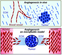
We developed a biomimetic microfluidic model to reproduce hallmark events of tumor-induced angiogenesis. The angiogenic capabilities of salivary gland adenoid cystic carcinoma (ACC) cells and oral squamous cell carcinoma (SCC) cells were assessed in this model. The traditional nude mouse xenograft model was used to investigate the physiological similarity of the microfluidic model to animal models, and the results showed that the angiogenic potential of ACC and SCC cells assessed by the microfluidic model was in agreement with the results obtained from the nude mouse model. The microfluidic model was subsequently used to evaluate the effect of antiangiogenic drugs on ACC- and SCC-induced angiogenesis. The antiangiogenic effect of anti-VEGF was further compared between the microfluidic and nude mouse models, and showed that it effectively inhibited tumor-induced angiogenesis in both the microfluidic model and the nude mouse model. Thus, tumor-induced angiogenesis reproduced in the microfluidic model may expand the capabilities of cell culture models, providing a low-cost, time-saving, and rapid alternative to animal models.

Introduction

Salivary gland adenoid cystic carcinoma (ACC) and oral squamous cell carcinoma (OSCC) are two common malignancies in the oral and maxillo-facial region.1,2 High microvessel density (MVD) has been found in both tumors and correlates with poor prognosis.3,4 Increased numbers of small blood vessels not only promote tumor growth by supplying oxygen and nutrients, but also facilitate tumor cell metastasis. Inhibition of blood vessel growth has therefore become a new strategy in anti-cancer therapy.

Commonly, new blood vessels within a tumor form by sprouting angiogenesis.5 Attracted by proangiogenic factors secreted by tumor cells, some endothelial cells from the pre-existing vasculature differentiate into tip cells with long filopodia and invade the extracellular matrix (ECM).6 Following tip cells, stalk cells extend fewer filopodia but establish a lumen and proliferate to support sprout elongation. A neoplastic mass cannot grow above a few millimeters in diameter without new vessel formation.7,8 Thus anti-angiogenic agents could offer novel therapeutic opportunities in cancer. The vascular endothelial growth factor (VEGF) signaling pathway is a key regulator governing initiation of tip cell differentiation and its direct invasion.6,9,10 VEGF has thus become the prime antiangiogenic drug target, with several VEGF (receptor)-based inhibitors approved by the US Food and Drug Administration for clinical use.11–13 An anti-VEGF antibody, bevacizumab, has been approved for the therapy of several advanced metastatic cancers, while SU5416, a long-term VEGFR2 kinase activity inhibitor, is undergoing preclinical trials.14 The phosphatidylinositol 3-kinase (PI3K)/Akt/mTOR signaling pathway also plays an important role in angiogenesis by regulating VEGF expression.15–17 A dual PI3K/mTOR ATP-binding inhibitor, PI-103, effectively blocks activation of both PI3K and mTOR in endothelial cells, resulting in increased inhibition of endothelial cell proliferation and survival in vitro and tumor growth in vivo.17

To elucidate the mechanism of angiogenesis and validate new antiangiogenic drugs, numerous cellular and animal models have been developed to reproduce the angiogenic cascade. To assess tumor-induced angiogenesis, the mouse xenograft model provides an important validation platform.18 The MVD of xenograft tissue is used to evaluate the angiogenic potential of tumor cells in vivo. However, the animal model is low throughput, high consumption and time-consuming. Consequently, to facilitate angiogenesis research, many in vitro models have been developed. The tube formation assay is the most popular in vitro model to evaluate angiogenesis.19 Endothelial cells are seeded onto the surface of Matrigel® and treated with certain stimuli that induce them to reorganize into multicellular cords that partially resemble vascular networks. However, tip cell differentiation and their directional invasion into the ECM cannot be reproduced using this model. Biomimetic models could bridge the gap between cultured endothelial cells and animal models. Microfluidic technology has been proven to be an ideal platform to culture mammalian cells with low consumption and high throughput.20–22 The accessibility for imaging and intervention makes the microfluidic model ideally suitable for research on angiogenesis, and some microfluidic models have been developed as in vitro platforms to study angiogenesis.23–29 Tumor-induced endothelial cell invasion into matrix and tube formation are represented among these microfluidic models.30–32 However, it is still unclear whether these new models could replace the in vivo animal model.

In this study, we developed a tumor-induced angiogenesis model based on the microfluidic technique. ACC- and OSCC-induced angiogenic processes, including tip cell differentiation, ECM invasion and capillary-like structure formation, were reproduced in this model. When compared with the nude mouse model, OSCC cells were found to exhibit stronger angiogenic capability than ACC cells in both microfluidic and animal models. Furthermore, when we used this microfluidic model to evaluate the effects of antiangiogenic drugs in the inhibition of ACC- and OSCC-induced angiogenesis, similar effects were observed in both the microfluidic model and nude mouse model.

Materials and methods

Microfluidic device fabrication

The microfluidic device is composed of a polydimethylsiloxane (PDMS) layer and a glass substrate. A master for casting the PDMS layer was constructed first. The master was fabricated by spin coating an SU8-2035 negative photoresist (Microchem Corp., Newton, MA, USA) onto a glass wafer, which was then patterned by photolithography. Sylgard 184 PDMS base and curing agent (Dow Corning Corp., Midland, MI, USA) were mixed thoroughly (10[thin space (1/6-em)]:[thin space (1/6-em)]1 by mass) and degassed under vacuum. The polymer curing process was carried out in an oven for 1 h. After cooling, the PDMS was peeled away from the master and trimmed to size. Inlet and outlet holes were created by punching through the PDMS with a razor-sharp punch. The PDMS layer was bonded to a glass slide after oxygen plasma treatment for 60 s. The device was sterilized with UV light for 2 h before use.

Characterization of concentration gradient in the angiogenesis channel

To characterize the diffusion of proangiogenic factors in the angiogenesis channel, FITC-dextran (20 kD; Invitrogen, Carlsbad, CA, USA) of a similar size to VEGF was used. First, Cultrex Basement Membrane Extract (BME, R&D chemistry, Westchester, NY, USA) was thawed overnight at 4 °C on ice and carefully injected into the angiogenesis channels of the microfluidic device. The device was then placed in a Petri dish and transferred to a 37 °C incubator for 20 min for BME gelling. Then 1 µmol L−1 FITC-dextran in medium was loaded into the cell culture chambers and control medium was loaded into the vessel channels. The time when FITC-dextran solution was loaded was set as t = 0. The images for the FITC distribution were taken using a fluorescent inverted microscope (Olympus IX71, Tokyo, Japan) every 30 min. The fluorescence intensity profile was analyzed by Image-pro plus 6.0.

General cell culture

Human umbilical vein endothelial cell (HUVEC) were purchased from ATCC (Rockville, MD, USA) and cultured at 37 °C with 5% CO2 and 95% relative humidity in endothelial cell medium (Cell Sciences, Canton, MA, USA) supplemented with 5% fetal bovine serum (FBS, Cell Sciences), 100 U mL−1 penicillin–streptomycin solution (Cell Sciences) and 1% endothelial cell growth supplement (Cell Sciences). The ACC cell line (ACC-M) was cultured in the same conditions as used in a previous study.33 The SCC cell line (UM-SCC6), a kind gift from Prof. Songling Wang (Capital Medical University, China), was cultured at 37 °C with 5% CO2 and 95% relative humidity in DMEM/high glucose (Hyclone, Logan, UT, USA) supplemented with 10% FBS and 100 U mL−1 penicillin–streptomycin solution.

Tumor-induced angiogenesis assay and inhibition of angiogenesis using the microfluidic model

BME was loaded in the angiogenesis channels to mimic the ECM between blood vessels and tumor cells. HUVECs were seeded onto the surface of the vessel channels. ACC-M or UM-SCC-6 cells (5000 cells per chamber) were seeded into the cell culture chambers to mimic primary tumors. To inhibit tumor-induced angiogenesis, medium supplemented with monoclonal VEGF antibody (100 ng mL−1; anti-VEGF; Sigma, St Louis, MO, USA), SU5416 (1000 nM; Calbiochem. Darmstadt, Germany) or PI-103 (1000 nM; Calbiochem.) was introduced into the vessel channels. A sequence of photomicrographs was taken to record the morphology and invasion of HUVECs in the angiogenesis channels using an inverted fluorescent microscope at different time points. At the end of each experiment, the cells were fixed with 3.7% formaldehyde solution in (phosphate buffered saline) PBS for 10 minutes at room temperature and stained with 5 µL 200 units per mL methanolic stock solutions of fluorescent phallotoxins (Invitrogen). To characterize the capillary-like structures, immunofluorescent staining against CD105 (1[thin space (1/6-em)]:[thin space (1/6-em)]15; Dako, Denmark) was performed. Finally, 4′, 6-diamidino-2-phenylindole (1[thin space (1/6-em)]:[thin space (1/6-em)]2000; DAPI, Roche Diagnostics, Basel, Switzerland) was used to stain nuclei for 5 min at room temperature. The invasion distance and area of the HUVEC cells into the BME were quantified using Image-pro plus 6.0.

In vivo tumor angiogenesis assay using a nude mouse model

BALB/c nude mice, 4–5 weeks old (male, Animal Experiment Center of Dalian Medical University) were used to assess the angiogenic capability of tumor cells in vivo. Mice were divided into four groups: the ACC-M group without bevacizumab treatment, the ACC-M group with bevacizumab treatment, the UM-SCC6 group without bevacizumab treatment, and the UM-SCC6 group with bevacizumab treatment, with four mice in each group. Cells were harvested and resuspended at a concentration of 1 × 10⁶ cells per mL (ACC-M) or 1 × 10⁷ cells per mL (UM-SCC6) in PBS, then 200 µL of the cell suspension was inoculated subcutaneously. The body weight of nude mice was measured once every five days. Bevacizumab (Roche Diagnostics) was administered at 5 mg kg−1·b.w. two times per week in the bevacizumab treatment groups. Control animals received an equivalent volume of sodium chloride injection (100 µL) two times per week. The first bevacizumab treatment started when the maximum diameter of the tumor was 5 mm. The animals were sacrificed 7 days after treatment. Tumors from every group were collected and processed for hematoxylin and eosin (HE) staining. Immunohistochemical staining was performed using a monoclonal antibody against CD34 (1[thin space (1/6-em)]:[thin space (1/6-em)]100; Abcam, Cambridge, UK) to determine the MVD of xenografts. The staining procedure and MVD evaluation were as described previously.34

Statistical analysis

Statistical analyses were performed using SPSS version 13.0 for Windows (SPSS Inc., Chicago, IL, USA). The Mann–Whitney U test was used to analyze comparisons of binary variables. Each experiment was conducted at least three times, and significance was identified as a P value of <0.05.

Results

Construction of the microfluidic model

Tumor cells in vivo secrete various proangiogenic factors that promote angiogenesis (Fig. 1A). To reproduce tumor-induced angiogenesis, a microfluidic-based model, reconstructing a primary tumor and its nearby pre-existing blood vessels, was developed in this study. The biomimetic model comprised six separate angiogenesis units and two parallel vessel channels (Fig. 1B–D). Each angiogenesis unit contained one opening cell culture chamber, 2 mm in diameter, and two angiogenesis channels located on either side of the cell culture chamber. Each angiogenesis channel is 800 micrometers in length, 400 micrometers in width, and 70 micrometers high. Each vessel channel is 25 millimeters in length, 500 micrometers in width, and 140 micrometers high. The height of the endothelial cell culture channels was designed to be higher than that of the angiogenesis channels to generate stop-flow junctions at the interface between these two types of channels. BME was used as the substitute ECM and was loaded into the angiogenesis channels through the cell culture chamber. Tumor cells were seeded into the cell culture chamber to mimic primary tumors. HUVEC were seeded onto the surface of vessel channels to mimic blood vessels (Fig. 1E). To characterize the gradient of proangiogenic factors generated by tumor cells in the angiogenesis channel, FITC-dextran with a similar size to VEGF was added into the cell culture chambers. A gradient from the cell culture chamber to the vessel channel was found to be established overtime (Fig. 2A and B).
image file: c6ra05645h-f1.tif
Fig. 1 A microfluidic device for studying tumor-induced angiogenesis. (A) The process of tumor-induced angiogenesis in vivo. (B) Chip design. The device includes six separate angiogenesis units and two parallel vessel channels. (C) Magnified illustration of the angiogenesis unit, including one opening cell culture chamber and two angiogenesis channels. (D) Photo of the microfluidic device. (E) Schematic illustration showing cell loading steps. (F) The angiogenesis channels (800 µm in length) were filled with BME. The dashed square indicates the location of fluorescence intensity measurement. (G) Plot of the fluorescence intensity profile across the angiogenesis channel. The fluorescence signal from the cell culture chamber to the HUVEC culture channel weakened gradually and formed a relatively stable concentration with time.

image file: c6ra05645h-f2.tif
Fig. 2 Tumor-induced angiogenesis assay on the microfluidic device. (A) ACC-M- and UM-SCC6-induced angiogenesis. The samples were stained with rhodamine-phalloidin after 24 h. Tip cells with long filopodia could be seen in the invasion front. (B) HUVEC formed tube-like structures in the angiogenesis channel after 48 h stimulation by ACC-M and UM-SCC6. The samples were stained for CD105 (green) and counterstained with DAPI (blue). (C) Invasion area of HUVEC induced by ACC-M and UM-SCC6 after 24 h stimulation. (D) Invasion distance of HUVEC induced by ACC-M and UM-SCC6 after 24 h stimulation. The invasion area and distance of HUVEC induced by UM-SCC6 were significantly higher than those induced by ACC-M. * indicates P < 0.05. Scale bar = 100 µm.

Modeling tumor-induced angiogenesis in the microfluidic model

The angiogenic capabilities of ACC and SCC cell lines, ACC-M and UM-SCC6, were assessed using the microfluidic model and further compared between them. We found that some endothelial cells in vessel channels extended long processes and invaded the ECM towards ACC-M and UM-SCC6 cells (Fig. 3A). The morphology of cells with long filopodia at the invasion front was consistent with tip cells. Tube-like structures positive for the capillary biomarker CD105 formed in the angiogenesis channel after 48 h stimulation of ACC-M or UM-SCC6 cells (Fig. 3B). Endothelial cells, visualized with green cytoplasm and blue nuclei, neatly arrayed in two rows with a hollow cavity in the middle. The capillary-like structures induced by ACC-M were relatively shorter than those induced by UM-SCC6. To further evaluate the differences in angiogenic potential between ACC-M and UM-SCC6 cells, the invasion distance and area of HUVEC were compared between the two groups. The results showed that both the invasion distance and area induced by ACC-M were significantly lower than those induced by UM-SCC6 (Fig. 3C and D). These results suggest that UM-SCC6 cells have a stronger angiogenic potential than ACC-M.
image file: c6ra05645h-f3.tif
Fig. 3 Tumor-induced angiogenesis assay in a nude mouse model. (A and B) HE staining of the xenografts of ACC-M and UM-SCC6. Scale bar = 100 µm. (C and D) CD34 positive microvessels (brown) in the xenografts of ACC-M and UM-SCC6. Scale bar = 50 µm. (E) Comparison of MVD between ACC-M and UM-SCC6 xenografts. The MVD index of UM-SCC6 was significantly higher than that of ACC-M. * indicates P < 0.05.

Comparison of tumor-induced angiogenesis between the microfluidic and nude mouse models

To further investigate the physiological similarity of the microfluidic model to animal models, we transplanted ACC-M and UM-SCC6 cells into nude mice subcutaneously and obtained xenograft tumors (Fig. 4A–C). The MVD of these xenograft tumors was evaluated (Fig. 4D and E). Small vessels positive to CD-43 could be identified in both ACC-M and UM-SCC6 xenografts, however the MVD of UM-SCC6 xenografts was significantly higher than that of ACC-M xenografts. This suggests thatUM-SCC6 cells have a greater ability to promote angiogenesis in vivo than ACC-M cells. This result was consistent with the results obtained using the microfluidic model.
image file: c6ra05645h-f4.tif
Fig. 4 Inhibition of ACC-M-induced angiogenesis in the microfluidic model. (A) Images of ACC-M-induced angiogenesis in the microfluidic model treated with anti-VEGF, PI-103 and SU5416 for 24 h. (B) Quantification of HUVEC invasion area. The invasion area decreased significantly following treatment with anti-VEGF, PI-103, and SU5416, compared to controls. (C) Quantification of HUVEC invasion distance. Invasion distance decreased significantly following treatment with anti-VEGF and PI-103, compared to controls. * indicates P < 0.05. Scale bar = 100 µm.

Validation of antiangiogenic drugs with the microfluidic model

To evaluate the suitability of the microfluidic model for testing the effect of antiangiogenic drugs on tumor-induced angiogenesis, we treated the microfluidic model with anti-VEGF, SU5416 or PI-103. As shown in Fig. 5A–C, anti-VEGF, PI-103 and SU5416 all caused a decrease in the ACC-M-induced ECM invasion by endothelial cells. Statistical analysis demonstrated that both anti-VEGF and PI-103 significantly reduced both invasion area and distance. However SU5416 only caused a significant inhibition of the ACC-M-induced HUVEC invasion area and could not significantly inhibit invasion distance. This suggests that anti-VEGF, PI-103 and SU5416 can all inhibit ACC-M-induced angiogenesis in the microfluidic model, but that anti-VEGF and PI-103 show a stronger antiangiogenic activity than SU5416.
image file: c6ra05645h-f5.tif
Fig. 5 Inhibition of UM-SCC6-induced angiogenesis in the microfluidic model. (A) Images of UM-SCC6-induced angiogenesis in the microfluidic model treated with anti-VEGF, PI-103 and SU5416 for 24 h. (B) Quantification of HUVEC invasion area. The invasion area decreased significantly following treatment with anti-VEGF, PI-103, and SU5416, compared to controls. (C) Quantification of HUVEC invasion distance. Invasion distance decreased significantly following treatment with anti-VEGF, PI-103, and SU5416, compared to controls. * indicates P < 0.05. Scale bar = 100 µm.

Next, the antiangiogenic effects of anti-VEGF, PI-103 and SU5416 on UM-SCC6-induced angiogenesis were assessed. The results showed that the three drugs all significantly inhibited endothelial invasion area and distance (Fig. 6A–C). This suggests that anti-VEGF, PI-103 and SU5416 can all inhibit UN-SCC6-induced angiogenesis in the microfluidic model.


image file: c6ra05645h-f6.tif
Fig. 6 Antiangiogenic effects of bevacizumab on ACC-M and UM-SCC6 in nude mice models. (A and B) ACC-M xenografts with or without bevacizumab treatment were immunohistochemically stained with an anti-CD34 antibody. (C) MVD of ACC-M xenografts decreased significantly following bevacizumab treatment. (D and E) UM-SCC6 xenografts with or without bevacizumab treatment were immunohistochemically stained with an anti-CD34 antibody. (F) MVD of UM-SCC6 xenograft decreased following bevacizumab treatment, but the change was not significant. * indicates P < 0.05.

Validation of bevacizumab in nude mouse models

In order to verify the results obtained from the microfluidic model, we used bevacizumab, a recombinant humanized anti-VEGF monoclonal antibody, to interfere with angiogenesis in the ACC-M and UM-SCC6 xenografts. The results showed that the MVD in the ACC-M xenografts treated with bevacizumab was significantly lower than that in the ACC-M xenografts without bevacizumab treatment (Fig. 6A–C). Similarly, the MVD of the UM-SCC6 xenografts with bevacizumab was lower than that in the UM-SCC6 xenografts without bevacizumab treatment, but not significantly (Fig. 6D–F). The results above suggest that bevacizumab can inhibit ACC-M- and UM-SCC6-induced angiogenesis in the nude mouse model. In addition, the same concentration of bevacizumab exerts a stronger anti-angiogenic effect on ACC-M than on UM-SCC6. We hypothesize that it might be due to differences between the cell lines, since UM-SCC-6 cells have a greater angiogenic capability than ACC-M cells.

Discussion

Approaches aimed at blocking angiogenic signals in cancer have become a new strategy in cancer therapy.5,6 However the gap between endothelial cell culture and in vivo models limits the ability to elucidate the molecular mechanisms of angiogenesis and to develop new anti-angiogenic agents. Biomimetic models, that can reproduce complex and integrated physiological and pathological events, present the potential to take the place of animal models in basic angiogenesis research and antiangiogenic drug development.

Tip cell differentiation, ECM invasion, and capillary-like structure formation are hallmark biological events during angiogenesis. A number of proangiogenic factor-induced angiogenic models based-on microfluidics have been developed.24,26,29 Typical features of in vivo angiogenesis, including directed invasion of tip cells and capillary-like structure formation, have been reproduced in these models. However no single pro-angiogenic factor has been found that can mimic the complex in vivo angiogenic process because tumor cells secrete a mixture of proangiogenic factors.35–39 Thus some researchers developed microfluidic chips to reconstruct tumor-induced angiogenesis.30,32 They co-cultured different tumor cells with endothelial cells on chips and observed endothelial cell invasion induced by tumor cells. However, neither tip cell differentiation nor capillary-like structure formation were reported in their studies. The microfluidic model developed in our study integrated six identical units that could mimic avascular primary tumors and their nearby pre-existing blood vessels. Pro-angiogenic factors secreted by tumor cells diffused through angiogenic channels and stimulated endothelial cells to differentiate into tip cells with long filopodia, invade ECM, and then form capillary-like structures. Thus all the hallmark features of angiogenesis were reproduced in this model. The endothelial invasion area and distance could be used as indicators to assess the angiogenic capability of different tumor cells in the microfluidic model. Furthermore, it is simple to compare the angiogenic capability of different tumors using this model because a fixed number of tumor cells could be easily seeded in the open cell culture chamber. The cell culture medium could be continuous flowing or static in different microfluidic-based angiogenic models. Usually, continuous flowing medium was employed to keep the gradient concentration when the effects of exogenous factors on angiogenesis were studied.27,40 By contrast, static condition was commonly used to study tumor-induced angiogenesis because endogenous proangiogenic factors secreted by tumor cells were few.25,30–32 Especially, the number of tumor cells cultured on microfluidic chips was very small. Similar to other tumor-induced angiogenic microfluidic models, the cell culture medium was kept static in our study.

Next, we wanted to determine if our microfluidic model had any physical similarity to traditional animal models such as the nude mouse. We compared the angiogenic capabilities of ACC-M and UM-SCC6 cells assessed by the microfluidic model with the traditional nude mouse model. The results demonstrated that UM-SCC6 cells exhibited greater angiogenic capability than ACC-M cells in both the microfluidic and animal models, strongly suggesting that the microfluidic model behaves similarly to the animal model. Compared to the animal model, the microfluidic model was simpler to operate, less expensive, and more time-saving, taking only 2 days to perform angiogenic tests. In contrast, it took several weeks to assess angiogenesis in xenografts using the animal models.

To demonstrate whether this microfluidic model could be used to rapidly and inexpensively screen drugs for their potential to inhibit angiogenesis, we used anti-VEGF, SU5416, and PI-103 to inhibit both ACC-M- and UM-SCC6-induced angiogenesis. All three drugs exhibited antiangiogenic effects against both ACC-M- and UM-SCC6-induced angiogenesis in the microfluidic model. Furthermore, we compared the antiangiogenic effects of anti-VEGF on tumor-induced angiogenesis assessed by both the microfluidic and animal models. Both models showed that anti-VEGF was able to significantly inhibit ACC-M-induced angiogenesis, while anti-VEGF significantly inhibited UM-SCC-6-induced angiogenesis in the microfluidic model and to a certain degree in mouse models. Thus, our new model introduced here provides a novel platform for anti-angiogenic drug screening.

Conclusions

In summary, the microfluidic model developed in this study reproduced tumor-induced tip cell differentiation, ECM invasion and capillary-like formation in vitro. The biomimetic model showed good physiological similarity to in vivo angiogenesis. It offers a novel platform to study the mechanism of angiogenesis and to rapidly and inexpensively screen drugs for their potential to inhibit angiogenesis.

Ethics statement

All procedures using mice were approved by the Ethical Committee of the Dalian Medical University and performed in accordance with the guidelines of the Animal Management Committee of Dalian Medical University.

Disclosure statement

The authors have no conflict of interest.

Contributions

Tingjiao Liu designed the study and wrote the manuscript. Lilu Liu and Zhaorong Xie performed experiments on the microfluidic model and analyzed the data. Wenyuan Zhang, Shimeng Fang, Jing Kong, Dong Jin, Jiao Li, Xiaojie Li performed animal experiments and analyzed the data. Yong Luo and Bingcheng Lin fabricated the microfluidic chips. All the authors read and approved the final manuscript.

Acknowledgements

This work was supported by National Natural Science Foundation of China (No. 81171425).

References

  1. S. Warnakulasuriya, Oral Oncol., 2009, 45, 309–316 CrossRef PubMed .
  2. C. A. Moskaluk, Head Neck Pathol., 2013, 7, 17–22 CrossRef PubMed .
  3. P. W. Kammerer, B. Al-Nawas, S. Kalkan, J. Liese, K. Fruth, B. Frerich and J. Brieger, J. Oral Pathol. Med., 2015, 44, 429–436 CrossRef PubMed .
  4. A. C. Faur, E. Lazar and M. Cornianu, Acta Pathol., Microbiol. Immunol. Scand., 2014, 122, 418–426 CrossRef CAS PubMed .
  5. G. Bergers and L. E. Benjamin, Nat. Rev. Cancer, 2003, 3, 401–410 CrossRef CAS PubMed .
  6. M. Potente, H. Gerhardt and P. Carmeliet, Cell, 2011, 146, 873–887 CrossRef CAS PubMed .
  7. J. Folkman, Nat. Med., 1995, 1, 27–31 CrossRef CAS PubMed .
  8. D. Hanahan, Science, 1997, 277, 48–50 CrossRef CAS PubMed .
  9. S. Ziyad and M. L. Iruela-Arispe, Genes Cancer, 2011, 2, 1085–1096 CrossRef PubMed .
  10. S. P. Herbert and D. Y. Stainier, Nat. Rev. Mol. Cell Biol., 2011, 12, 551–564 CrossRef CAS PubMed .
  11. Y. Kubota, Keio J. Med., 2012, 61, 47–56 CrossRef CAS PubMed .
  12. T. Y. Seiwert, D. J. Haraf, E. E. Cohen, K. Stenson, M. E. Witt, A. Dekker, M. Kocherginsky, R. R. Weichselbaum, H. X. Chen and E. E. Vokes, J. Clin. Oncol., 2008, 26, 1732–1741 CrossRef CAS PubMed .
  13. R. K. Jain, D. G. Duda, J. W. Clark and J. S. Loeffler, Nat. Clin. Pract. Oncol., 2006, 3, 24–40 CrossRef CAS PubMed .
  14. D. B. Mendel, R. E. Schreck, D. C. West, G. Li, L. M. Strawn, S. S. Tanciongco, S. Vasile, L. K. Shawver and J. M. Cherrington, Clin. Cancer Res., 2000, 6, 4848–4858 CAS .
  15. B. H. Jiang, J. Z. Zheng, M. Aoki and P. K. Vogt, Proc. Natl. Acad. Sci. U. S. A., 2000, 97, 1749–1753 CrossRef CAS PubMed .
  16. Y. Alvarez, O. Astudillo, L. Jensen, A. L. Reynolds, N. Waghorne, D. P. Brazil, Y. Cao, J. J. O'Connor and B. N. Kennedy, PLoS One, 2009, 4, e7867 Search PubMed .
  17. R. Marone, V. Cmiljanovic, B. Giese and M. P. Wymann, Biochim. Biophys. Acta, 2008, 1784, 159–185 CrossRef CAS PubMed .
  18. J. Bailey, ATLA, Altern. Lab. Anim., 2009, 37, 399–416 CAS .
  19. Y. Kubota, H. K. Kleinman, G. R. Martin and T. J. Lawley, J. Cell Biol., 1988, 107, 1589–1598 CrossRef CAS PubMed .
  20. J. El-Ali, P. K. Sorger and K. F. Jensen, Nature, 2006, 442, 403–411 CrossRef CAS PubMed .
  21. D. Huh, G. A. Hamilton and D. E. Ingber, Trends Cell Biol., 2011, 21, 745–754 CrossRef CAS PubMed .
  22. B. Xiong, K. Ren, Y. Shu, Y. Chen, B. Shen and H. Wu, Adv. Mater., 2014, 26, 5525–5532 CrossRef CAS PubMed .
  23. H. Lee, S. Kim, M. Chung, J. H. Kim and N. L. Jeon, Microvasc. Res., 2014, 91, 90–98 CrossRef CAS PubMed .
  24. D. H. Nguyen, S. C. Stapleton, M. T. Yang, S. S. Cha, C. K. Choi, P. A. Galie and C. S. Chen, Proc. Natl. Acad. Sci. U. S. A., 2013, 110, 6712–6717 CrossRef CAS PubMed .
  25. L. L. Bischel, E. W. Young, B. R. Mader and D. J. Beebe, Biomaterials, 2013, 34, 1471–1477 CrossRef CAS PubMed .
  26. B. M. Baker, B. Trappmann, S. C. Stapleton, E. Toro and C. S. Chen, Lab Chip, 2013, 13, 3246–3252 RSC .
  27. I. Barkefors, S. Thorslund, F. Nikolajeff and J. Kreuger, Lab Chip, 2009, 9, 529–535 RSC .
  28. R. Sudo, S. Chung, I. K. Zervantonakis, V. Vickerman, Y. Toshimitsu, L. G. Griffith and R. D. Kamm, FASEB J., 2009, 23, 2155–2164 CrossRef CAS PubMed .
  29. C. Kim, J. Kasuya, J. Jeon, S. Chung and R. D. Kamm, Lab Chip, 2015, 15, 301–310 RSC .
  30. C. Zheng, L. Zhao, G. Chen, Y. Zhou, Y. Pang and Y. Huang, Anal. Chem., 2012, 84, 2088–2093 CrossRef CAS PubMed .
  31. L. E. Dickinson, C. Lutgebaucks, D. M. Lewis and S. Gerecht, Lab Chip, 2012, 12, 4244–4248 RSC .
  32. S. Chung, R. Sudo, P. J. Mack, C. R. Wan, V. Vickerman and R. D. Kamm, Lab Chip, 2009, 9, 269–275 RSC .
  33. T. Liu, B. Lin and J. Qin, Lab Chip, 2010, 10, 1671–1677 RSC .
  34. H. Guan, J. Tan, F. Zhang, L. Gao, L. Bai, D. Qi, H. Dong, L. Zhu, X. Li and T. Liu, Histopathology, 2015, 66, 781–790 CrossRef PubMed .
  35. N. Ferrara and T. Davis-Smyth, Endocr. Rev., 1997, 18, 4–25 CrossRef CAS PubMed .
  36. A. Stratmann, W. Risau and K. H. Plate, Am. J. Pathol., 1998, 153, 1459–1466 CrossRef CAS PubMed .
  37. T. Etoh, H. Inoue, S. Tanaka, G. F. Barnard, S. Kitano and M. Mori, Cancer Res., 2001, 61, 2145–2153 CAS .
  38. S. Suzuki, Y. Dobashi, Y. Hatakeyama, R. Tajiri, T. Fujimura, C. H. Heldin and A. Ooi, BMC Cancer, 2010, 10, 659 CrossRef CAS PubMed .
  39. I. Vlodavsky, Y. Friedmann, M. Elkin, H. Aingorn, R. Atzmon, R. Ishai-Michaeli, M. Bitan, O. Pappo, T. Peretz, I. Michal, L. Spector and I. Pecker, Nat. Med., 1999, 5, 793–802 CrossRef CAS PubMed .
  40. J. W. Song and L. L. Munn, Proc. Natl. Acad. Sci. U. S. A., 2011, 108, 15342–15347 CrossRef CAS PubMed .

Footnote

Co-first authors.

This journal is © The Royal Society of Chemistry 2016
Click here to see how this site uses Cookies. View our privacy policy here.