Enhanced photoelectrochemical performance of electrodeposited hematite films decorated with nanostructured NiMnOx

Nimai Bhandaryab, Aadesh P. Singh*c, Pravin P. Ingolea and Suddhasatwa Basu*b
aDepartment of Chemistry, Indian Institute of Technology Delhi, New Delhi-110016, India
bDepartment of Chemical Engineering, Indian Institute of Technology Delhi, New Delhi-110016, India. E-mail: sbasu@iitd.ac.in; Fax: +91-11-26581120; Tel: +91-11-26591035
cDepartment of Physics, Indian Institute of Technology Delhi, New Delhi-110016, India. E-mail: aadshp1982@gmail.com

Received 13th February 2016 , Accepted 30th March 2016

First published on 1st April 2016


Abstract

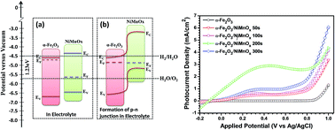
In the present work, we report a novel nickel-manganese oxide (NiMnOx) decorated hematite (α-Fe2O3) photoanode for efficient water splitting in a photoelectrochemical (PEC) cell. The photoanodes are prepared by a two step electrodeposition process. NiMnOx loading on the hematite surface is varied by changing the electrodeposition time. The NiMnOx loaded α-Fe2O3 photoanodes are characterized by X-ray diffraction, Raman spectroscopy, X-ray photoelectron spectroscopy, field emission scanning electron microscopy, UV-Vis absorption and photoluminescence spectroscopy. The results demonstrate that the NiMnOx decorated α-Fe2O3 photoanode exhibits excellent photoelectrochemical activity. The α-Fe2O3/NiMnOx photoanode, where NiMnOx was coated for 200 s, shows a maximum photocurrent of 2.35 mA cm−2 at an applied potential of 0.23 V vs. the Ag/AgCl reference electrode. There is a large shift in the onset potential towards the cathodic region also observed. The photoconversion efficiency is calculated which is around 0.85% at 0.23 V vs. Ag/AgCl. The superior PEC performance of NiMnOx decorated α-Fe2O3 photoanodes can be explained by a combined effect of better water oxidation on the hematite surface and efficient separation of photogenerated electron–hole pairs on its surface due to NiMnOx modification.


Introduction

Photoelectrochemical (PEC) splitting of water appears to be the most promising, economically viable and sustainable way for the production of clean solar fuel i.e. hydrogen.1 Since its first report in 1972 by Fujishima and Honda, many metal oxide semiconductor catalysts have been explored for water splitting like α-Fe2O3, BiVO4, WO3, ZnO, Cu2O etc.1–6 However, at present, the solar-to-hydrogen conversion efficiency through the PEC route is too low for the technology to be economically feasible. A nanostructured hematite (α-Fe2O3) thin film for water-splitting in photoelectrochemical (PEC) cells has great potential in the design of low-cost, environment friendly solar-hydrogen production. Hematite (α-Fe2O3) is an attractive material for its use in PEC cell for the generation of hydrogen because of its suitable band gap (2.2 eV), abundance, stability and cost-effectiveness.7–10 But, its performance in a PEC cell is limited by high rate of charge recombination, unfavorable band edge position with respect to water redox level and short hole diffusion length.11–16 To effectively separate electron–hole pairs the hematite nanostructured has been modified by doping with other metal ions and nanostructuring the material by various techniques to enhance the PEC performance by increasing carrier density and semiconductor–electrolyte interface areas.17–20 A number of dopants such as Ti, Zn, Zr, Sn, Pt etc. have shown enormous effect with enhanced PEC performance in hematite due to higher donor density and improved electronic conductivity.21 Wang et al. reported Ti doped hematite show enhanced photocurrent due to increasing donor density and reducing electron hole recombination.22 Doping with silicon in hematite also show the better PEC performance in terms of higher photocurrent density in mesostructured hematite.23

Recently, it is observed that the slow water oxidation kinetics on hematite surface, which limits the PEC performance, can be overcome by surface treatment with oxygen evolving catalyst. A thin secondary metal oxide layer of ZnO, TiO2, Al2O3, In2O3, Ga2O3 over hematite has been reported to accelerate the solar water oxidation by passivating surface states of α-Fe2O3.24–26 The surface modifications by oxygen evolving catalyst generally improve water oxidation kinetics and effectively separate the photogenerated electron–hole pair which leads to better PEC performance. A series of oxygen evolving catalysts such as IrOx, Co-Pi, Ni(OH)2, NiFeOx, FeOOH, NiOOH etc. over α-Fe2O3 surface have also been reported for better PEC performance.27–33 The enhancement due to Co-Pi on α-Fe2O3 is attributed to fast water oxidation kinetics and effective separation of photogenerated holes.34 Du et al. reported that α-Fe2O3 nanotube decorated with a thin layer of NiFeOx catalyst showed a 280% enhancement in photoconversion efficiency as compared to pure hematite.31 This is due to the promoted charge migration, faster oxygen evolution and inhibited recombination at semiconductor–electrolyte junction.

Herein, we propose surface modification of hematite with an oxygen evolving catalyst (OEC) NiMnOx which is synthesized by electrodeposition technique. It is expected that NiMnOx loading on pristine hematite surface facilitate the charge separation and refrain the electron hole recombination. Furthermore a lower over-potential for PEC performance observed due to the oxidizing capability of the said catalyst. PEC measurement shows that NiMnOx loading on pristine α-Fe2O3 improves photocurrent density as well as a large cathodic shift in onset potential for photoelectrochemical water splitting compared to pure hematite.

Experimental section

Material preparation

All chemicals used in the experiment were of analytical grade and used without any further purification. Electrodeposition of α-Fe2O3 thin film were carried out using three electrode assembly where indium doped tin oxide (SnO2:In, ITO) glass as working electrode, Pt wire as counter electrode and Ag/AgCl saturated with KCl as reference electrode were used. The electrodeposition solution consisted of 5 mM FeCl3 + 5 mM KF + 0.1 M KCl + 1 M H2O2 in Milli-Q water. Cyclic voltammetry were performed in the potential range 0 V to −0.3 V vs. Ag/AgCl (sat. KCl) with a sweep rate of 10 mV s−1 for 30 cycles at room temperature to deposit β-FeOOH film on ITO glass. Thus obtained yellowish coloured β-FeOOH film was air annealed 500 °C for 2 hours to get reddish coloured α-Fe2O3 thin film. Further, to modify the α-Fe2O3 thin film, an oxygen evolution catalyst, NiMnOx, was deposited on annealed α-Fe2O3 film via electrodeposition technique in second step. Here α-Fe2O3 deposited thin film on ITO substrate was used as working electrode, Pt wire as counter and Ag/AgCl (sat. KCl) as reference electrode. For NiMnOx deposition, 20 mM NiSO4·6H2O and 10 mM Mn(CH3COO)2·4H2O in Milli-Q water was used as precursor solution and a constant potential of 1.2 V vs. Ag/AgCl (sat. KCl) was applied for various time duration of 50, 100, 200 and 300 seconds to deposit different amount of NiMnOx over the α-Fe2O3 surface. These obtained films were washed with deionized water and heat treated at 300 °C for 2 hours using muffle furnace.

Material characterization

Phase of the material prepared was determined by X-ray diffraction (XRD) analysis using Rigaku Miniflex 600 X-ray diffractometer using Cu Kα (λ = 0.15418 nm) radiation and further confirmed by Raman spectra which was recorded using a Horiba Xplora micro-Raman (model: WITec alpha300M) spectroscope equipped with the 514 nm line of Ar+ ion laser as the excitation source. Surface morphology and elemental composition of the synthesized photoanodes were investigated by Field-Emission Scanning Electron Microscopy (FE-SEM) were performed on a FEI QUANTA 200F operating at an accelerating voltage of 10 kV. Elemental dispersive analysis (EDS) was performed using TEM-EDX (FEI-Technai-G20) set up by transferring the samples on Cu-grid. The optical properties of all samples were analyzed by UV-Vis spectroscopy recorded on a LAMBDA L602008 spectrometer over a wavelength range of 200–800 nm. The photoluminescence (PL) spectra of the as-synthesized samples in the powder form were investigated at room temperature on a Fluoromax-4 fluorescence spectrophotometer (Horiba Jobin Yvon Japan) with an excitation wavelength (λexcitation) of 514 nm and the width of the excitation and emission slit were 2 and 5 nm, respectively. The emission spectrum was monitored over a wavelength range of 450–650 nm.

Electrochemical characterization

For electrochemical measurements, thin films of all the samples were converted into the electrodes with an active area of about 1 × 1 cm2. For this purpose an ohmic electrical contacts were made using silver paste and copper wire from the exposed area of conducting glass substrate and later the exposed area was covered with non-transparent and non-conducting epoxy resin. The electrochemical measurements were conducted in a three-electrode photoelectrochemical cell to obtain the current–voltage (IV) characteristics, Mott–Schottky and electrochemical impedance spectroscopy (EIS) measurements. The photoelectrochemical cell was controlled using CIMPS-2 (controlled intensity modulated photospectroscopy) system consisting of Zennium Electrochemical Workstation (X-Pot Potentiostat). Linear sweep voltammetry scans under dark and visible light illumination from a 150 W xenon lamp that fitted with a filter that cuts light with wavelengths ≥380 nm and having output illumination intensity of 100 mW cm−2. And Mott–Schottky measurement in dark condition were carried out in the potential range −1.0 to +1.0 V versus Ag/AgCl with a scan rate of 20 mV s−1 in 1 M NaOH electrolyte (pH = 13.6). The electrochemical impedance spectroscopy (EIS) measurements were carried out in the frequency range of 100 kHz to 100 mHz with AC signal amplitude of 10 mV under open bias condition. Chronoamperometric measurements were carried to check out the stability of the material in 1 M NaOH under visible light irradiation.

Result and discussion

XRD measurements were carried out to investigate the phase structures of electrodeposited NiMnOx on hematite thin films and are shown in Fig. 1. Initially, the as prepared electrodeposited films are reported to be β-FeOOH35 and after annealing at 500 °C for 2 h in air, the β-FeOOH is completely changed to reddish coloured hematite (α-Fe2O3) phase of iron oxide. The XRD pattern of pure hematite sample exhibits the diffraction peaks at 2θ values 24.13, 33.1, 35.6, 40.9, 49.4, 54.0, 58.3, 62.3 and 63.7 which refers to the (012), (104), (110), (113), (024), (116), (122), (214) and (300) planes of the hematite phase [JCPDS card no. 33-0664]. However with NiMnOx modified samples, no additional peaks for the NiMnOx were observed in the XRD pattern, which can be explained due to the presence of small amount and high dispersion on hematite surface. In the XRD pattern, other than hematite peaks few strong peaks are observed, which corresponds to background of ITO substrate. XRD analysis confirms hematite and NiMnOx modified hematite thin films display similar pattern. To check the presence of NiMnOx on the surface of α-Fe2O3, elemental composition of surface modified films were determined by the EDX analysis. As shown in Fig. 2b, the EDX image confirms the presence of Ni and Mn on hematite surface where NiMnOx deposited for 200 s on hematite.
image file: c6ra03984g-f1.tif
Fig. 1 XRD pattern of for (a) pristine α-Fe2O3 and NiMnOx loaded α-Fe2O3 for different loading time (b) 50 s (c) 100 s (d) 200 s and (e) 300 s by electrodeposition technique.

image file: c6ra03984g-f2.tif
Fig. 2 EDX analysis of (a) pristine α-Fe2O3; and (b) 200 s NiMnOx loaded α-Fe2O3.

To further characterize the pristine and NiMnOx modified hematite for phase formation, Raman spectra was carried out and shown in Fig. 3. The Raman spectroscopy is attributed to the slight change in bond and distortion in crystal lattice. The typical band of A1g mode was observed at 225 and 477 cm−1 and the Eg modes were observed at 247, 295, 410 and 610 cm−1 in the Raman spectra of pure hematite. Upon NiMnOx deposition small shift in the Raman spectra is noted. This is may be due to surface of hematite is covered by NiMnOx layer which changes the polarizability of the surface that effects the Raman shift.


image file: c6ra03984g-f3.tif
Fig. 3 Raman spectra for (a) pristine α-Fe2O3 and (b) 200 s NiMnOx deposition on α-Fe2O3 by electrodeposition technique.

A field emission scanning electron microscope (FE-SEM) was employed to observe the surface morphology and microstructures of pristine α-Fe2O3 and NiMnOx deposited α-Fe2O3 samples. Fig. 4 shows the surface morphology of pristine and 200 s NiMnOx deposited on α-Fe2O3 films on ITO substrate. It can be seen that pristine α-Fe2O3 thin film is homogeneously grown on ITO glass surface with regular size of particle morphology (Fig. 4a). In Fig. 4b, NiMnOx deposition on α-Fe2O3 can be noticed clearly as there is some dense and hazy morphology throughout over the α-Fe2O3 surface. This morphology indicates NiMnOx is homogenously decorated on hematite surface.


image file: c6ra03984g-f4.tif
Fig. 4 FE-SEM images of (a) pristine α-Fe2O3; and (b) 200 s NiMnOx loaded α-Fe2O3.

Fig. 5 shows the UV-Vis optical absorption spectra of pristine and NiMnOx loaded hematite thin film. Hematite shows good absorption in the visible region as it absorbs 40% of visible light from the sun whereas surface modification with NiMnOx further improves the visible light absorption activity.36 The visible light absorption increases with deposition time upto 200 s but there is a decrease in intensity is observed with further deposition time for 300 s. Results, clearly indicates the NiMnOx deposited samples has much more visible light absorption capability than pristine hematite thin film.


image file: c6ra03984g-f5.tif
Fig. 5 Optical absorption spectra for pristine and NiMnOx loaded α-Fe2O3 for different electrodeposition time.

Surface X-ray Photoelectron Spectroscopy (XPS) measurements were performed on SPECS spectrometer using a standard Mg Kα radiation, at a working pressure lower than 10−9 mbar. The reported binding energies (BEs, standard deviation = ±0.2 eV) were corrected for charging by assigning to the adventitious C 1s signal a BE of 284.8 eV. X-ray photoelectron spectra of the as-prepared hematite film confirmed the presence of Fe and O. However, the NiMnOx loaded hematite film confirmed the presence of Mn and Ni along with Fe and O. The Fe 2p3/2 peak in both the samples obtained at a binding energy of ∼710.2 eV (Fig. 6a) was consistent with the typical values observed for hematite. Also the energy separation between Fe 2p3/2 and Fe 2p1/2 (Δ = 13.5 eV) clearly supported the formation of pure iron(III) oxide,37 without any evidence of Fe(II) species. Fig. 6b shows the O 1s XPS spectra of the hematite films before and after NiMnOx deposition. The O 1s surface photoelectron peak was decomposed by means of two components located at BE = 530.1 and 532.0 eV, respectively. The main peak was at 530.1 eV consistent with surface O2− possessed for iron(III) oxide.38 Fig. 6c shows typical XP spectra of Mn 2p. The surface Mn peak was characterized by two well-evident components located at BE values of 641 and 652 eV corresponding to the 2p1/2 and 2p3/2 peaks, respectively. The peaks with higher binding energy above the main peaks as well as the splitting of the main peaks were observed in the film, which indicates the existence of Mn(IV) state.39 The Ni peaks at around 854 and 872 eV (Fig. 6d) corresponded to NiO.39 As the concentration of Ni in the 200 s NiMnOx loaded hematite film is low, therefore, the XPS spectra for Ni 2p is relatively noisy. However, in Ni 2p spectra, the Ni 2p3/2 peak is clearly visible at 854 eV and we assumed that Ni 2p1/2 peak at 872 eV is immersed in the noise level. Hence from XPS analysis, it is confirmed that there is presence of binary mixed metal oxide in the form of NiO and MnO2.


image file: c6ra03984g-f6.tif
Fig. 6 The core level XPS spectra of (a) Fe 2p (b) O 1s (c) Mn 2p and (d) Ni 2p for pristine and 200 s NiMnOx deposited α-Fe2O3 samples.

Photoelectrochemical measurements

To observe the photoelectrochemical performance of these photoanodes, the linear-sweep-voltammetry (LSV) for pure α-Fe2O3 and NiMnOx deposited α-Fe2O3 photoanode were performed using a three electrode PEC cell in 1 M NaOH solution both under dark and under light illumination with output intensity 100 mW cm−2. The photocurrent density calculated by subtracting the dark current from the current under illumination after divided by the electrode area is plotted against applied potential (Fig. 7). In all the samples the dark current from 0 V to 0.95 V (vs. Ag/AgCl) was almost zero. The photocurrent onset potential for pristine α-Fe2O3 was obtained at 0.86 V and the photocurrent intensity was 1.35 mA cm−2 at a potential of 1.0 V vs. Ag/AgCl reference electrode. It also provides good evidence that the electrodeposited nanostructures of α-Fe2O3 can be used for photoelectrochemical water spitting. The photoelectrochemical response of NiMnOx deposited α-Fe2O3 photoanode shows significant enhancement as compared to pristine α-Fe2O3 and exhibited superior PEC performance in terms of lower onset potential and higher photocurrent density. The result indicates that the NiMnOx loading has played a very important role in enhancing the photocatalytic reaction. The photocurrent onset potential of α-Fe2O3 is shifted to more negative from 0.86 to −0.1 V with 200 s NiMnOx loading on hematite which is comparable to previously reported work by Li et al.33 where hematite surface was modified with NiO and there enhancement in photoelectrochemical performance was observed. As compared to α-Fe2O3 photoanode, the 200 s NiMnOx loaded α-Fe2O3 photoanode showed a pronounced photocurrent of 2.35 ± 0.1 mA cm−2 at a potential of 0.23 V, which is much higher than other loading amount of NiMnOx, suggesting that an optimal loading of NiMnOx on α-Fe2O3 photoanode promoted the light harvesting. Further with 300 s NiMnOx modification, there is lowering of photocurrent is noticed. This can be explained by considering the effect of MnO2 on α-Fe2O3 surface. MnO2 is electrically conducting but bulk MnO2 is insulating.40 Therefore with higher loading of NiMnOx, the presence of high concentration of bulk MnO2, most of active sites of α-Fe2O3 are blocked. Also, the NiMnOx loaded samples exhibit poor optical transparency due to the dark blackish colour. Therefore the light reaching to α-Fe2O3 surface is absorbed by the catalyst and reduces the performance of α-Fe2O3. Hence, due to of these two major factors, the PEC performance of 300 s NiMnOx loaded α-Fe2O3 decreases. There are few other co-catalyst reported to decorate the α-Fe2O3 photoanode for photoelectrochemical water splitting application; the photocurrent density and photocurrent onset of our NiMnOx loaded α-Fe2O3 is significantly higher than other catalysis such as Ni(OH)2,27 NiFeOx,31 Co-Pi,34 NiO33 etc. with different morphologies reported previously under similar measurement conditions. A low photocurrent onset and higher photocurrent are extremely important to achieve a higher efficiency of PEC hydrogen generation.
image file: c6ra03984g-f7.tif
Fig. 7 Photoelectrochemical performances in term of current–potential curves for α-Fe2O3 and NiMnOx deposited α-Fe2O3 photoanodes under dark and visible light illumination in 1 M NaOH (pH = 13.6) solution.

Additionally, we have calculated the solar to hydrogen (STH) conversion efficiency for the water splitting reaction for all the samples with visible light source of irradiance 100 mW cm−2 using the equation:18

 
image file: c6ra03984g-t1.tif(1)
where, Jp is the photocurrent density (mA cm−2) at the measured applied bias, Io is the incident light intensity of 100 mW cm−2 and Vapp is the applied potential to the PEC cell with reference to standard reversible hydrogen electrode (RHE) potential. Again, Vapp = VmeaVaoc, where Vmea is the electrode potential (versus RHE) of the working electrode at which the photocurrent was measured under illumination and Vaoc is the electrode potential (versus RHE) of the same working electrode at open circuit condition under similar illumination conditions and in the same electrolyte. In 1 M NaOH electrolyte, the RHE potential can be converted from the Ag/AgCl reference electrode as: ERHE = E0Ag/AgCl + EAg/AgCl + 0.059pH. The solar to hydrogen conversion efficiency is plotted vs. applied potential (V vs. Ag/AgCl) and has been shown in Fig. 8. The 200 s NiMnOx loaded α-Fe2O3 photoanode exhibit an optimal conversion efficiency of ∼0.85% at 0.23 V vs. Ag/AgCl, whereas the pristine α-Fe2O3 exhibit the efficiency of ∼0.003% at a low bias of 0.23 V vs. Ag/AgCl.


image file: c6ra03984g-f8.tif
Fig. 8 Photoconversion efficiency versus applied potential curves for α-Fe2O3 and NiMnOx deposited α-Fe2O3 photoanodes.

Further the catalyst was checked for its stability by chronoamperometry in 1 M NaOH for long term application in PEC cell under illumination condition. Once the light was turned on, a spike in the photoresponse can be observed because of the rapid effect upon power excitation, but the photocurrent quickly turned back to a steady state within few seconds. Fig. 9 shows the total current under illumination of α-Fe2O3/NiMnOx-200 s over a long time to confirm the stability of the prepared catalyst. It showed that under illumination with visible light, the photocurrent of α-Fe2O3/NiMnOx-200 s was steady during 3 h, and the photocurrent almost 2.3 mA cm−2. The achieved high efficiency and good stability indicate that the 200 s NiMnOx loaded α-Fe2O3 photoanode retain the original structure after long time operation of PEC water splitting.


image file: c6ra03984g-f9.tif
Fig. 9 Chronoamperometry plots for α-Fe2O3 and NiMnOx depositedα-Fe2O3 photoanodes at 0.23 V vs. Ag/AgCl under visible light illumination.

The Mott–Schottky curves (1/C2 versus electrode potential) were obtained under dark conditions to estimate the donor density (Nd), flatband potential (VFB) and depletion layer width of thin films in the same three-electrode configuration. The flat band potential of a semiconductor can be obtained from Mott–Schottky plot using the equation:37

 
image file: c6ra03984g-t2.tif(2)
where, ε is the dielectric constant of the semiconductor, ε0 is the permittivity of the vacuum, Nd is the donor density, Vapp is the applied potential, VFB is the flat band potential and kT/q is the temperature-dependent term in the Mott–Schottky equation. The intercept of linear plot at 1/C2 = 0 gives the flat band potential. The Mott–Schottky plot for pristine α-Fe2O3 and α-Fe2O3/NiMnOx modified hematite photoanodes are shown in Fig. 10. The pristine α-Fe2O3 sample showed a positive slope in the Mott–Schottky plots with flat band potential −0.6 V vs. Ag/AgCl reference electrode, which indicated that α-Fe2O3 is an n-type semiconductor with electrons as the majority carriers. However, Mott–Schottky analysis for α-Fe2O3/NiMnOx photoanodes exhibit the p–n junction feature as shown in the Fig. 10, where the co-existence of positive and negative slopes with two flat band potential values are observed.33 This suggests that NiMnOx act as p-type material in PEC cell. The negative flat band potential for Fe2O3/NiMnOx with 200 s deposition time of NiMnOx is relatively more negative, −0.94 V, as compared to Fe2O3/NiMnOx with 300 s deposition time of NiMnOx. This exhibits that deposition of NiMnOx for 200 s provide the optimal loading for NiMnOx to shift the flat band potential at higher cathodic side.


image file: c6ra03984g-f10.tif
Fig. 10 Mott–Schottky plots of pristine α-Fe2O3 and NiMnOx loaded α-Fe2O3 photoanode for various electrodeposition times under dark measured at 1 kHz frequency in 1 M NaOH.

Moreover, it is proposed that the presence of NiMnOx catalyst decreased the reaction barriers for water oxidation and facilitated the holes transfer to the electrode/electrolyte interface, accounting for great improvement in PEC performance. It is commonly known that the electrochemical impedance spectroscopy (EIS) analysis provides information about the interfacial properties of electrode. The diameter of the semicircle in EIS equals the electron transfer resistance, which subjects the electron transfer kinetics of the redox probe at the electrode interface.41 Fig. 11 shows the typical EIS curve for pristine α-Fe2O3 photoanode and α-Fe2O3/NiMnOx photoanode measured in 1 M NaOH solution. It is evident from the figure that the hematite has the higher impedance while there is decrease in the impedance with NiMnOx modification. Pure hematite has an impedance of 25 ohm while 200 s NiMnOx decorated hematite shows 10 ohm which is the minimum under experimental condition. Further addition of NiMnOx, for 300 s leads to increase the impedance as it is not conducting in bulk which also reflects in the photocurrent plot. EIS measurements suggest that the resistance at α-Fe2O3/NiMnOx/electrolyte interface is much smaller than the α-Fe2O3/electrolyte interface. This might be due to conducting effect of the NiMnOx, which would decrease the charge transfer barrier at the electrode interface. As a result, the oxygen evolution reaction barrier is reduced thereby water oxidation is easier to occur.


image file: c6ra03984g-f11.tif
Fig. 11 Nyquist plots of electrochemical impedance spectra of pristine α-Fe2O3 and NiMnOx loaded α-Fe2O3 photoanode for various deposition time under visible light illumination.

To confirm efficient charge separation by NiMnOx co-catalyst deposited on α-Fe2O3 thin films, photoluminescence (PL) spectroscopy was used. Fig. 12 shows the PL spectra of hematite and 200 s NiMnOx loaded α-Fe2O3 thin film. Although it looks quite noisy, the broad-band emission around 540 nm (excited at 514 nm) can be assigned to the recombination of photoexcited holes with electrons occupying the singly ionized oxygen vacancies in α-Fe2O3.42 As shown in Fig. 12, a strong intense peak in pristine α-Fe2O3 is observed in PL spectra. However, the PL intensity of NiMnOx loaded α-Fe2O3 photoanode largely decreased, indicating that the recombination of electron–hole pairs has been reduced. The 200 s NiMnOx loaded α-Fe2O3 photoanode shows minimum intense PL spectra. The effective lowering of PL intensity suggests the surface modified hematite films inhibit significant recombination of photogenerated electrons–hole pairs.


image file: c6ra03984g-f12.tif
Fig. 12 Photoluminescence spectra of (a) pristine α-Fe2O3 and (b) 200 s NiMnOx deposited α-Fe2O3 photoanode.

From Mott–Schottky analysis we found that a p–n junction was formed between α-Fe2O3 and NiMnOx in the 1 M NaOH solution (pH = 13.6). The strategy of constructing p–n junction has been widely used to improve the separation of photo-generated holes and electrons.43,44 The adsorbed OH and H+ on the oxide semiconductor surface have significant effect on the Fermi level (EF), conduction band (EC) and valence band (EV) of the semiconductor.44 Based on these results, we have proposed a mechanism of electron transfer between the α-Fe2O3/NiMnOx/electrolyte interface and as shown in Scheme 1. From Scheme 1a and b, the flatband potential (VFB) values of α-Fe2O3 and NiMnOx extrapolated from Mott–Schottky plots in 1 M NaOH solution (pH = 13.6) are −0.85 V vs. Ag/AgCl and −0.02 V vs. Ag/AgCl, respectively. The EF values of α-Fe2O3 and NiMnOx in 1 M NaOH solution (pH = 13.6) calculated from VFB are −4.65Vvacuum and −5.52Vvacuum (Scheme 1a) according to the relation between EF, EFB and RHE.45,46 As a result, the p-type NiMnOx and n-type α-Fe2O3 form a p–n junction (Scheme 1b) in the electrolyte, and an internal built-in electric field directing from the n-type α-Fe2O3 to p-type NiMnOx is generated. Under visible light illumination, the photo-generated holes in α-Fe2O3 transfer to the surface of NiMnOx along the built-in electric field, and subsequently utilized in water oxidation. Thus NiMnOx acts as hole extractor to separate photo-generated holes and electrons. Hence reducing the recombination of electron–hole pairs, NiMnOx modified photoanodes enhance the activity of the photocatalyst.


image file: c6ra03984g-s1.tif
Scheme 1 Schematic representation of energy level band alignments between α-Fe2O3 and NiMnOx in (a) electrolyte and (b) the formation of p–n junction in electrolyte.

Conclusion

The present study showed the successful use of NiMnOx co-catalysts to improve the photoelectrochemical performance of electrodeposited α-Fe2O3 thin film. The photoelectrochemical study clearly shows an improvement in photocurrent density upto 2.3 mA cm−2 at 0.23 V vs. Ag/AgCl for α-Fe2O3/NiMnOx with 200 s coating of NiMnOx sample under visible light illumination with output intensity 100 mW cm−2. EIS and Mott–Schottky analysis also support the enhancement in photocurrent density. Photoluminescence spectra also support that there is significant inhibition of recombination of electron–hole pairs as lowering of intensity is observed. NiMnOx modified hematite exhibit excellent photoelectrochemical activity which is a promising in application in field of solar cell, hydrogen production for commercial purpose.

Acknowledgements

Authors gratefully acknowledge the financial support provided by the Department of Science and Technology, New Delhi India under DST-UKERI program. N. B. is thankful to IIT Delhi for fellowship. A. P. S. is grateful to Department of Science & Technology, New Delhi, India for financial support in terms of INSPIRE Faculty award No. IFA12-PH-16.

References

  1. A. Fujishima and K. Honda, Nature, 1972, 238, 37–38 CrossRef CAS PubMed.
  2. M. Gratzel, Nature, 2001, 414, 338–344 CrossRef CAS PubMed.
  3. G. Wang, Y. Ling, X. Lu, F. Qian, Y. Tong and J. Z. Zhang, J. Phys. Chem. C, 2013, 117, 10957–10964 CAS.
  4. X. Liu, F. Wang and Q. Wang, Phys. Chem. Chem. Phys., 2012, 14, 7894–7911 RSC.
  5. K. S. Ahn, Y. Yan, S. Shet, K. Jones, T. Deutsch, J. Turner and M. Al-Jassim, Appl. Phys. Lett., 2008, 93, 163117 CrossRef.
  6. Z. Zhonghai and W. Peng, J. Mater. Chem., 2012, 22, 2456–2464 RSC.
  7. C. Grimes, O. K. Varghese and S. Ranjan, Light, Water, Hydrogen, Springer, 2007 Search PubMed.
  8. O. Khaselev and J. A. Turner, Science, 1998, 280, 425–427 CrossRef CAS PubMed.
  9. R. M. Navarro Yerga, M. C. Álvarez Galván, F. del Valle, J. A. Villoria de la Mano and J. L. G. Fierro, ChemSusChem, 2007, 2, 471–485 CrossRef PubMed.
  10. A. Kudo and Y. Miseki, Chem. Soc. Rev., 2009, 38, 253–278 RSC.
  11. T. Lindgren, H. Wang, N. Beermann, L. Vayssieres, A. Hagfeldt and S.-E. Lindquist, Sol. Energy Mater. Sol. Cells, 2002, 71, 231–243 CrossRef CAS.
  12. U. Bjorksten, J. Moser and M. Gratzel, Chem. Mater., 1994, 6, 858–863 CrossRef.
  13. A. G. Joly, J. R. Williams, S. A. Chambers, G. Xiong, W. P. Hess and D. M. Laman, J. Appl. Phys., 2006, 99, 053521 CrossRef.
  14. B. M. Klahr and T. W. Hamann, J. Phys. Chem. C, 2011, 115, 8393–8399 CAS.
  15. T. W. Hamann, Dalton Trans., 2012, 41, 7830–7834 RSC.
  16. C. Y. Cummings, F. Marken, L. M. Peter, A. A. Tahir and K. G. U. Wijayantha, Chem. Commun., 2012, 48, 2027–2029 RSC.
  17. B. D. Alexander, P. J. Kulesza, I. Rutkowska, R. Solarska and J. Augustynski, J. Mater. Chem., 2008, 18, 2298–2303 RSC.
  18. K. Rajeshwar, J. Appl. Electrochem., 2007, 37, 765–787 CrossRef CAS.
  19. F. E. Osterloh, Chem. Mater., 2008, 20, 35–54 CrossRef CAS.
  20. R. van de Krol, Y. Liang and J. Schoonman, J. Mater. Chem., 2008, 18, 2311–2320 RSC.
  21. S. Shen, J. Mater. Res., 2014, 29, 29–46 CrossRef CAS.
  22. G. Wang, Y. Ling, D. A. Wheeler, K. E. N. George, K. Horsley, C. Heske, J. Z. Zhang and Y. Li, Nano Lett., 2011, 11, 3503–3509 CrossRef CAS PubMed.
  23. I. Cesar, A. Kay, J. A. Gonzalez Martinez and M. Gratzel, J. Am. Chem. Soc., 2006, 128, 4582–4583 CrossRef CAS PubMed.
  24. F. L. Formal, N. Tetreault, M. Cornuz, T. Moehl, M. Gratzel and K. Sivula, Chem. Sci., 2011, 2, 737–743 RSC.
  25. M. Barroso, C. A. Mesa, S. R. Pendlebury, A. J. Cowan, T. Hisatomi, K. Sivula, M. Gratzel, D. R. Klug and J. R. Durrant, PNAS, 2012, 109, 15640–15645 CrossRef CAS PubMed.
  26. X. Yang, R. Liu, C. Du, P. Dai, Z. Zheng and D. Wang, ACS Appl. Mater. Interfaces, 2014, 6, 12005–12011 CAS.
  27. G. Wang, Y. Ling, X. Lu, T. Zhai, F. Qian, Y. Tong and Y. Li, Nanoscale, 2013, 5, 4129–4133 RSC.
  28. S. D. Tilley, M. Cornuz, K. Sivula and M. Grätzel, Angew. Chem., 2010, 122, 6549–6552 CrossRef.
  29. B. Klahr, S. Gimenez, F. Fabregat-santiago, J. Bisquert and T. W. Hamann, J. Am. Chem. Soc., 2012, 134, 16693–16700 CrossRef CAS PubMed.
  30. D. K. Zhong and D. R. Gamelin, J. Am. Chem. Soc., 2010, 132, 4202–4207 CrossRef CAS PubMed.
  31. C. Du, X. Yang, M. T. Mayer, H. Hoyt, J. Xie, G. McMahon, G. Bischoping and D. Wang, Angew. Chem., Int. Ed., 2013, 52, 12692–12695 CrossRef CAS PubMed.
  32. S. C. Riha, B. M. Klahr, E. C. Tyo, S. Seifert, S. Vajda, M. J. Pellin, T. W. Hamann and A. B. F. Martinson, ACS Nano, 2013, 7, 2396–2405 CrossRef CAS PubMed.
  33. J. Li, F. Meng, S. Suri, W. Ding, F. Huang and N. Wu, Chem. Commun., 2012, 48, 8213–8215 RSC.
  34. D. K. Zhong and D. R. Gamelin, J. Am. Chem. Soc., 2010, 132, 4202–4207 CrossRef CAS PubMed.
  35. R. S. Schrebler, L. Ballesteros, A. Burgos, E. C. Munoz, P. Grez, D. Leinen, F. Martin, J. Ramon, R. Barrado and E. A. Dalchielec, J. Electrochem. Soc., 2011, 158(8), D500–D505 CrossRef CAS.
  36. (a) H. Pan, X. Meng, J. Cai, S. Li and G. Qin, RSC Adv., 2015, 5, 19353–19361 RSC; (b) Z. Zhang, M. F. Hossain and T. Takahashi, Appl. Catal., B, 2010, 95, 423–429 CrossRef CAS.
  37. Y. Liu, Yu-Xiang Yu and Wei-De Zhang, Electrochim. Acta, 2012, 59, 121–127 CrossRef.
  38. J. D. Desai, H. M. Pathan, S. K. Min, K. D. Jung and O. S. Joo, Appl. Surf. Sci., 2005, 252, 1870–1875 CrossRef CAS.
  39. Hae-Min Lee, K. Lee and Chang-Koo Kim, Materials, 2014, 7, 265–274 CrossRef.
  40. D. A. Tompsett and M. S. Islam, J. Phys. Chem. C, 2014, 118, 25009–25015 CAS.
  41. (a) J. F. Zang, S. J. Bao, C. M. Li, H. J. Bian, X. Q. Cui, Q. L. Bao, C. Q. Sun, J. Guo and K. R. Lian, J. Phys. Chem. C, 2008, 112, 14843–14847 CrossRef CAS; (b) A. D. Fabio, A. Giorgi, M. Mastragostino and F. Soavi, J. Electrochem. Soc., 2001, 148, A845 CrossRef.
  42. L. Yuan, R. Cai, J. I. Jang, W. Zhu, C. Wang, Y. Wang and G. Zhou, Nanoscale, 2013, 5, 7581–7588 RSC.
  43. E. S. Kim, N. Nishimura, G. Magesh, J. Y. Kim, J.-W. Jang, H. Jun, J. Kubota, K. Domen and J. S. Lee, J. Am. Chem. Soc., 2013, 135, 5375–5383 CrossRef CAS PubMed.
  44. W. J. Chun, A. Ishikawa, H. Fujisawa, T. Takata, J. N. Kondo, M. Hara, M. Kawai, Y. Matsumoto and K. Domen, J. Phys. Chem. B, 2003, 107, 1798–1803 CrossRef CAS.
  45. D. E. Scaife, Sol. Energy, 1980, 25, 41–54 CrossRef CAS.
  46. Y. Matsumoto, J. Solid State Chem., 1996, 126, 227–234 CrossRef CAS.

This journal is © The Royal Society of Chemistry 2016
Click here to see how this site uses Cookies. View our privacy policy here.