
Enhancement of emission intensity of Sr2Si5N8:Eu2+ red-emitting phosphor by localized surface plasmon resonance of Ag nanoparticles with different morphologies
Hao Zhonga,
JiaYe Tanga,
LuYuan Haoa,
Xin Xu*a and
Simeon Agathopoulosb
aChinese Academy of Sciences Key Laboratory of Materials for Energy Conversion, Department of Materials Science and Engineering, University of Science and Technology of China, Hefei, Anhui 230026, People's Republic of China. E-mail: xuxin@ustc.edu.cn; Fax: +86-551-63601592; Tel: +86-551-63600824 Tel: +86-186-55117978
bMaterials Science and Engineering Department, University of Ioannina, GR-451 10 Ioannina, Greece
First published on 18th May 2016
Abstract
The emission intensity of commercial red Sr2Si5N8:Eu2+ phosphor, which is used in white light-emitting diodes (LEDs), was enhanced by coupling with the localized surface plasmon (LSP) oscillation of nano-structured (nano-spheres and nano-rods) Ag particles produced by a seed growth method. Coatings of Ag nanoparticles mixed with phosphors on an epoxy substrate were prepared. The experimental results showed that spectral overlap occurs between the LSP resonance band of Ag nanoparticles and the excitation and emission spectra of the phosphor. The emission intensity was enhanced (∼25%) by the synergistic effect of Ag nano-spheres and nano-rods mixed in optimal proportions. The produced phosphors improved the quantum efficiency of red phosphors, which is a very important feature in white LED systems.
Introduction
The new generation of luminescence and display devices, including white light-emitting diodes (LEDs), is a rapidly developing field in both science and technology1–3 because of their remarkable advantages such as low energy consumption, high quantum efficiency, long lifetime, excellent color quality, and high display rendering index as well as their environmentally friendly materials.4,5The most convenient method of obtaining white light is to combine LED chips with down-conversion phosphors, which can convert a portion of the light of the LED chip to light with a different spectrum in visible light. The phosphors used in white LED systems are always inorganic materials, such as garnets, silicates, oxynitrides, and nitrides.6 In particular, the currently used white LEDs are based on In-doped blue GaN LED chips and yellow YAG (Y3Al5O12:Ce3+) phosphors. When the LED chips work, the combination of the blue light from the LED chip and the yellow light from the phosphor results in white light.7,8 However, this white light is called cold white light because there are no red components. Thus, red phosphors are greatly needed to produce warm light, specifically to enrich the spectrum in the range of 600–650 nm. Moreover, a red phosphor may achieve high quantum efficiency in the entire white LED system.9
However, the currently available commercial red phosphors show the largest technical gap in the white LED techniques, because they have an intrinsically low quantum efficiency, which seriously jeopardizes the color quality and the energy efficiency of the entire LED system.10,11 In order to enhance the emission efficiency of the currently available red phosphors, many methods have been proposed, which mainly focus on developing new materials and new production techniques.11–15
A promising alternative approach that is progressively gaining interest is where an enhancement of emission intensity is achieved by utilizing localized surface plasmon resonance (LSPR). The term LSPR defines the electromagnetic oscillation of surface electrons in nano-sized metal particles, induced by the absorption of light with a specific wavelength that matches the surface plasmon (SP) resonance that is inherent to the nano-particles. This can enhance the radiative recombination of the excited photoluminescence materials by reducing the non-radiative recombination and can improve the light absorption of a phosphor film. The wavelength selected depends on the materials, size, and shape of the metal nanostructure. The LSPR of the metal nanostructure allows the easy tuning of the SP resonance and the enhancement of luminescence intensity. This method has already been applied, and higher performance was obtained in high harmonic lasers, display and luminescence devices, bio sensors, and solar cells.16,17
This paper presents the fabrication of thin films of the commercial red emitting Sr2Si5N8:Eu2+ phosphors with different amounts of Ag nanoparticles with different morphologies. The influence of Ag concentration and morphology on the SP resonance of Ag nanoparticles in the emission intensity of the phosphors is experimentally investigated and discussed.
Materials and experimental procedure
All the chemical reagents reported in this section were purchased from Sinopharm Chemical Reagent Co. (Shanghai, China). Their purity is stated in parentheses.Preparation of Ag seed
First, nano-seeds of Ag (with a diameter of ∼ 4 nm) were prepared as follows.18,19 To an aqueous solution of 0.25 mM AgNO3 (99.8%) and 0.25 mM tri-sodium citrate (99%), under vigorous stirring, 0.6 ml of 10 mM NaBH4 (96%) solution was added rapidly (i.e. at once) and the stirring was stopped after 30 s. The sodium citrate was used to suppress particle growth.Procedure for Ag nanoparticles
The obtained solution of Ag nano-seeds was added to a solution made of 16 ml of 0.1 M soft template cetyltrimethylammonium bromide (CTAB, 99%), 0.5 ml of 10 mM AgNO3, and 1 ml of 0.1 M ascorbic acid (99.7%). Then, 1 ml of 0.2 M NaOH (96%) was added and the solution was stirred for a certain time. The color of solution changed from golden to red and then to green, indicating the formation of Ag nanoparticles. The Ag nanoparticles were separated from the solution by centrifugation, and then dried in a vacuum oven.Different amounts of Ag nano-seeds were tested to produce Ag nanoparticles. These samples synthesized by seed growth method are hereafter termed sample 1. For comparison purposes, commercial Ag nanoparticles were also used (purchased from Hangzhou Wan Jing New Materials Co.) and these samples are termed sample 2.
Separation method
As shown in the next section of the results, two different morphologies of Ag nanoparticles were obtained via this method, nano-spheres and nano-rods, which were separated by centrifugation. The solution was first centrifuged at 2000 rpm for 10 min. The solid contained mostly nano-rods while the supernatant contained mostly smaller nano-spheres. Thus, the supernatant was centrifuged at 10![[thin space (1/6-em)]](https://www.rsc.org/images/entities/char_2009.gif) 000 rpm for 15 min to get the nano-spheres.
000 rpm for 15 min to get the nano-spheres.
Fabrication of phosphor film
A commercial red-emitting Sr2Si5N8:Eu2+ phosphor applied in LEDs (purchased from Beijing Nakamura-Yuji Science and Technology Co.) was selected because its excitation spectrum covers a broad range from ultraviolet to visible light. To fabricate the phosphor films, a paste (suspension) of phosphor with poly-methyl-methacrylate (PMMA) in a toluene solution was prepared, and different amounts of Ag nanoparticles were added to it. The mixture was ultrasonically agitated to obtain a homogeneous suspension. Then, a coating of this suspension was applied onto the surface of an epoxy substrate with a doctor blade; the coating paste was naturally dried and a uniform coating layer of 200 μm was finally obtained. For comparison purposes, Ag-free coatings were also prepared as reference samples.Characterization techniques
The emission spectra were obtained under excitation at 420 nm with a blank sample as reference, using a fluorescence spectrophotometer (Model F-4600, Hitachi, Tokyo, Japan). The decay time spectra were recorded on a spectrophotometer (FLS920, Edinburgh Instruments Ltd.). The morphology of the products was observed by transmission electron microscopy (model JSM-6390LA, JEOL, Tokyo, Japan). The preparation of the samples was a follows: the Ag nanoparticles were well dispersed in deionized water and then a drop of this suspension was put gently on a carbon-coated copper grid and dried in a vacuum oven at 60 °C for 20 min. The absorption spectra were recorded by a UV-vis spectrophotometer (model UV759, Shanghai Precision & Scientific Instruments Co.).At this point, it is worth noting that special care is needed when producing the suspension of the PMMA/Ag/phosphor and when applying the coating because the LSPR of Ag nano-particles is a very sensitive feature. Thus, in this study, the methods were precisely and carefully followed for the preparation of all samples in order to be sure that the emission enhancement of the phosphors would be truly detected. In particular, direct contact between Ag nanoparticles and phosphor usually causes a decrease in emission intensity. Thus, the distance between Ag and phosphor particles must be 10–100 nm to achieve local field enhancement.20
Results and discussion
The photoluminescence spectrum of the Sr2Si5N8:Eu2+ phosphor and its body color under visible light and excitation at 365 nm are presented in Fig. 1. The excitation spectrum covers a broad range from ultraviolet to visible light with a maximum at 420 nm, where a single intense red emission band peaks at 620 nm. The absorption spectra (SP-resonance band) of the two Ag samples, 1 and 2, dispersed in water solution are shown in Fig. 2. The spectrum of the synthesized Ag nanoparticles (sample 1) has two peaks at 425 and 611 nm, whereas the spectrum of the commercial Ag nanoparticles (sample 2) has only one peak at 412 nm. Thus, the absorption spectrum of the synthesized Ag nanoparticles (sample 1) overlaps the emission spectrum and the excitation spectrum of the red phosphor. | ||
| Fig. 1 Photoluminescence spectrum of the commercial red emitting Sr2Si5N8:Eu2+ phosphor. The insets show the body color of the phosphor under visible light and excitation at 365 nm. | ||
 | ||
| Fig. 2 Absorption spectra of synthesized Ag nanoparticles by seed growth method (sample 1, red line) and commercial Ag nanoparticles (sample 2, black line), dispersed in water. | ||
Fig. 3 shows the emission spectra of the phosphors with Ag nanoparticles from samples 1 (Fig. 3a) and 2 (Fig. 3b) and the enhancement ratio (with respect to the Ag-free films) at the peak of the emission band for different contents of Ag nanoparticles (weight ratio: Ag nanoparticles/phosphor) in the range between 5 and 20% under excitation at 420 nm (which is the wavelength applied in the LED chips). In both cases, there is no influence on the wavelength of the emission peak (at ∼620 nm). This suggests that the incorporation of Ag nanoparticles does not influence the color quality of the Sr2Si5N8:Eu2+ phosphors.
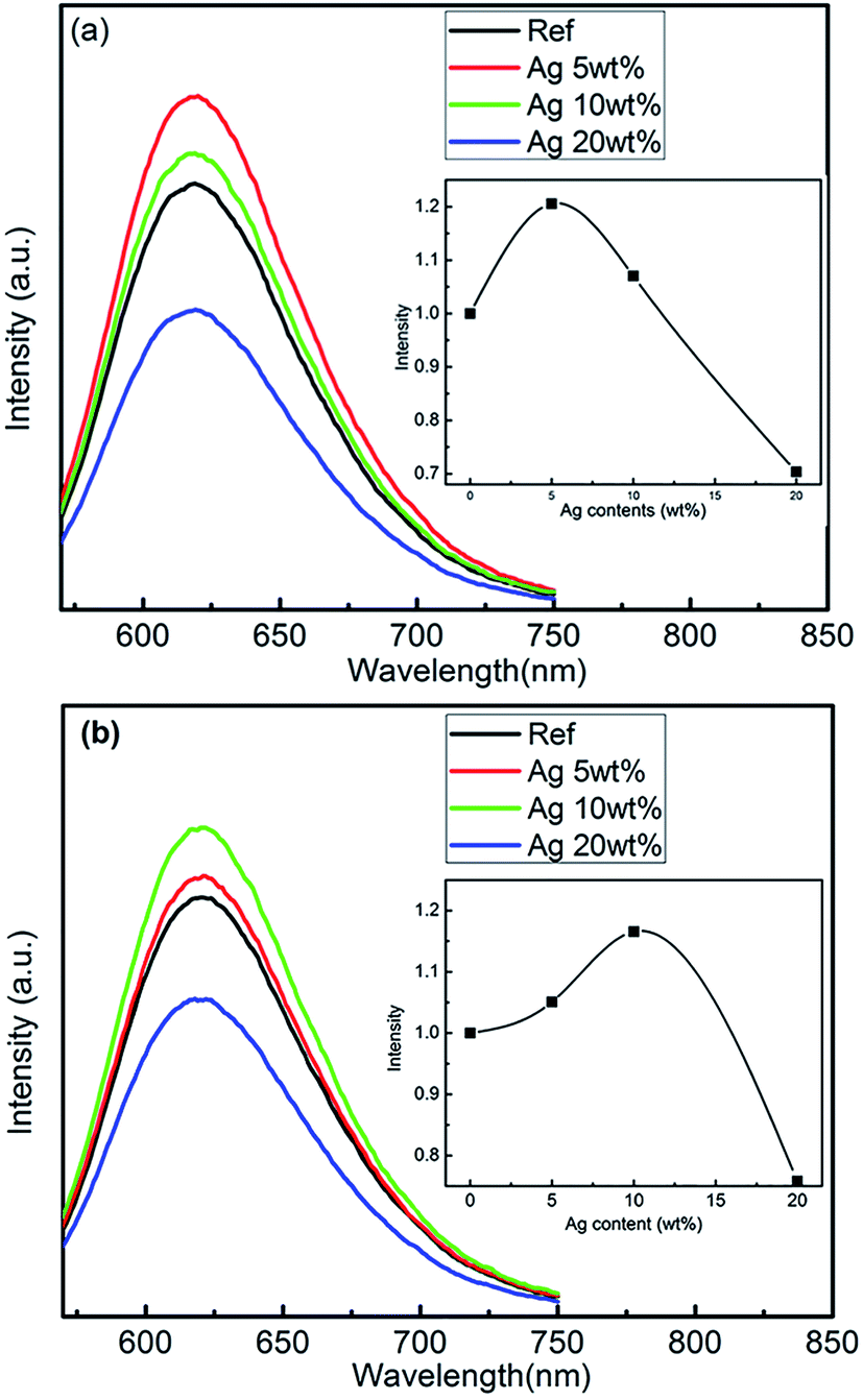 | ||
| Fig. 3 Emission spectra of phosphor films with different Ag contents under excitation at 420 nm: (a) Ag sample 1 (synthesized), and (b) Ag sample 2 (commercial). The insets show the influence of the enhancement ratio (with respect to the reference: Ag-free films) on Ag content. (wt% refers to the weight ratio of Ag nanoparticles/phosphor). | ||
However, the amount of Ag nanoparticles affects the intensity of the emission band. This is more pronounced in the phosphors with Ag from sample 1 than sample 2: phosphors with Ag from sample 1 reach higher values of the enhancement ratio (1.21) for a lower amount of Ag nanoparticles (5%) than the phosphors with Ag from sample 2 (1.17 and 10%, respectively). The increase of the emission intensity with the increase of Ag nanoparticles content is attributed to the LSPR of Ag nanoparticles. However, in both phosphors (with Ag samples 1 and 2), a further increase of the Ag nanoparticles content beyond that of the maximum emission intensity causes a dramatic decrease of emission intensity (for 20 wt% Ag content, the decrease is ∼30% with respect to the reference). This suggests that the Ag nanoparticles act as a luminescence quencher when their concentration exceeds a certain level in the layer. Apparently, a very high amount of Ag nanoparticles in the coating affects the light absorption and energy transition process between the phosphor and Ag nanoparticles.21 Accordingly, Ag sample 1 has a better SP resonance enhancement than the commercial Ag sample 2. This suggests that the plasmon effect of Ag nanoparticles synthesized by the seed growth method on the emission enhancement is better.
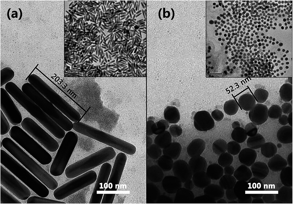
TEM images of the Ag particles in samples 1 and 2 are shown in Fig. 4a and b, respectively. Fine and uniform Ag nano-particles in the form of nano-spheres and nano-rods were synthesized by the seed growth method (Fig. 4a). After centrifugation, the fraction of the nano-spheres was separated from the fraction of nano-rods and these two morphologies are shown separately in the TEM images in Fig. 5. It is observed that the Ag nano-rods have a longitudinal axis of ∼200 nm (Fig. 5a). The average aspect ratio was calculated as 4.06 ± 1.09 and this is a result from the aspect ratios of more than 250 different nano-rods observed in TEM images. It is worthy to note that the aspect ratio can be controlled by the amount of Ag seeds and can govern the absorption band of Ag nanoparticles.18,19 On the other hand, the Ag nano-spheres have a diameter of 40–70 nm (Fig. 5b), which is similar to that of the spherical particles observed in sample 2 of commercial Ag (Fig. 4b). However, the inset of Fig. 4b shows that the Ag nanoparticles of sample 2 form agglomerates, while both nano-rods and nano-spheres in sample 1 feature uniform size and morphology, as shown in the insets of Fig 5. This poorer uniformity of sample 2 seemingly affected the SP effect of the Ag nanoparticles and impaired the emission intensity of the phosphors.22 Furthermore, the aggregations of Ag nanoparticles should make no contribution to the LSPR, resulting in a higher concentration of quenching for Ag sample 2 than for the Ag sample 1, as can be seen in the insets of Fig. 3.
 | ||
| Fig. 4 TEM images of the Ag nanoparticles (a) synthesized by the seed growth method, and (b) commercial. The inset shows the agglomerate of the commercial Ag nanoparticles. | ||
 | ||
| Fig. 5 TEM images of the two morphologies of Ag nanoparticles synthesized by the seed growth method: (a) Ag nano-rods, and (b) Ag nano-spheres. The insets of (a) and (b) show the wider field TEM images of Ag nano-rods and Ag nano-spheres, respectively. | ||
According to earlier studies, the two morphologies of the Ag nanoparticles of Fig. 5 are expected to have different absorption characteristics.23–25 Thus, the effect of LSPR on the emission enhancement of the phosphors with Ag nano-spheres and Ag nano-rods was investigated.
The absorption spectra of Ag nano-spheres and Ag nano-rods dispersed in water are plotted in Fig. 6. The absorption spectrum of Ag nano-spheres has only one peak at 408 nm, which could be assigned to the dipole resonance of Ag nano-spheres, while that of Ag nano-rods has two peaks at 431 and 620 nm. The resonances for light polarized in the longitudinal direction (i.e. along the long axis of the particle) and in transverse direction are different for ellipsoidal nanoparticles. Therefore, the 431 nm peak corresponds to the transverse mode resonance and the 620 nm peak to longitudinal resonance, which is very sensitive to the aspect ratio R.
 | ||
| Fig. 6 Absorption spectra of the Ag nano-spheres and Ag nano-rods, synthesized by the seed growth method. | ||
The PL enhancement of molecular near nano-sized metal particles is due to two contributions.20,25 First, the optical intensity incident on the molecule can be enhanced, and hence the excitation rate through near field enhances when the excitation wavelength is close to the LSPR band. Second, the radiative decay rate in the molecule is modified by the LSP resonant coupling of the nano-sized metal particles when the emission wavelength of the molecule is close to the plasmon resonance wavelength.
It is clearly seen that the absorption spectrum of the Ag nano-spheres merely overlaps the excitation band of the phosphor (Fig. 1), while that of the Ag nano-rods overlaps both the excitation and the emission spectrum. This may explain the better enhancement of emission intensity achieved with Ag sample 1 than with the commercial Ag in sample 2 (Fig. 3).25,26
The emission intensity can be further optimized through the synergistic effect of the two contributions. The effect of the ratio of nano-rods to nano-spheres on emission intensity was investigated. The enhancement (with respect to the Ag-free phosphor) of the emission intensity in phosphors with 5 wt% loading of Ag nanoparticles (which was the optimum loading, according to Fig. 3) that had different ratios of nano-rods to nano-spheres is shown in the plot in Fig. 7. It is clearly seen that the presence of Ag nanoparticles, whether nano-rods or nano-spheres, increases the emission intensity, but the pure Ag nano-rods have a stronger effect than the pure Ag nano-spheres, due to the existence of both contributions to excitation and emission. However, the maximum is reached for a certain value (∼50%) of this ratio. These features suggest that the two absorption peaks, which are overlapped with the excitation and emission spectra of the phosphor, have an enhancement effect due to different LSPRs. If this ratio exceeds that certain value, the intensity decreases; for instance, in the case of a ratio of 25/75, the excitation energy should be transferred directly to the Ag nano-spheres, resulting in the emission quenching of the phosphor.
 | ||
| Fig. 7 Enhancement (with respect to Ag-free phosphors) of the emission intensity of Sr2Si5N8:Eu2+ phosphors with different ratios of Ag nano-rods/nano-spheres (synthesized by the seed growth method). Ag-content was 5 wt%. | ||
Fig. 8 shows the time resolved decay of Sr2Si5N8:Eu2+ with and without Ag nanoparticles. The best enhanced sample (ratio 50%) as a representative is compared to the Ag-free sample. The decay curves fit well to a double-exponential function as
 | (1) |
 | ||
| Fig. 8 Decay curves of Sr2Si5N8:Eu2+ phosphor with and without Ag nanoparticles. | ||
The lifetimes can be calculated from the formula for τ
 | (2) |
The calculated lifetimes of phosphor with and without Ag nanoparticles are 1.05 μs and 0.65 μs, respectively. The increased decay rate of Sr2Si5N8:Eu2+ with Ag nanoparticles confirms the coupling of the phosphor with SPs in the Ag nanoparticles.27 The coupling through the close proximity of phosphor to Ag nanoparticles can lead to increased radiative decay rates and also enhanced fluorescence intensities.27,28
Conclusions
The visible emission of Sr2Si5N8:Eu2+ phosphor can be enhanced by coupling electric transition of localized surface plasmon oscillation of Ag nanoparticles, which specifically occurs due to the interactions between phosphors and SPs, with the synergistic effect of Ag nanospheres and nono-rods synthesized by the seed growth method. The visible light emission mainly occurs at the surface of phosphors and the local field enhancement is due to LSPR of Ag nanoparticles. Thus, a specific ratio of phosphor and Ag nanoparticles ensures the best results and was found to be 5 wt% in the investigated system. The morphologies and the SP resonance bands of the Ag nanoparticles can be tuned by adjusting the amount of Ag nano-seeds. The absorption spectrum of Ag nano-spheres merely overlaps the excitation spectrum of the phosphor, whereas that of Ag nano-rods overlaps both the excitation and the emission spectrum of the phosphor. Both types of particles favor enhancement of the emission intensity of the phosphors. However, the highest enhancement of emission intensity was achieved by mixing the two types of Ag nano-particles at a certain ratio.Acknowledgements
This research was supported by the National Natural Science Foundation of China (Grant No. 51372238), the National Basic Research Program of China (973 Program, 2012CB922004), and the CNPC-CAS Strategic Cooperation Research Program (2015A-4812).References
- S. Nakamura, S. Pearton and G. Fasol, The blue laser diode: the complete story, Springer Science & Business Media, 2013 Search PubMed.
- Y. Narukawa, M. Ichikawa, D. Sanga, M. Sano and T. Mukai, J. Phys. D: Appl. Phys., 2010, 43, 354002 CrossRef.
- S. Ye, F. Xiao, Y. X. Pan, Y. Y. Ma and Q. Y. Zhang, Mater. Sci. Eng., R, 2010, 71, 1–34 CrossRef.
- V. Bachmann, C. Ronda and A. Meijerink, Chem. Mater., 2009, 21, 2077–2084 CrossRef CAS.
- P. F. Smet, A. B. Parmentier and D. Poelman, J. Electrochem. Soc., 2011, 158, R37 CrossRef CAS.
- R.-J. Xie and N. Hirosaki, Sci. Technol. Adv. Mater., 2007, 8, 588–600 CrossRef CAS.
- I. Ahemen, K. De Dilip and A. N. Amah, Appl. Phys. Res., 2014, 6, 95 Search PubMed.
- C. C. Lin and R. S. Liu, J. Phys. Chem. Lett., 2011, 2, 1268–1277 CrossRef CAS PubMed.
- N. C. George, K. A. Denault and R. Seshadri, Annu. Rev. Mater. Res., 2013, 43, 481–501 CrossRef CAS.
- G.-H. Lee and S. Kang, J. Lumin., 2011, 131, 2582–2588 CrossRef CAS.
- Y. Hu, W. Zhuang, H. Ye, D. Wang, S. Zhang and X. Huang, J. Alloys Compd., 2005, 390, 226–229 CrossRef CAS.
- H. Zhu, C. C. Lin, W. Luo, S. Shu, Z. Liu, Y. Liu, J. Kong, E. Ma, Y. Cao, R. S. Liu and X. Chen, Nat. Commun., 2014, 5, 4312 CAS.
- G. R. Dillip and B. Deva Prasad Raju, J. Alloys Compd., 2012, 540, 67–74 CrossRef CAS.
- T. Takahashi and S. Adachi, J. Electrochem. Soc., 2008, 155, E183 CrossRef CAS.
- K. Uheda, N. Hirosaki, Y. Yamamoto, A. Naito, T. Nakajima and H. Yamamoto, Electrochem. Solid-State Lett., 2006, 9, H22 CrossRef CAS.
- J. A. Schuller, E. S. Barnard, W. Cai, Y. C. Jun, J. S. White and M. L. Brongersma, Nat. Mater., 2010, 9, 193–204 CrossRef CAS PubMed.
- D. K. Gramotnev and S. I. Bozhevolnyi, Nat. Photonics, 2010, 4, 83–91 CrossRef CAS.
- C. J. Murphy and N. R. Jana, Adv. Mater., 2002, 14, 80–82 CrossRef CAS.
- N. R. Jana, L. Gearheart and C. J. Murphy, Chem. Commun., 2001, 617–618, 10.1039/b100521i.
- H. Chen, L. Shao, Q. Li and J. Wang, Chem. Soc. Rev., 2013, 42, 2679–2724 RSC.
- J. H. Song, T. Atay, S. Shi, H. Urabe and A. V. Nurmikko, Nano Lett., 2005, 5, 1557–1561 CrossRef CAS PubMed.
- Y. Chen, K. Munechika and D. S. Ginger, Nano Lett., 2007, 7, 690–696 CrossRef CAS PubMed.
- A. Jakab, C. Rosman, Y. Khalavka, J. Becker, A. Trugler, U. Hohenester and C. Sonnichsen, ACS Nano, 2011, 5, 6880–6885 CrossRef CAS PubMed.
- S. Mazzucco, N. Geuquet, J. Ye, O. Stephan, W. Van Roy, P. Van Dorpe, L. Henrard and M. Kociak, Nano Lett., 2012, 12, 1288–1294 CrossRef CAS PubMed.
- Z. Buch, V. Kumar, H. Mamgain and S. Chawla, Chem. Commun., 2013, 49, 9485–9487 RSC.
- F. Tam, G. P. Goodrich, B. R. Johnson and N. J. Halas, Nano Lett., 2007, 7, 496–501 CrossRef CAS PubMed.
- M.-K. Kwon, J.-Y. Kim, B.-H. Kim, I.-K. Park, C.-Y. Cho, C. C. Byeon and S.-J. Park, Adv. Mater., 2008, 20, 1253–1257 CrossRef CAS.
- M. H. Chowdhury, K. Ray, C. D. Geddes and J. R. Lakowicz, Chem. Phys. Lett., 2008, 452, 162–167 CrossRef CAS PubMed.
| This journal is © The Royal Society of Chemistry 2016 |