Selective concentration-dependent manipulation of intrinsic fluorescence of plasma proteins by graphene oxide nanosheets

Kenryabc, Kian Ping Lohbd and Chwee Teck Lim*bce
aNUS Graduate School for Integrative Sciences and Engineering, National University of Singapore, Singapore 117456, Singapore
bCentre for Advanced 2D Materials and Graphene Research Centre, National University of Singapore, Singapore 117543, Singapore. E-mail: ctlim@nus.edu.sg
cDepartment of Biomedical Engineering, National University of Singapore, 9 Engineering Drive 1, Singapore 117576, Singapore
dDepartment of Chemistry, National University of Singapore, Singapore 117543, Singapore
eMechanobiology Institute, National University of Singapore, Singapore 117411, Singapore

Received 25th February 2016 , Accepted 27th April 2016

First published on 28th April 2016


Abstract

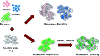
We investigate the molecular interactions between graphene oxide (GO) and blood plasma proteins, in particular, the influence of GO on the intrinsic fluorescence of these proteins. We observe that GO acts as an efficient quencher of the intrinsic fluorescence of albumin, globulin, and fibrinogen. Interestingly, we also note for the first time that, in addition to the robust fluorescence quenching, GO is capable of selectively amplifying the fluorescence emission of fibrinogen up to approximately 30% or 1.3 fold under certain concentrations but not those of albumin and globulin. We suggest that GO may possibly play a dual role in controlling the intrinsic fluorescence emission of the plasma proteins. Furthermore, this role switching may be influenced by the competition between the aggregation and encapsulation effects. We propose that the GO-induced intrinsic fluorescence quenching is driven by the physical encapsulation of the plasma proteins by GO nanosheets. Contrastingly, the GO-mediated fluorescence amplification is promoted by an aggregation of fibrinogen.


Introduction

Graphene oxide (GO), the oxygenated derivative of graphene, is a two-dimensional single layer of carbon atoms containing the hydroxyl and epoxy groups on its basal planes as well as the carboxyl groups at its periphery.1,2 These hydrophilic functional groups coupled with the aromatic domains provide GO with an appropriate platform for interactions with a multitude of biomolecules, such as proteins, peptides, and amino acids.3,4 Amino acids, including tryptophan, tyrosine, and phenylalanine, are capable of associating with GO through aromatic π–π stacking or electrostatic interaction.5 Based on these unique interactions, GO has been actively pursued as a nanoplatform for various advanced optoelectronics and biosensing applications.6,7 In fact, GO has been widely demonstrated to effectively quench the fluorescence of various emission-active fluorophores, including small molecular dyes, dye-labeled peptides, and conjugated polymers, due to energy transfer or non-radiative dipole–dipole coupling interaction induced by strong π–π interaction between GO and these fluorophores.8,9 Capitalizing on this attractive fluorescence quenching characteristic, numerous GO-based optical sensors have been developed for highly sensitive biomolecule detections.10–12 Despite the promising outlooks, further exploration and understanding on the GO-biomolecule interplays are necessary and fundamentally significant, especially on the relationship between GO and those drug- and disease-related proteins and biomolecules. To date, there are still limited studies reporting on these interactions,13,14 particularly those involving GO and plasma proteins, such as albumin, globulin, and fibrinogen. Besides, a large number of these studies on the interactions between GO and biomolecules have been performed in the presence of fluorescent dye-labeled probes.

With a molecular weight of 65 kDa, albumin is one of the smallest and most abundant proteins in the circulatory system. The globular albumin is generally known as a multifunctional transporter molecule and it constitutes about 55% of the plasma proteins in blood. Structurally, albumin comprises three homologous domains in which each domain may be further classified into two sub-domains.15 Globulin is another vital globular protein responsible for numerous physiological functions, such as molecular transport and maintenance of immune system. With a molecular weight of approximately 92 to 120 kDa and constituting roughly 38% of the plasma proteins, globulin is the second most abundant plasma protein. Fibrinogen, on the other hand, is a large protein with a molecular weight of approximately 340 kDa. It is a macromolecular plasma protein which plays an active and essential role in the blood coagulation process. The native structure of fibrinogen is of triglobular configuration which consists of a small central domain connected to two terminal globular regions by the α–helical coiled-coil domains.16 In addition, a single fibrinogen molecule possesses a length of about 45 to 50 nm.16,17

Generally, blood plasma proteins possess three aromatic residues, i.e., tryptophan, tyrosine, and phenylalanine, that contribute differently to their intrinsic fluorescence emissions.18,19 Out of these amino acid residues, tryptophan is recognized as the dominant source of UV absorption and intrinsic fluorescence emission of proteins at around 280 nm and 350 nm, respectively. The tryptophan fluorescence is much stronger than those displayed by the other two aromatic amino acids. In fact, the fluorescence intensity of tryptophan is approximately eight and 140 times higher than those of tyrosine and phenylalanine, respectively. Although the quantum yields of tryptophan and tyrosine are similar, the fluorescence of tyrosine is frequently quenched in the native proteins probably as a result of its energy transfer to tryptophan or because of the tyrosine–peptide chain interactions.20 On the other hand, the intrinsic fluorescence of phenylalanine is negligible as it possesses low absorptivity and low quantum yield. The intrinsic tryptophan fluorescence emission of a protein, interestingly, is strongly influenced and highly sensitive to its local microenvironment. For example, when tryptophan is buried within the hydrophobic core of the protein, it possesses a high quantum yield and displays strong fluorescence intensity. Conversely, its quantum yield decreases and tryptophan exhibits weak fluorescence emission when it is exposed to a hydrophilic solvent.

Here, we examine the GO–plasma protein interaction, in particular, the effect of GO on the intrinsic fluorescence of the three plasma proteins of albumin, globulin, and fibrinogen. We observe that GO quenches the intrinsic tryptophan fluorescence of all plasma proteins efficiently. Remarkably, we also demonstrate that, besides the robust fluorescence quenching, GO is capable of selectively amplifying the intrinsic tryptophan fluorescence of fibrinogen up to about 1.3 fold under specific low concentrations but not those of albumin and globulin. To the best of our knowledge, this is the first study reporting on the novel GO-induced enhancement of the intrinsic fluorescence of fibrinogen. In light of our experimental observations, we propose that GO can possibly play a dual role in controlling the intrinsic fluorescence of the plasma proteins. This fluorescence manipulation may be dictated by the direct competition between the aggregation and encapsulation effects.

Results and discussion

First, we characterized the surface morphology and lateral size of GO nanosheets. GO solution was dropped onto a freshly cleaved mica and the morphology and thickness of the individual GO nanosheets were examined using the tapping mode of an atomic force microscope (AFM) (Fig. 1a). To acquire the statistical data on the lateral size and its distribution of these GO nanosheets, we evaluated more than 400 GO nanosheets (Fig. 1b). Based on this, we noted that GO nanosheets possessed an average size of approximately 0.9 μm. In addition, they had a thickness of approximately 1.2 nm, indicating that they were of monolayer (inset of Fig. 1b). This was in close agreement with the previous reported thickness of single-layer GO.21,22 Next, the chemical environment of these GO nanosheets was evaluated utilizing the X-ray photoelectron spectroscopy (XPS) (Fig. 1c). As shown, GO exhibited a highly asymmetrical C1s peak. Various oxygen functional groups, such as epoxy, hydroxyl, and carbonyl, could be distinguished based on the different chemical bonds of C–C (50.72% at 284.5 eV), C–O (34.34% at 286.7 eV), and C[double bond, length as m-dash]O (14.94% at 288.0 eV). We further characterized the optical UV-Vis absorbance of the single layer GO nanosheets (Fig. 1d). We noted that GO nanosheets exhibited a maximum absorption at approximately 234 nm because of the π–π* transition of the aromatic C[double bond, length as m-dash]C bonds as well as a weak shoulder at about 300 nm as a result of the n–π* transition of the C[double bond, length as m-dash]O bonds. Again, this observation was similar to the previously published absorption spectrum of GO.9,23 In addition to GO, we performed optical characterizations on the three plasma proteins, specifically on their aromatic amino acid tryptophan (Fig. 1e–g). The plasma proteins were prepared at a concentration of 1.1 μM and their absorbance and intrinsic tryptophan fluorescence emission were probed at an excitation wavelength of 280 nm. We observed that albumin exhibited peak absorption and fluorescence emission at approximately 278 nm and 342 nm, respectively (Fig. 1e). In contrast, the optical absorbance/fluorescence of both globulin and fibrinogen peaked at roughly 282/330 nm (Fig. 1f) and 278/336 nm (Fig. 1g), respectively.
image file: c6ra04978h-f1.tif
Fig. 1 Characterizations of GO and plasma proteins. (a) Representative surface morphology of GO nanosheets dropped on freshly cleaved mica obtained using the tapping mode of an AFM. Scale bar represents 1 μm. (b) Lateral size distribution of GO nanosheets. Approximately 400 GO nanosheets were evaluated to obtain the lateral size distribution. Inset depicts the sectional profiles of GO nanosheets (i.e., S1 and S2 in (a)) showing a thickness of about 1.2 nm. This suggests that GO nanosheets were of monolayer. (c) XPS C1s peak of GO nanosheets depicting the contribution of various oxygen functionalities (i.e., C–C bond at 50.72%, C–O bond at 34.34%, and C[double bond, length as m-dash]O bond at 14.94%) (d) Normalized UV-Vis absorbance of GO illustrating a maximum absorption at around 234 nm. (e–g) Normalized UV-Vis absorbance and intrinsic fluorescence spectra of 1.1 μM: (e) albumin, (f) globulin, and (g) fibrinogen. Excitation wavelength was set at 280 nm. Each spectrum was obtained as an average of three independent readings.

Subsequent to the optical characterizations of both GO and plasma proteins, we started investigating the GO–plasma protein interactions by evaluating the absorbance characteristic of these proteins in the presence of GO nanosheets (Fig. 2). We observed that with the plasma proteins fixed at 1.1 μM, the addition of GO nanosheets in the system induced changes in the characteristic absorption spectra of all plasma proteins. Specifically, we noted diminished 280 nm absorption bands coupled with a corresponding increase in the absorption bands between 230 nm and 260 nm (Fig. 2a–c). In fact, the absorption spectra of the three proteins gradually adopted the shape of that of pure GO nanosheets as their concentration in the system increased. This demonstrates the strong molecular interactions between GO nanosheets and blood plasma proteins as well as possibly, a subtle change in the structures of the amino acid chains of the plasma protein molecules.


image file: c6ra04978h-f2.tif
Fig. 2 UV-vis absorbance of plasma proteins in the presence of GO nanosheets with increasing concentration. UV-Vis absorption spectra of: (a) albumin, (b) globulin, and (c) fibrinogen, in the absence and presence of GO nanosheets with increasing concentration. Each spectrum was obtained as an average of three independent readings.

Next, we sought to investigate the effect GO nanosheets would induce on the intrinsic fluorescence emission of the three plasma proteins (Fig. 3). The intrinsic fluorescent probes of the three plasma proteins were excited at 280 nm and their fluorescence emissions were recorded from 310 to 420 nm. Fig. 3a–d show the evolutions of the intrinsic fluorescence of 1.1 μM albumin and globulin in the presence of GO nanosheets with increasing concentration, respectively. As anticipated, the emission intensity of both albumin and globulin decreased progressively with a corresponding increase in the GO concentrations from 0.5 to 25 μg mL−1. Strong interactions between GO nanosheets and albumin and globulin were noted according to the efficient quenching of the protein fluorescence emission. Importantly, this experimental data coincided with conventional observations in which GO serves as an effective fluorescent quencher for amino acids, proteins, and other fluorescent materials.8,9


image file: c6ra04978h-f3.tif
Fig. 3 Fluorescence emission of plasma proteins in the presence of GO nanosheets with increasing concentration. (a and c) Fluorescence spectra of: (a) albumin and (c) globulin, in the presence of GO nanosheets with increasing concentrations from 0 to 25 μg mL−1 (b and d) Fluorescence intensity ratio I/I0 as a function of GO concentration where I0 and I are the emission intensities of: (b) albumin and (d) globulin, in the absence and presence of GO, respectively. (e) Representative fluorescence spectra of fibrinogen in the presence of GO nanosheets with increasing concentrations from 0 to 3 μg mL−1. Fluorescence amplification occurred at low GO concentrations of up to 3 μg mL−1. (f) Representative fluorescence spectra of fibrinogen in the presence of GO nanosheets with increasing concentrations from 3.5 to 25 μg mL−1. Fluorescence quenching took place at high GO concentrations beyond 3 μg mL−1. (g) Fluorescence intensity ratio I/I0 as a function of GO concentration where I0 and I are the emission intensities of fibrinogen in the absence and presence of GO, respectively. Inset shows the enhancement of the fluorescence intensity ratio of fibrinogen I/I0 in the presence of GO nanosheets with low concentrations ranging from 0.5 to 3 μg mL−1. Data are presented as mean ± standard error. Each spectrum was obtained as an average of three independent readings.

In addition to those of albumin and globulin, we examined the evolution of the fluorescence emission of 1.1 μM fibrinogen in the presence of GO nanosheets with various concentrations (Fig. 3e–g). Intriguingly, in our system, besides the commonly recognized fluorescence quenching (Fig. 3f and g), we observed an amplification of the intrinsic fluorescence of fibrinogen in the presence of GO nanosheets with low concentrations, ranging from 1 μg mL−1 to approximately 3 μg mL−1 (Fig. 3e–g). Particularly, the intrinsic fluorescence of fibrinogen increased up to around 1.3 fold or 30% with the addition of 2 to 2.5 μg mL−1 GO (inset of Fig. 3g). Nevertheless, the low GO concentration-induced intrinsic fluorescence amplification was not detected from the fluorescence spectra of albumin and globulin. Beyond these GO concentrations, the fluorescence intensity of fibrinogen was gradually quenched and the emission reduced with no shift in its peak wavelength (Fig. 3f).

To shed light on the possible physical mechanisms underlying the manipulation of the intrinsic fluorescence of albumin, globulin, and fibrinogen by GO nanosheets, we sought to characterize the structural morphologies of the plasma protein–GO complexes upon the addition of GO with a range of concentrations (Fig. 4 and 5). As control, the surface morphologies of freshly cleaved mica (i.e., substrate on which the plasma proteins and GO–protein complexes were deposited and imaged) (Fig. 4a) and 1.1 μM albumin (Fig. 4b) and globulin (Fig. 4d) were examined using the tapping mode of an AFM. As observed, the albumin and globulin molecules possessed a regular circular shape and an approximate size of a few to tens of nm. Sectional profiles revealed that albumin and globulin displayed similar heights of about a few nm (Fig. 4b(ii and iii)) to 10 nm (Fig. 4d(ii and iii)), respectively. Contrastingly, the GO–albumin and GO–globulin complexes adopted irregular shapes (Fig. 4c and e). Furthermore, the topographical sectional features of the GO–protein complexes demonstrated that they had a height of approximately 20 nm (Fig. 4c(ii and iii) and e(ii and iii)) which was significantly larger than those of the albumin and globulin molecules. Importantly, upon a closer examination of the structural morphologies of the complexes, it is interesting to note that apparently, the GO–albumin and GO–globulin complexes were formed from the encapsulation or wrapping of individual protein molecules or aggregates within GO nanosheets. This, in fact, provides an important clue to the physical mechanisms driving the GO-induced fluorescence quenching.


image file: c6ra04978h-f4.tif
Fig. 4 Surface morphologies of the GO–albumin and GO–globulin complexes as characterized using the tapping mode of an AFM. Surface morphologies of: (a) freshly cleaved mica, (b) 1.1 μM albumin, (c) GO–albumin complex at albumin concentration of 1.1 μM and GO concentration of 25 μg mL−1, (d) 1.1 μM globulin, and (e) GO–globulin complex at globulin concentration of 1.1 μM and GO concentration of 25 μg mL−1: (i) 2D representative AFM amplitude images, (ii) 2D representative AFM topographical images with (iii) the corresponding sectional profiles (i.e., S1 and S2 in (ii)), and (iv) 3D representative AFM topographical images. All scale bars represent 1 μm.

image file: c6ra04978h-f5.tif
Fig. 5 Surface morphologies of the GO–fibrinogen complexes as characterized using the tapping mode of an AFM. Surface morphologies of: (a) 1.1 μM fibrinogen, (b) GO–fibrinogen complex with an amplified intrinsic fluorescence at fibrinogen concentration of 1.1 μM and low GO concentration of 2.5 μg mL−1 (i.e., Fb + low [GO]), and (c) GO–fibrinogen complex with a quenched intrinsic fluorescence at fibrinogen concentration of 1.1 μM and high GO concentration of 25 μg mL−1 (i.e., Fb + high [GO]): (i) 2D representative AFM amplitude images, (ii) 2D representative AFM topographical images with (iii) the corresponding sectional profiles (i.e., S1 and S2 in (ii)), and (iv) 3D representative AFM topographical images. All scale bars represent 1 μm.

Subsequent to characterizing the structural morphologies of the GO–albumin and GO–globulin complexes, we performed similar morphology characterization on the GO–fibrinogen complexes, especially, when the fluorescence emissions were amplified and quenched at GO concentrations of 2.5 μg mL−1 and 25 μg mL−1, respectively (Fig. 5). As control, 1.1 μM fibrinogen was deposited on mica and surface-characterized using AFM (Fig. 5a). As depicted in the AFM topographical images, the fibrinogen molecules adopted an elongated spheroid shape and possessed height of roughly five to 10 nm. When GO of low concentration of 2.5 μg mL−1 was mixed with fibrinogen, unlike albumin and globulin which were directly encapsulated or wrapped by GO nanosheets, it was highly likely that fibrinogen adsorbed onto the GO surface and aggregated into spherical-shaped structures, forming the GO–fibrinogen complex (Fig. 5b). Sectional profiles revealed that the fibrinogen aggregates possessed height of up to approximately 50 to 60 nm, depending on the size of the aggregates (Fig. 5bii and iii). However, when a higher concentration of GO of 25 μg mL−1 was added, fibrinogen was observed to form large spherical- and ellipsoid-shaped aggregates with heights of up to around 160 nm (Fig. 5c). Moreover, it was clearly evident that some fibrinogen aggregates were covered, encapsulated, or fully wrapped by multiple layers of large GO nanosheets, leading to the formation of large GO–fibrinogen complexes. In light of all the obtained data, we proposed the possible mechanisms driving the observed manipulation of the intrinsic fluorescence of the three plasma proteins (Fig. 6). In fact, the requirement for a particular low GO concentration for the fluorescence amplification of fibrinogen suggests that GO might play a dual role in controlling the fluorescence emission of fibrinogen.


image file: c6ra04978h-f6.tif
Fig. 6 Proposed mechanisms of the manipulation of the intrinsic fluorescence of the plasma proteins by GO nanosheets. Schematic illustration showing the proposed mechanisms underlying the amplification/quenching of the intrinsic fluorescence of albumin, globulin, and fibrinogen in the presence of GO nanosheets with increasing concentration.

In general, albumin and globulin are globular proteins with small physical sizes and molecular weights as compared to the large fibrinogen macromolecules. Upon interactions with GO nanosheets, it was highly likely that the GO–albumin and GO–globulin complexes formed immediately through the physical encapsulation or wrapping of the individual protein molecules by GO nanosheets (Fig. 4c and e). As these molecular interactions occurred, there would be energy transfer between GO nanosheets and plasma proteins as a result of the π–π stacking and interaction. This would eventually promote the quenching of the intrinsic fluorescence of both albumin and globulin. Also, as the physical distance separating GO nanosheets and individual plasma protein molecules reduced due to the formation of the GO–protein complexes and a progressive increase in the GO concentration in the system, a resultant increase in the π–π interaction and energy transfer would follow, leading to a corresponding increase in the GO-induced fluorescence quenching efficiency.

Fibrinogen, on the other hand, is a large elongated macromolecular structure.24 At low GO concentrations, fibrinogen and its hydrophobic tryptophan residue might bind non-specifically to GO nanosheets through hydrophobic interactions. Subsequently, the molecular motion and structural flexibility of fibrinogen decreased, leading to a corresponding decrease in the collision-induced non-radiative energy loss and a slight increase in the intrinsic fluorescence of fibrinogen. Recent study has reported that GO itself is capable of promoting and accelerating protein aggregation.25 In addition, it was possible that fibrinogen adsorbed on both sides of GO nanosheets, increasing the likelihood of the formation of more fibrinogen aggregates. Also, several studies have recently demonstrated the enhancement of the aggregation-induced fluorescence emission of active fluorophores by GO under certain concentrations.26,27

Here, it is noteworthy that under a similar low GO concentration range, the intrinsic fluorescence emissions of albumin and globulin were not amplified. Irrespective of the GO concentration in the system, the fluorescence emissions of both proteins were continuously quenched. As such, we suggest that the absence of the amplification of the fluorescence of albumin and globulin might be attributed to the synergistic effect brought about by the lower likelihood of both proteins to form aggregates as well as their rapid physical encapsulations by GO nanosheets. It was highly possible that the aggregates of the large macromolecular fibrinogen could be formed more easily as compared to those of the small albumin and globulin. This is because during the folding and/or unfolding processes, there was a higher probability for the hydrophobic backbone of fibrinogen to be exposed to external microenvironments as compared to those of albumin and globulin, triggering intermolecular aggregation.28,29 Moreover, the fibrinogen molecules comprise more domains than the albumin and globulin molecules. As a consequence, the interdomain surface contacts of fibrinogen might occur more frequently and at a higher rate than those of the other two proteins, initiating more aggregations. Finally, fibrinogen might experience more competing aggregation reactions due to the slower rate of refolding of its unfolded conformers. Intriguingly, the lower probability of albumin and globulin to form aggregates might also be ascribed to the rapid and facile physical encapsulations of these two plasma proteins due to their small sizes and molecular weights.

When the concentration of GO nanosheets increased beyond 3 μg mL−1, an opposite effect in the fluorescence emission of fibrinogen could be observed. Instead of fluorescence amplification, the intrinsic fluorescence of fibrinogen was quenched steadily in the presence of additional GO nanosheets. Interestingly, here, we observed that fibrinogen aggregates were also formed in the presence of increasing concentration of GO nanosheets (Fig. 5c). In fact, our data on the formation of a high number of fibrinogen aggregates in spite of the presence of hydrophilic GO corroborated with the observation of an earlier study,25 further confirming the capability of GO to induce protein aggregation despite its hydrophilic nature. Also, it is important to highlight that the intrinsic fluorescence quenching occurred although more fibrinogen aggregates were generated. Apparently, at high GO concentration, fibrinogen aggregation no longer played a dominant role in influencing fluorescence emission.

Alternatively, it was highly likely that an increase in the number of multilayer GO nanosheets led to an encapsulation or full wrapping of the fibrinogen aggregates (Fig. 5c), similar to that experienced by the albumin and globulin molecules. At the same time, with more GO nanosheets, the molecular interactions between fibrinogen and two or more overlapping and stacked GO nanosheets would increase proportionately as a result of the shortening of the physical distance separating the protein molecules from GO. With a significant increase in the GO–fibrinogen interactions, more energy transfer would occur between fibrinogen and GO due to a corresponding increase in the π–π stacking and interaction. Moreover, fibrinogen might experience a conformational change with increasing interactions with GO. Consequently, the shielded tryptophan residue which was previously hidden in the internal hydrophobic parts of fibrinogen would be exposed. This ultimately resulted in the dwarfing of the aggregation-induced effects for a robust quenching of the intrinsic fluorescence of fibrinogen. In short, we suggest that the fibrinogen aggregation and encapsulation effects contributed synergistically to the intrinsic fluorescence amplification and quenching of fibrinogen, correspondingly (Fig. 6).

Conclusions

In summary, we probed the molecular interactions between GO nanosheets and blood plasma proteins. We noted that GO quenched the intrinsic fluorescence of albumin, globulin, and fibrinogen. Interestingly, we also observed, for the first time, the concentration-dependent amplification of the fluorescence of fibrinogen by GO nanosheets. In fact, besides the effective fluorescence quenching, we demonstrated that GO was able to selectively enhance the intrinsic fluorescence emission of fibrinogen up to roughly 30% under low concentrations but not those of albumin and globulin. In light of these observations, we proposed that GO might potentially play a twofold role in controlling the fluorescence emission of the plasma proteins. The switching between these roles might be dependent on the competition between aggregation (i.e., fluorescence amplification) and wrapping (i.e., fluorescence quenching) effects. More specifically, we believe that the GO-induced fluorescence quenching was caused by the physical wrapping of the plasma proteins by GO nanosheets. On the other hand, the distinct GO-promoted intrinsic fluorescence amplification might be ascribed to the GO-induced aggregation of fibrinogen. Overall, we anticipate that this study will provide a new avenue for the exploration and development of novel GO-based nanomaterials for label-free fluorescence “turn-on” sensing of chemical and biomolecular targets as well as for general biological and biomedical research.

Experimental section

Materials

Plasma protein powders of albumin, globulin, and fibrinogen were purchased (Sigma-Aldrich, St. Louis, MO) and used directly without further purification. All proteins were prepared in phosphate-buffered saline (PBS 1×) and fixed at a concentration of 1.1 μM. GO nanosheets were prepared based on the Hummer's method and its concentration was varied from 0 to 25 μg mL−1.

Surface morphology characterization

GO nanosheets were deposited on a freshly cleaved mica. Surface morphology of the as-deposited GO nanosheets was then characterized using the tapping mode of an atomic force microscope (AFM) (Bruker, Billerica, MA) in air under room temperature. Lateral size distribution of GO nanosheets was evaluated based on the obtained AFM images using the ImageJ software (NIH, US). Approximately 400 GO nanosheets were measured to derive their lateral size distribution. Surface morphology of the GO–fibrinogen complexes was also obtained based on the tapping mode AFM under ambient conditions.

Optical characterization assays

XPS C1s spectrum of GO nanosheets was recorded utilizing an unmonochromated A1 Kα X-ray source at 1486.6 eV (Thermo VG Scientific, UK) equipped with a Phobios 100 electron analyzer (SPECS GmbH, Germany). Absorption spectra of albumin, globulin, fibrinogen, and GO nanosheets were obtained using a UV-Vis spectrophotometer (NanoDrop 2000, Thermo Scientific). Intrinsic fluorescence of the plasma proteins in the absence and presence of GO nanosheets was measured through fluorescence intensity scan using a fluorescence microplate reader (Infinite M200, Tecan) in a black 96-well microplate (Nunc MicroWell, Thermo Scientific). Fluorescence excitation was set at 280 nm. Fluorescence emission spectra were recorded from 310 to 420 nm. Each spectrum was obtained as an average of at least three independent readings under room temperature.

References

  1. M. J. Allen, V. C. Tung and R. B. Kaner, Chem. Rev., 2010, 110, 132–145 CrossRef CAS PubMed.
  2. Kenry, K. P. Loh and C. T. Lim, Small, 2015, 11, 5105–5117 CrossRef CAS PubMed.
  3. D. R. Dreyer, S. Park, C. W. Bielawski and R. S. Ruoff, Chem. Soc. Rev., 2010, 39, 228–240 RSC.
  4. A. M. H. Ng, Kenry, C. Teck Lim, H. Y. Low and K. P. Loh, Biosens. Bioelectron., 2015, 65, 265–273 CrossRef CAS PubMed.
  5. M. Zhang, B.-C. Yin, X.-F. Wang and B.-C. Ye, Chem. Commun., 2011, 47, 2399–2401 RSC.
  6. K. P. Loh, Q. Bao, G. Eda and M. Chhowalla, Nat. Chem., 2010, 2, 1015–1024 CrossRef CAS PubMed.
  7. Q. Bao and K. P. Loh, ACS Nano, 2012, 6, 3677–3694 CrossRef CAS PubMed.
  8. X. Ling, L. Xie, Y. Fang, H. Xu, H. Zhang, J. Kong, M. S. Dresselhaus, J. Zhang and Z. Liu, Nano Lett., 2010, 10, 553–561 CrossRef CAS PubMed.
  9. S. Li, A. N. Aphale, I. G. Macwan, P. K. Patra, W. G. Gonzalez, J. Miksovska and R. M. Leblanc, ACS Appl. Mater. Interfaces, 2012, 4, 7069–7075 CAS.
  10. S. He, B. Song, D. Li, C. Zhu, W. Qi, Y. Wen, L. Wang, S. Song, H. Fang and C. Fan, Adv. Funct. Mater., 2010, 20, 453–459 CrossRef CAS.
  11. W. Wu, H. Hu, F. Li, L. Wang, J. Gao, J. Lu and C. Fan, Chem. Commun., 2011, 47, 1201–1203 RSC.
  12. E. Morales-Narváez and A. Merkoçi, Adv. Mater., 2012, 24, 3298–3308 CrossRef PubMed.
  13. Z. Ding, H. Ma and Y. Chen, RSC Adv., 2014, 4, 55290–55295 RSC.
  14. Z. Ding, Z. Zhang, H. Ma and Y. Chen, ACS Appl. Mater. Interfaces, 2014, 6, 19797–19807 CAS.
  15. S. Curry, P. Brick and N. P. Franks, Biochim. Biophys. Acta, Mol. Cell Biol. Lipids, 1999, 1441, 131–140 CrossRef CAS.
  16. N. Hassan, J. Maldonado-Valderrama, A. P. Gunning, V. J. Morris and J. M. Ruso, J. Phys. Chem. B, 2011, 115, 6304–6311 CrossRef CAS PubMed.
  17. K. L. Marchin and C. L. Berrie, Langmuir, 2003, 19, 9883–9888 CrossRef CAS.
  18. C. A. Royer, Chem. Rev., 2006, 106, 1769–1784 CrossRef CAS PubMed.
  19. J. Kim, H. Song, I. Park, C. R. Carlisle, K. Bonin and M. Guthold, Microsc. Res. Tech., 2011, 74, 219–224 CrossRef PubMed.
  20. A. Ghisaidoobe and S. Chung, Int. J. Mol. Sci., 2014, 15, 22518–22538 CrossRef CAS PubMed.
  21. H. Wang, Q. Zhang, X. Chu, T. Chen, J. Ge and R. Yu, Angew. Chem., Int. Ed., 2011, 50, 7065–7069 CrossRef CAS PubMed.
  22. M. Li, X. Yang, J. Ren, K. Qu and X. Qu, Adv. Mater., 2012, 24, 1722–1728 CrossRef CAS PubMed.
  23. D. Li, M. B. Muller, S. Gilje, R. B. Kaner and G. G. Wallace, Nat. Nanotechnol., 2008, 3, 101–105 CrossRef CAS PubMed.
  24. G. S. Retzinger, A. P. DeAnglis and S. J. Patuto, Arterioscler., Thromb., Vasc. Biol., 1998, 18, 1948–1957 CrossRef CAS.
  25. M. Zhang, X. Mao, C. Wang, W. Zeng, C. Zhang, Z. Li, Y. Fang, Y. Yang, W. Liang and C. Wang, Biomaterials, 2013, 34, 1383–1390 CrossRef CAS PubMed.
  26. X. Qi, H. Li, J. W. Y. Lam, X. Yuan, J. Wei, B. Z. Tang and H. Zhang, Adv. Mater., 2012, 24, 4191–4195 CrossRef CAS PubMed.
  27. J. Geng, L. Zhou and B. Liu, Chem. Commun., 2013, 49, 4818–4820 RSC.
  28. A. Mitraki and J. King, Nat. Biotechnol., 1989, 7, 690–697 CrossRef CAS.
  29. R. Seckler and R. Jaenicke, FASEB J., 1992, 6, 2545–2552 CAS.

This journal is © The Royal Society of Chemistry 2016