Morphology controllable syntheses of micro- and nano-iron pyrite mono- and poly-crystals: a review

Haiyang Xian abc, Jianxi Zhu *ab, Xiaoliang Liang ab and Hongping He ab
aKey Laboratory of Mineralogy and Metallogeny, Guangzhou Institute of Geochemistry, Chinese Academy of Sciences, Guangzhou 510640, Guangdong, People's Republic of China. E-mail: zhujx@gig.ac.cn
bGuangdong Provincial Key Laboratory of Mineral Physics and Materials, Guangzhou 510640, Guangdong, People's Republic of China
cUniversity of Chinese Academy of Sciences, Beijing 100049, People's Republic of China

Received 24th February 2016 , Accepted 19th March 2016

First published on 22nd March 2016


Abstract

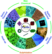
Synthesis of iron pyrite with defined morphology has long been actively pursued, due to the strong size and shape dependence of their chemical and physical properties. This review provides comprehensive information outlining current knowledge regarding the morphology controllable syntheses of micro- and nano-iron pyrite mono- and poly-crystals. The wet-chemical methods are summarized as the controllable syntheses, including the hydrothermal, solvothermal, hot-injection and heating-up methods, sulphidation and methods with other relatively high efficiencies. The present study reveals the discussion of relationship between the morphologies and major controlling factors, the temperature, precursor chemicals, solvents and surfactants. The existing challenges for future fine tuning of iron pyrite facets are also proposed for improving the performance of iron pyrite based materials.


1. Introduction

Iron pyrite is a common type of sulphide mineral and is also known as “fool's gold”; however, its various applications make it a promising material in the energy storage and environmental protection areas. It is a very attractive candidate for the next generation photovoltaic material, due to its high absorption coefficient and its energy band gap (Eg ≈ 0.95 eV), which is appropriate for photovoltaic energy conversion.1–8 Not only could pyrite affect the environment in which it is present through its surface reactivity,9 but it has also been widely used in environmental applications for heavy metal adsorption, reduction or related processes.10–20

The atomic structure of iron pyrite (Fig. 1) is well-known and was first determined by Bragg21 with his new X-ray diffraction system in 1914. The structure of iron pyrite is an analogy of the NaCl-type structure. The disulphide dumbbells S22− groups are situated at the Cl positions, i.e. at the cube centre and the midpoints of cube edges, and the ferrous ion atoms at the Na+ positions, i.e. the corners and face centres. Regarding the arrangement of the disulphide dumbbells, the symmetry of pyrite structure, Pa3, is lower than that of NaCl-type structure, Fm3m. The space group of pyrite determines its crystal habit, leading to the most common shapes of its crystals are {100}, {111} and {210} facets in nature.22


image file: c6ra04874a-f1.tif
Fig. 1 Representation of the iron pyrite atomic structure.

Chemical reactions always occur on the surfaces, and different shapes of crystals present different surfaces, therefore, the synthesis of transition metal dichalcogenides MX2 (M = Fe, Co, Ni, Cu, Zn; X = S, Se) with the pyrite structure and defined morphology has long been actively pursued.23 For iron pyrite, the morphology is significant to its surface reactivity or potential applications, e.g. solar cells and lithium batteries, and environmental protection. This is attributed to the fact that all these applications are mainly based on the surface characteristics of iron pyrite, which are determined by its morphology.

The consideration of morphology controllable syntheses of iron pyrite has been brought into attention for nearly two decades. Many synthetic routes to iron pyrite have been suggested since the first synthesis of iron pyrite, which were designed to investigate the formation of iron pyrite through the hydrothermal method in 1912.24 So far, hydrothermal, solvothermal, hot-injection, heating-up, low temperature aqueous, sulphidation and vapour growth syntheses are the main synthetic routes to iron pyrite. Among these methods, the hydrothermal, solvothermal, hot-injection, heating-up and low temperature aqueous routes could be classified into one group, namely the wet-chemical routes. Compared to others, the wet-chemical and sulphidation routes are much more suitable to materialize the morphological controlling of synthetic iron pyrite.

To the best of our knowledge, however, the experimental syntheses of iron pyrite framboids have only been examined in one review study,25 the purpose of which was to determine the formation mechanism of iron pyrite framboid texture. There has been no comprehensive review which would particularly discuss about the controllable syntheses of iron pyrite. In this review study, it was attempted to link the reported literature sources and draw the outline of current knowledge about morphology controllable syntheses of micro-nano iron pyrite mono- and poly-crystals. This review will not focus on the syntheses of polycrystalline thin films, their properties or applications, as these features are being rapidly developing and would constitute a much larger review paper. This review focuses on the progress made in the field of morphology controllable syntheses of iron pyrite mono- and poly-crystals.

2. Wet-chemical syntheses of shape-controlled iron pyrite

2.1 Hydrothermal synthesis

The hydrothermal method can be defined as a method for the synthesis of crystals that depends on solubility of minerals in hot water, at a temperature always higher than 100 °C, under high pressure.26 The synthesis is always performed in an apparatus consisting of a steel vessel, which is always termed as an autoclave. In the autoclave, some certain nutrient, which always contains all the elements for a certain kind of desired crystal, is supplied along with water. A temperature gradient is maintained between the dissolution and growth zones of the designed crystals. At the end with the higher temperature the nutrient solute dissolves, while at the cooler end it nucleates and grows to crystals.

During the past decade, many hydrothermal routes, based on the polysulphide synthesis pathway, have been carried out to synthesize iron pyrite crystals. Many types of chemicals, such as FeSO4,27–30 FeCl3,8,31,32 FeCl2,33,34 Na2S,31,32 Na2S2O3[thin space (1/6-em)]28,30 and S,28,29 were used as precursors. In general, the Fe and S based sources came from different precursors, but some chemicals, such as iron diethyldithiocarbamate (Fe(S2CNEt2)3),35 contain both S and Fe were considered as the single source of Fe and S for synthesizing iron pyrite. All the published hydrothermal experiments of the past decade are summarized in Table 1. It can be observed that the Fe source usually comes from one chemical, while the S source from more than one, in the majority of the previous studies.

Table 1 Iron pyrite from reported hydrothermal synthesis
Reactant Surfactant T & t Shape Ref
a As polyvinylpyrrolidone. b As hexadecyltrimethylammonium bromide. c As polyethylene glycol. d As polyvinyl alcohol. e As 1-octylamine. f As ethanol. g As ethanolamine.
FeSO4·7H2O + thiourea PVPa 200 °C for 40 h Particle 27
FeSO4·7H2O + Na2S2O3 + S 200 °C for 24 h Framboid 28
Fe(S2CNEt2)3 180 °C for 12 h Cube 35
FeSO4 + Na2S2O3 + S 200 °C for 24 h Polyhedron 29
Fe foil + S 160 °C for 12 h Nanosheet 36
[(C2H5O)2P(S)S]3Fe CTABb 200 °C for 12 h Quasi-cube 37
FeCl3·6H2O + Na2S·9H2O + 5S PEG-400c 180 °C for 20 h Polyhedron 31
FeCl2·4H2O + S PVPa + PVAd 453 K for 12 h Cube 33
FeCl2·4H2O + S PVPa + PVAd 453 K for 12 h Octahedron 33
FeSO4·7H2O + Na2S2O3 + S 200 °C for 24 h Particle 30
FeSO4·7H2O + Na2S2O3 + S CTABb 200 °C for 24 h Sphere-like 30
FeCl2·4H2O + 2S PVPa 200 °C for 24 h Cube 34
FeCl3·6H2O + SDS + Na2S·9H2O 180 °C for 18 h Flower like 8
FeCl3·6H2O + S + Na2S + iron foil 160 °C for 6 h Cube 32
Fe(acac)3 + Na2S2O3·5H2O + S OTAe + EAf 220 °C for 12 h Ellipsoid-like 38
Nano-Fe3O4 + Na2S2O3·5H2O + S OTAe + EAf 220 °C for 12 h Ellipsoid-like 38
FeSO4·7H2O + Na2S·9H2O + S 160 °C for 3–24 h Cube 39 and 40
FeSO4 + Na2S2O3 180 °C for 4 h Spherical shape 41
FeCl3 + Na2S + S PEG-400c 120 °C for 48 h Coagulation 42
Fe + S CTABb 200 °C for 24 h Spheroid shape 43
Fe(NO3)3 + L-cysteine ETAg[thin space (1/6-em)]:[thin space (1/6-em)]H2O = 8[thin space (1/6-em)]:[thin space (1/6-em)]1 200 °C for 48 h Cube 44
Fe(NO3)3 + L-cysteine ETAg[thin space (1/6-em)]:[thin space (1/6-em)]H2O = 1[thin space (1/6-em)]:[thin space (1/6-em)]1 200 °C for 48 h Flake-like 44
Fe(NO3)3 + L-cysteine ETAg[thin space (1/6-em)]:[thin space (1/6-em)]H2O = 1[thin space (1/6-em)]:[thin space (1/6-em)]8 200 °C for 48 h Tetrakaidecahedron 44
FeCl2·4H2O + S Gelatin 200 °C for 48 h Particle 45


Different starting materials may lead to different shapes of the synthetic iron pyrite crystals. From the published reports, it can be observed that iron pyrite single crystals with certain facets could not always be obtained, when Na2S2O3 and elemental sulphur were selected as the S precursors. However, certain conditions, faceted iron pyrite crystals were able to be synthesized when the precursors were without Na2S2O3. Thus, it can be concluded that both the precursor and the hydrothermal conditions can control the shape of iron pyrite.

From Table 1, it can also be observed that two sulphur-containing chemicals have always been preferred as the S sources to synthesize iron pyrite. One is elemental sulphur and the other is Na2S2O3 or Na2S. Wu et al.,28 Feng et al.,29 Zhang et al.30 and Xia et al.38 used Na2S2O3 and elemental sulphur as the S precursors to synthesize iron pyrite at hydrothermal conditions. The iron pyrite samples they obtained contained irregular particles or aggregation of polyhedrons. Zou et al.,31 Middya et al.42 and Yang et al.39,40 used Na2S and elemental sulphur as the S precursors. Particularly at Yang et al.'s work,39,40 they initially prepared Fe solution with FeSO4·7H2O + Na2S·9H2O and S solution with Na2S·9H2O + S; the S solution was heated to 100 °C until no residual sulphur was remaining, and then mixed the prepared solutions into an autoclave to perform the hydrothermal experiments. The iron pyrite they obtained was of cubic shape, however, the surface of the cube was relatively rough. Kush et al.8 used FeCl3·6H2O, sodium dodecyl sulfonate and Na2S·9H2O as precursor and flower-like iron pyrite particles were obtained. Qiu et al.32 prepared a precursor solution, mixed with FeCl3·6H2O, S powder and Na2S, and the resulting suspension reacted with a piece of clean iron foil in a hydrothermal system. The products they obtained were uniformly distributed cubes with smooth surfaces (Fig. 2a).


image file: c6ra04874a-f2.tif
Fig. 2 FESEM images of (a) cubic iron pyrite film obtained in ref. 32 and (b) thin iron pyrite nanosheets transformed into flower-like in ref. 36. (a) is reprinted from ref. 32. Copyright 2013 Royal Society of Chemistry; (b) is reprinted with permission from ref. 36. Copyright 2008 American Chemical Society.

When comparing the number of S sources to synthesize iron pyrite, the one S source may be much easier to operate in the hydrothermal experiments, versus two S sources. Hu et al.36 and Wang et al.33 only used one S chemical, i.e. elemental sulphur, during the hydrothermal process and their results appear more interesting than those employed two S sources. For instance, Hu et al.36 synthesized FeS2 nanosheet films on iron substrates through a one-step hydrothermal treatment of iron foil and sulphur powder. Fig. 2b reveals that the thin iron pyrite nanosheets transformed into flower-like and the thicknesses of the nanosheets were approximately 30 nm. Wang et al.33 obtained cube-shaped and octahedron-shaped iron pyrite crystals (Fig. 3) by employing polyvinylpyrrolidone (PVP) and polyvinyl alcohol (PVA) as surfactant and by simply adjusting the NaOH concentration. And the size of the product can be adjusted by varying the reaction parameters.


image file: c6ra04874a-f3.tif
Fig. 3 SEM (a and c) and TEM (b and d) images of the cube-shaped and octahedron-shaped iron pyrite crystallites.33 Reprinted with permission from ref. 33. Copyright 2010 Royal Society of Chemistry.

Cubic iron pyrite crystals could be synthesized via a single-source approach. Chen et al.35 synthesized cubic FeS2 crystals by using iron diethyldithiocarbamate as precursor. Wadia et al.37 approached their synthesis procedure by beginning with the formation of the single source molecular precursor iron(III) diethyl dithiophosphate ([(C2H5O)2P(S)S]3Fe) in aqueous solution through the reaction between iron(III) chloride (FeCl3) and diethyl dithiophosphate ammonium salt ((C2H5O)2P-(S)SNH4). Following, the single source precursor, with the addition of hexadecyltrimethylammonium bromide (CTAB) acting as a surfactant, undergoes thermal decomposition by a hydrothermal reaction. The prepared pyrite had the morphology of quasi-cubic nanocrystal agglomerations.

Surfactants always play an important role in shape controlling during crystal growth. It is well established that the shape evolution of crystals during growth in a given environment is largely driven by the inherent necessity of minimizing the total surface energy.46 The computed relaxed surface energies of pyrite (100), (111), (210) and (110) surfaces are 1.06, 1.40, 1.50 and 1.68 J m−2, respectively.47,48 Since the surface energy of the (100) is the lowest, the most common naturally and synthetically occurring pyrite surface is (100). The surface energy may decrease with the selective adsorption of appropriate molecules and/or ions on certain surfaces. It consequently leads to the growth rate of different surfaces can be controlled by using different adsorbates.

Although various types of surfactants have been supplied in synthesis of pyrite,27,30,31,33,34,37,38,42,43,45 only a few obtained the {100} and/or {111} facets.33,34 No facets have been synthesized by employing CTAB and all the pyrite crystals synthesized with CTAB tend to form aggregations.30,37,43 However, regarding PVP, it can be used as a surface capping agent to bind the iron atoms at the (111) surface and direct the formation of {100} facets. Compared to the amount of previous studies in order to synthesize other synthetic crystals with specific facets,49 it is relatively feebler to work for the synthesis of iron pyrite. Hence, some appropriate capping agents should be carried out to synthesize iron pyrite with specific facets and this should be emphasized in the future research on iron pyrite synthesis.

The pH may be another important factor in controlling the iron pyrite shape during the hydrothermal processes. However, limited studies have been focused on pH.33,38,39,42 From these studies, it can be easily found that iron pyrite crystals could easier form regular shapes in an acidic environment38 than in alkaline.33,39 Combined with the surfactant of PVP and PVA, the iron pyrite shape can be controlled by only adjusting the NaOH concentration, i.e. the pH.33 However, the mechanism that describes the effect of pH on the synthesis of iron pyrite is still unknown.

2.2 Solvothermal synthesis

Solvothermal synthesis is similar to hydrothermal synthesis, where the synthesis is also conducted in a stainless steel autoclave. The only difference between both routes could be that the precursor solution of the solvothermal method is usually an organic solvent instead of aqueous, as in hydrothermal method. Table 2 summarizes the synthesis of iron pyrite from reported solvothermal syntheses. It can be observed that ethylenediamine, benzene, ethanediol and ethanol are usually used as the precursor organic solvent. To produce iron pyrite by the solvothermal synthesis method, E'jazi and Aghaziarati50 used various solvents including ethanol, 1-propanol, 2-propanol, 1-butanol and ethylenediamine. Their results showed that ethanol and 1-butanol were fairly appropriate to prepare iron pyrite.
Table 2 Iron pyrite from reported solvothermal syntheses
Reactant Solvent & surfactant T & t Shape Ref
a As FeSO4·7H2O, Fe(NO3)3·9H2O, FeCl3·6H2O, thiourea, Na2S and Na2SO3. b As polyvinylpyrrolidone. c As Triton X-100. d As dimethyl formamide. e As ethanol, 1-propanol, 2-propanol, 1-butanol and ethylenediamine. f As oleylamine. g As octadecylamine, toluene, dimethylformamide, octadecylxanthate, dodecylamine, oleylamine and trioctylphosphine oxide.
FeSO4·7H2O + Na2S3 Ethylenediamine 130 °C, 8–12 h Nanorod 51
Benzene Nanoparticle
FeSO4·7H2O + thiourea Ethylenediamine 180–230 °C, 12 h Nano rod 52
FeCl3 + thiourea Ethylenediamine Micro rod
Fe(NO3)3·9H2O + thiourea Ethylenediamine Nanowire
FeSO4 + thiourea Alcohol–water + PVPb 200 °C, 36 h Nanoparticle 57
Fe(NO3)·9H2O + Na2S Ethylenediamine 180 °C, 12 h Nanowire 53
FeCl2·4H2O + S Ethylene glycol + TX-100c 180 °C, 12 h Cube 58
Ethylene glycol + PVPb Octahedron
FeSO4·7H2O + thiourea Ethanol–water + PVPb 180 °C, 36 h Polyhedron 59
Fe(NO3)3·9H2O + thiourea Ethanediol 200 °C, 24 h Sphere 55
FeCl3·6H2O + S Ethylene glycol + DMFd 160 °C, 12 h Sphere 56
Variousa Variouse 160–200 °C, 5 h Irregular 50
Potassium ferrocyanide + S Alcohol + PVPb 200 °C, 20 h Particle 60
FeCl2 + S OLAf + 1,2-dodecanediol 180 °C, 18 h Cube 61
FeE3 + S Variousg 190 °C, 3–22 h Nano particle 62
Fe2O3 + S 1-Octylamine + 1-octanol 260 °C Spheroidal 54
1-Octylamine Nanocube
1-Octanol Microsphere


Various types of solvents are chosen as the reaction media, which is due to the fact that the precursor organic solvent could be the shape controller during the solvothermal synthesis of iron pyrite. Qian et al.51 synthesized nano rod-like and spherical iron pyrite crystals by using ethylenediamine (EDA) and benzene, respectively, as the precursor solvent during solvothermal processes. When EDA is employed as the reaction media, regardless the precursor chemicals or the reaction parameters used, iron pyrite tends to grow into one-dimensional shape, nano wires and nano rods, which is affirmed by Kar et al. (Fig. 4).52,53 To perform selective synthesis of air-stable, phase-pure iron pyrite nano cubes, spheroidal nanocrystals and microspheres, Yu et al.54 utilized 1-octylamine and 1-octanol as the precursor solvent. Nano cubes and nano- and micro-spheres were obtained. Furthermore, when ethanediol55 and ethylene glycol56 were used, the shape of iron pyrite had the morphology of polyhedrons and spheres, but the surface index of the polyhedron was hard to identify.


image file: c6ra04874a-f4.tif
Fig. 4 Nanocrystalline FeS2 with different morphologies, synthesized by using Fe(NO3)3·9H2O at 180 °C with different molar concentration of the precursors: (a) less amount (half) of the precursor; (b) higher molar concentration (double) of the precursors.52 Reprinted with permission from ref. 52. Copyright 2004 Elsevier.

However, the mechanism of the organic solvent effect on the morphology of iron pyrite is still unknown, although many experiments show that the organic solvent could control the shape. It is only empirically evident that some kinds of solvent are suitable for certain morphologies. Therefore, before performing the substantial morphology controlling of iron pyrite through the organic solvent route, additional research studies should be carried out to ascertain the mechanism.

Not only could the precursor solvent affect the shape of products from the solvothermal routes, but also other parameters, such as surfactant, pH, Fe precursor, could control the growth of iron pyrite crystals. Similar to the hydrothermal processes, surfactant is also an important factor in the iron pyrite growth processes. Octadecylamine (ODA), octadecylxanthate (ODX), dodecylamine (DDA), oleylamine (OLA) and trioctylphosphine oxide (TOPO) were employed as surfactants,62 which have been confirmed to be suitable for obtaining iron pyrite colloid. PVP,57–60,63 Triton X-100,58 dimethyl formamide (DMF)56 and 1,2-dodecanediol61 were also proposed to control the shape of iron pyrite. Liu et al.60 and Liu et al.61 obtained iron pyrite cubic and irregular particles by using 1,2-dodecanediol and PVP as surfactant, respectively. Wang et al.56,58 successfully synthesized cubic, octahedron and spherical iron pyrite by using Triton X-100, PVP and DMF as surfactant, respectively. However, regular iron pyrite crystals were not only obtained due to the surfactant, but also NaOH concentration played a significant role.58

2.3 Hot-injection synthesis

The first hot-injection synthesis for cadmium chalcogenide nanocrystals was reported by Murray et al.64 in 1993. Later, this hot-injection method was extended to the synthesis of nanocrystals for various materials,65 including some metal sulphides (PbS, ZnS, CdS and MnS).66 However, regarding iron pyrite synthesis, the first attempt to use this method was reported 18 years later, in 2011.2,5,67

The hot-injection process involves two stages, the nucleation and the kinetically controlled growth. The quick injection of the precursor leads to a large supersaturation degree in the growth solution, which results in the formation of nuclei, releasing the excess of free energy present in the system. The nuclei formation in the growth solution reduces the supersaturation degree and thus the precursor concentration below a critical value, at which the nucleation stage is completed and the crystal growth stage begins. The rapidly injection process leads to the formation and crystals growth of all the nuclei almost at the same conditions, therefore, the hot-injection synthesis could lead to the monodisperse crystals.68

In a typical synthesis of iron pyrite by hot-injection method, the experiments are always carried out under inert atmosphere (usually Ar or N2), which is performed by standard Schlenk line techniques.2,5 The whole process mainly includes four steps.67–69 First, the sulphur and iron precursor solution is prepared in separate vessel, at certain temperature. Second, one of the prepared solutions of the precursor are rapidly injected into the other at the designated temperature (always higher than 100 °C). Third, after the injection, the reaction system is heated up to a constant temperature for different aging times, which always ranges from several minutes to hours. Finally, following cooling of the reaction system to ambient temperature, the precipitate is washed and collected via centrifugation. Table 3 summarizes the reported hot-injection syntheses of iron pyrite.

Table 3 Iron pyrite syntheses from reported hot-injection synthesis
Reaction systema Atm T, tb Shape Ref
a Sign on the left of the arrow stands for the injection solution; sign on the right hand of the arrow, in the parenthesis, stands for the injected solution and the injection temperature, and surfactant, out of the parenthesis. b T and t are aging temperature and time after injection. c As oleylamine. d As diphenyl ether. e As octadecylamine. f As 1-hexadecanesulfonic acid sodium salt. g As dodecylamine. h As ethanolamine. i As paraffin liquid. j As trioctylphosphine oxide. k As 1,2-hexanediol. l As 1,2-hexadecanediol. m As octadecene.
S(OLAc) → FeCl2(OLAc, 170 °C) + TOPOj N2 220 °C, 2 h Nanocube 2
S(OLAc) → FeCl2(OLAc, 100 °C) N2 220 °C, 20 min Nanodendrite; nanocube 67
S(DEd) → FeCl2(ODAe, 120 °C) Ar 220 °C, 3 h Nanoparticle 5
S(DEd) → FeCl2(ODAe, 100 °C) N2 200 °C, 1 h Nanocube 69
Fe(CO)5 → S(OLAc, 120 °C) Ar 120–240 °C, 4 h Nanoplate 70
S(OLAc) → FeCl2(OLAc + HDDOl + ODEm, 100 °C) N2 240 °C, 1 h Nanoparticle 74
S(DEd) → FeCl2(ODAe, 120 °C) Ar 220 °C, 2 h Nanocube 73
    145 °C, 2 h Nanosheet 73
S(OLAc) → FeCl2(OLAc, 150 °C) N2 250 °C, 30 min Nanoparticle 75
S(DEd) → FeCl2(ODAe, 120 °C) Ar 220 °C, 90 min Cube 76
S(DEd) → FeCl2(ODAe, 170 °C) Ar 220 °C, 90 min Popcorn-shaped 76
S(DEd) → FeCl2(ODAe, 220 °C) Ar 220 °C, 90 min Nanosphere 76
S(DEd) → Fe(acac)2(ODAe, 220 °C) Ar 220 °C, 90 min Hexagonal sheet 76
S(DEd) → Fe(CO)5(ODAe, 120 °C) Ar 220 °C, 90 min Hexagonal sheet 76
S(OLAc + HSASf) → FeCl3(OLAc, 120 °C) N2 220 °C, 10 min Cube 72
S(OLAc) → FeCl2(OLAc, 115 °C) + TOPOj N2 230 °C, 2 h Nanoparticle 4
S(OLAc) → FeCl2(OLAc, 110 °C) N2 180 °C, 5 min to 6 h Nanoparticle; nanoplate 77
S(DDAg) → FeCl2(DDAe, 95 °C) Ar 215 °C, 1 h Nanoparticle 78
S(DEd) → FeCl2(ELAh, 115 °C) N2 210 °C, 1 h Particle husk 79
S(OLAc) → FeCl2(OLAc, 115 °C) N2 230 °C, 2 h Cube 3
S(DEd) → FeCl2(ODAe + PLi, 220 °C) N2 205 °C, Nanocube 80
S(OLAc) → FeCl2(OLAc, 100 °C) N2 220 °C, 3 h Nanoparticle 81
S(DEd/OLAc) → FeCl2(ODAe/OLAc, 120 °C) N2 120 °C, 30 min Nanorod 82
    220 °C, 2 h Quasi-cube 82
S(OLAc) → FeBr2(OLAc, 120 °C) + TOPOj/HDOk N2 220 °C, 2 h Cube 71
S(OLAc) → FeCl2(OLAc, 90 °C) N2 180–260 °C, 1–24 h Cube 83
S(DEd) → FeCl2(ODAe, 120 °C) Ar 200 °C for 3 h Rod 84
S(DEd) → FeCl2(ODAe/OLAc, 120 °C) N2 220 °C, 2 h Nanoparticle 85


From Table 3, it can be observed that the injection processes could be divided into two classes, one involving injecting the sulphur solution into the iron solution, while the other requires injecting the iron solution into the sulphur solution. However, the majority of the reported processes are classified into the first class, while only one case70 is classified to the second. It can also be observed that elemental sulphur and FeCl2 were employed as the sulphur and iron precursors, in the majority of the reported experiments. Only some cases used iron pentacarbonyl70 and FeBr2 (ref. 71) as iron precursors. In most cases, OLA is often chosen as the reaction media due to its higher boiling point, commercial availability, and relatively low cost, compared to other alkylamines. Except for OLA, some other types of organic solvent, such as diphenyl ether (DE) or ODA, were also used in the iron pyrite hot-injection synthesis.

Combined with organic solvent, surfactants could affect the shape and size of iron pyrite. TOPO was firstly employed as surfactant to control the shape of iron pyrite in hot-injection syntheses. The prepared iron pyrite has cubic shape. The size of iron pyrite nanocrystals can be set between 60 and 200 nm by adjusting the amount of TOPO.2 In addition to TOPO, 1,2-hexanediol71 and 1-hexadecanesulfonate72 could be employed as surfactants to control the shape of iron pyrite. According to Bhandari et al.'s work,71 compared to TOPO, when 1,2-hexanediol is employed as capping ligands, the cubic iron pyrite could also be obtained. Lucas et al.72 ascertained 1-hexadecanesulfonate as an efficient ligand to synthesize cubic iron pyrite nanocrystals, by attempting phenyl diamines and high molecular weight surfactants, such as PVP and Triton X-100. However, neither shape nor size controlling were achieved.

With the hot-injection method, different shapes of iron pyrite can be obtained, such as cubes, dendrites, sheets or plates, all in the nanoscale. Following injection, the initial iron pyrite monomer concentration is an important role in controlling the shape of iron pyrite. Li et al.67 obtained iron pyrite nanocubes and nanodendrites by adjusting the precursor concentration, which results in different monomer concentration. Ge et al.77 also concluded that increasing the monomer concentration changes the shape of the final particles from nanocubes to nanoplates and finally to spheres.

The thermodynamic conditions are also a significant factor for synthesizing iron pyrite by the hot-injection method. Kirkeminde et al.76 reported the thermodynamically controlled synthesis of FeS2 nanocrystals, depending on the reaction temperature and chemical precursors. Similarly, Gong et al.73 also obtained iron pyrite cubes and nanosheets by changing the injection temperature. Fig. 5a–h displays a sequence of TEM images for the formation process of iron pyrite cubes and sheets. Kirkeminde et al.76 proposed an energy model for the formation of two different crystal faces for iron pyrite, the {100} and {111}. This model explains the energy aspect of the formation of iron pyrite at the {100} and {111}. However, it could not provide further information on how the iron pyrite nanocubes or nanospheres form. Hence, Oriented Attachment (OA), which appears to be a suitable mechanism during the development of nanoscale materials, was introduced in iron pyrite crystalline growth by Gong et al.73 and Zhu et al.85 They utilized the OA mechanism (Fig. 5i) to interpret the iron pyrite cubes and spheres73 and the anisotropic growth of iron pyrite,85 respectively.


image file: c6ra04874a-f5.tif
Fig. 5 Sequences of TEM images, showing the detail of the attachment process: (a) FeS2 QD seeds; (b) seed collision; (c) seed coalescence; (d) recrystallization process from polycrystal to monocrystal. (e–h) FeS2 seeds evolved into single crystal nanosheet by coalescence and recrystallization process and (i) schematic illustration of the cubic (pathway A) and sheet (pathway B) formation of FeS2 nanocrystals.73 Reprinted from ref. 73. Copyright 2013 Macmillan Publishers Limited.

However, the driving force of the OA mechanism for iron pyrite synthesis is still unknown. It would be interesting to know how the different facets combined with each other and does the driving force come from the surfactants. If so, the thing more interesting is how the surfactants work on the surface. Therefore, there is a long way to go into the mechanism of iron pyrite synthesis by hot-injection routes.

2.4 Heat-up synthesis

The heat-up synthesis is also an important method for the synthesis of iron pyrite nanocrystals. Compared to hot-injection synthesis, the heat-up method appears much more practical. The reaction system is heated up directly from room temperature to reaction temperature. In the reaction system, many types of organic solvents, which are also used in the hot-injection synthesis, are employed.

There are limited reports about the iron pyrite synthesis from the heat-up method, which are summarized in Table 4. It can be observed that a variety of chemicals are employed as the precursor materials. Although the number of studies on the heat-up synthesis of iron pyrite are fewer than that of hot-injection synthesis, the selection range of the iron precursor, from FeCl2 (ref. 7, 86 and 88) to Fe2O3 (ref. 87) and FeSO4,89 in the heat-up method is wider than that in the hot-injection method. Iron pyrite with various shapes, such as nanocubes, nanowires or nanosheets, can be obtained from the heat-up synthesis. Macpherson et al.7 obtained iron pyrite nanocubes by employing the heat-up synthesis method with a second growth method. Bai et al.88 managed to control the shape of iron pyrite form nanoparticles to nanowires and nanosheets by changing the molar ratio of the precursor Fe2+ and stabilizer, thioglycolic acid (TGA), from 1[thin space (1/6-em)]:[thin space (1/6-em)]4 to 1[thin space (1/6-em)]:[thin space (1/6-em)]3 or 1[thin space (1/6-em)]:[thin space (1/6-em)]2. Furthermore, Xu et al.89 synthesized mesoporous (Fig. 6a and c) and solid microspherical (Fig. 6b and d) iron pyrite, by adjusting the reaction time from 4 h to 20 h.

Table 4 Iron pyrite from reported heat-up syntheses
Reactants Solvent T, t Shape Ref
a As oleylamine. b As hexadecylamine. c As oleic acid. d As hexadecyltrimethylammonium bromide. e As dimethyl sulfoxide. f As thioglycolic acid. g As ethylenediamine. h As triethyleneglycol.
FeCl2(100 °C) + S OLAa 180 °C, 1 h Nanoparticle 86
FeCl2 + S HDAb + OLAa 250 °C, 3 h + 200 °C, 9 h Cube 7
Fe2O3 + S OLAa + OAc + CTABd 290 °C, 1 h Cube 87
FeCl2 + Na2S2O3 DMSOe + TGAf + EDAg 139 °C, 2–12 h Nanoparticle; nanowire; nanosheet 88
FeSO4 + S TEGh 200 °C, 4 h Mesoporous microsphere 89
200 °C, 20 h Solid microsphere 89


image file: c6ra04874a-f6.tif
Fig. 6 SEM images of the honeycomb-like (a and c) and solid (b and d) FeS2 microspheres.89 Reprinted with permission from ref. 89. Copyright 2014 John Wiley & Sons, Inc.

3. Sulphidation synthesis of iron pyrite with different morphologies

Sulphidation synthesis is widely used to convert precursor materials, such as metal and oxides, to sulphides, which is significant to the formation of sulphide minerals.90 It is also called as thermal sulphuration, sulphurization or sulphurating in some studies; sulphidation is the term that will be used during this review study. In a typical process, the precursor materials are initially prepared and, following that, thermal sulphidation is usually carried out at a certain temperature (between 200 and 600 °C), for a certain period of time (between several minutes to hours), under a sulphur or hydrogen sulphide (H2S) atmosphere, using a tube reactor equipped with gas flow and pressure control options.

The synthesis of iron pyrite by thermal sulphidation has been popular since the 1980s, which was used for preparing iron pyrite for the manufacturing of solar devices. Many types of precursor materials have been employed in the reported studies, including iron films,91–99 iron oxide films,100–105 iron sulphide films106,107 and other typical chemicals.1,108–117 The terminal morphology of iron pyrite is determined by the precursor materials. When the precursor materials are films, nanotubes or nanowires, so are the terminal pyrite samples. Therefore, the previous studies could be classified into two classes, according to the morphology of pyrite. One would focus on iron pyrite films, while the other on nano iron pyrite with different morphology. However, only the latter is discussed below since the purpose of this review.

Table 5 summarizes the different morphologies, of nano iron pyrite, as reported in previous studies, synthesised by thermal sulphidation. It can be observed that the morphology of iron pyrite depends on the precursor. Some precursors were sulphured directly without any treatment. Amorim et al.116 and Caban-Acevedo et al.117,118 used Fe2O3, low-carbon steel and FeCl2·4H2O/FeBr2 powder as precursors, which were sulphured directly, to prepare iron pyrite spheres, nanowires, nanobelts and nanoplates. Some precursors were also prepared by various methods, including template,119–121 solution synthesis122 and anodization.123

Table 5 Nano iron pyrite with different morphology from that of the reported thermal sulphidation synthesis
Precursor Method Sulphidation conditions Morphology Ref
a As electrodeposition. b As nanowire. c As nanotube. d As nanorod. e As nanobelt. f As nanoplate.
Iron films No Ar plasma-assisted in S nrd 124
Fe nwb EPa + AAO template 300 to 450 °C for 8 h in S nwb arrays 119
Fe2O3 No 400–500 °C in S Quasi sphere 116
Low-carbon steel No 350 °C for 20 h in S/H2S nwb 118
FeCl2·4H2O/FeBr2 powder No 425 °C for 45 min in S nrd, nbe, npf 117
FeF3·3H2O nwb Solution synthesis 500 °C for 2 h in S nwb 122
Fe2O3 nwb AAO template 500 °C for 1 h in S nwb, ntc 120
Fe(OH)3 ntc ZnO template 350 °C for 3 h in S nrd arrays 121
Fe3O4 ntc Anodization 400 °C for 5 h in S ntc 123


When the precursors are prepared by a specific method, the products usually have the same or similar morphology with their precursors. For instance, Li et al.122 used FeF3·3H2O nanowire, as precursor to prepare iron pyrite, prepared by solution synthesis, and the morphology of the products after sulphidation was also nanowires (Fig. 7a and b). In Wang et al.'s report,121 ZnO nanorod arrays were used as the initial template to produce Fe(OH)3 nanotube arrays and then the Fe(OH)3 nanotube arrays were used as a template to produce the final iron pyrite nanorod arrays (Fig. 7c and d).


image file: c6ra04874a-f7.tif
Fig. 7 SEM images of the NWs before (a) and after sulphidation (b),122 and FeS2 nanorod arrays: (c) top view and (d) cross-sectional view;121 (e and f) NR, (g) NB and (h) nanoplate.117 Reprinted with permission from ref. 117. Copyright 2013 American Chemical Society and ref. 121 and 122. Copyright 2014 Royal Society of Chemistry.

When the precursors are sulphured without any further treatment, the morphology of products is usually different to that of their precursors. Cabán-Acevedo et al.117,118 used low-carbon steel and FeCl2·4H2O or FeBr2 powder as precursors to prepare iron pyrite. The morphology of the products was amazingly different to that of the precursors: nanowire, nanorod, nanobelt and nanoplate were able to be obtained (Fig. 7e–h).

The existing literature about this method can be considered as the beginning to the new-fangled iron pyrite nanomaterials world. The morphology of iron pyrite, prepared from sulphidation synthesis, can be controlled by adjusting different precursors or different preparation methods. This method could be an important method for the preparation of novel iron pyrite nanomaterials.

4. Synthesis of iron pyrite with other methods of relatively high efficiency

Except for the above methods to synthesize iron pyrite with the desired morphologies, the synthesis of iron pyrite with other, of relatively higher efficiency methods, were also carried out. The sputtering deposition,125–129 microwave,130–132 plasma,91,124 magnetic field,133 electrochemical deposition,134 pulsed electron ablation135 and mechanical milling136 were employed to synthesize iron pyrite. However, not all of these highly efficient methods could be the appropriate ones to synthesize iron pyrite crystals with the designed morphologies.

A special morphology could be obtained when the microwave or magnetic field methods are employed. Kim and Batchelor,130 successfully synthesized iron pyrite within a few minutes, through the reaction of ferric iron and hydrogen sulphide, under the influence of irradiation by a conventional microwave oven. Aided by microwaves, monodisperse iron pyrite microspheres131 and cubic iron pyrite crystals (Fig. 8)132 could be obtained. Aided by magnetic field, Wei et al. obtained iron pyrite sponge-like nanochained networks.


image file: c6ra04874a-f8.tif
Fig. 8 (a) Indexed X-ray diffraction of synthetic cubic-FeS2. (b) FESEM image of synthetic cubic-FeS2 that confirms cubic structure with 2–3 μm cubes.132 Reprinted with permission from ref. 132. Copyright 2012 John Wiley & Sons, Inc.

5. Summary and outlook

As a conclusion, monodisperse iron pyrite crystals, spheres and nano arrays could be obtained by employing the summarized methods above. Merits could be found in these synthetic routes. To fabricate micro- or nano-iron pyrite mono-crystals with monomorphous facets, the wet-chemical routes, i.e. the hydrothermal, solvothermal, hot-injection and heat-up methods, are appropriate for controlling the size and shape. Most particularly, the hot-injection method is efficient for monodisperse narrow size distribution. If attempting to do so with any other methods of higher efficiency, it may be more helpful to tune the facets. Compared to the wet-chemical routes, the sulphidation method would be easier to operate and synthesize polycrystalline iron pyrite with diverse morphologies. However, it is hard to synthesize iron pyrite mono crystals from sulphidation. Due to the lowest surface energy, the cubic iron pyrite planes of {100} are much easier synthesized and more regular than other synthetic facets, like the octahedral {111}. To obtain iron pyrite polycrystals with different morphologies, such as iron pyrite nanoarrays or nanowires, a designed precursor template should be employed for the sulphidation method.

Compared to other semiconductor photocatalysts, such as TiO2,46 the relative research studies on crystal facet engineering of iron pyrite is at early stages. Nowadays, the crystal facet engineering of semiconductors has become an important strategy for defining the physicochemical properties and thus optimizing the reactivity and selectivity of photocatalysts,49 the crystal facet engineering of iron pyrite could become a valid strategy for fine-tuning of the photovoltaic and environmental properties. Therefore, the new target area of iron pyrite syntheses should be focused on the iron pyrite crystals with tailored facets.

Although some facets, such as the cubic {100} and the octahedral {111}, of iron pyrite have been synthesized in previous studies, some challenges are still not overcome, for well tuning the crystal facets of iron pyrite. Some challenges and probable solutions are listed below: (i) the interactive mode and force between the surfactants or solvents with iron pyrite crystalline facets are not clear. Computational simulations should be performed to provide guidelines to the choice of suitable surfactants or solvents. (ii) The suitability of inorganic ions for capping agents, like F for anatase {100} facets,137 to achieve various shapes should be examined. Only when a significant volume of experiments has been carried out, should this issue be resolved roundly. (iii) The driving force and acting form of the OA mechanism in iron pyrite crystal growth processes are also other issues that remain to be clarified. (iv) The relationship between the precursor materials and pH with the evolution of iron pyrite shape is another issue which should be disclosed, based on well-designed experiments. This may be a potential approach to transform the synthetic iron pyrite from octahedral {111} to a more regular.

Acknowledgements

This work was supported by the National Natural Science Foundation of China (Grant No. 41573112) and Youth Innovation Promotion Association CAS (Grant No. 2014324). This is a contribution No. IS-2210 from GIGCAS. We thank the anonymous referees for their heroic efforts and constructive comments on an earlier version of this paper, which did much to improve the manuscript.

References

  1. S. Nakamura and A. Yamamoto, Sol. Energy Mater. Sol. Cells, 2001, 65, 79–85 CrossRef CAS.
  2. Y. Bi, Y. Yuan, C. L. Exstrom, S. A. Darveau and J. Huang, Nano Lett., 2011, 11, 4953–4957 CrossRef CAS PubMed.
  3. M. A. Khan, J. C. Sarker, S. Lee, S. C. Mangham and M. O. Manasreh, Mater. Chem. Phys., 2014, 148, 1022–1028 CrossRef.
  4. S. C. Mangham, M. A. Khan, M. Benamara and M. O. Manasreh, Mater. Lett., 2013, 97, 144–147 CrossRef CAS.
  5. J. Puthussery, S. Seefeld, N. Berry, M. Gibbs and M. Law, J. Am. Chem. Soc., 2011, 133, 716–719 CrossRef CAS PubMed.
  6. P. Namanu, M. Jayalakshmi and K. U. Bhat, J. Mater. Sci.: Mater. Electron., 2015, 26, 8534–8539 CrossRef CAS.
  7. H. A. Macpherson and C. R. Stoldt, ACS Nano, 2012, 6, 8940–8949 CrossRef CAS PubMed.
  8. P. Kush, N. C. Mehra and S. Deka, Sci. Adv. Mater., 2013, 5, 788–795 CrossRef CAS.
  9. R. Murphy and D. R. Strongin, Surf. Sci. Rep., 2009, 64, 1–45 CrossRef CAS.
  10. T. H. Chen, J. Z. Wang, J. Wang, J. J. Xie, C. Z. Zhu and X. M. Zhan, Int. J. Environ. Sci. Technol., 2015, 12, 885–892 CrossRef CAS.
  11. C. Kantar, C. Ari and S. Keskin, Water Res., 2015, 76, 66–75 CrossRef CAS PubMed.
  12. C. Kantar, C. Ari, S. Keskin, Z. G. Dogaroglu, A. Karadeniz and A. Alten, J. Contam. Hydrol., 2015, 174, 28–38 CrossRef CAS PubMed.
  13. L. H. Kong, X. Y. Hu and M. C. He, Environ. Sci. Technol., 2015, 49, 3499–3505 CrossRef CAS PubMed.
  14. H. F. Liu, T. W. Qian and M. G. Zhang, Spectrosc. Spectral Anal., 2015, 35, 543–546 CAS.
  15. P. Pourghahramani and B. N. Akhgar, J. Ind. Eng. Chem., 2015, 25, 131–137 CrossRef CAS.
  16. S. Ammar, M. A. Oturan, L. Labiadh, A. Guersalli, R. Abdelhedi, N. Oturan and E. Brillas, Water Res., 2015, 74, 77–87 CrossRef CAS PubMed.
  17. L. Labiadh, M. A. Oturan, M. Panizza, N. B. Hamadi and S. Ammar, J. Hazard. Mater., 2015, 297, 34–41 CrossRef CAS PubMed.
  18. L. Reyes-Bozo, M. Escudey, E. Vyhmeister, P. Higueras, A. Godoy-Faundez, J. L. Salazar, H. Valdes-Gonzalez, G. Wolf-Sepulveda and R. Herrera-Urbina, Miner. Eng., 2015, 78, 128–135 CrossRef CAS.
  19. M. Sanchez-Arenillas and E. Mateo-Marti, Chem. Phys., 2015, 458, 92–98 CrossRef CAS.
  20. Y. Q. Zhang, H. P. Tran, I. Hussain, Y. Q. Zhong and S. B. Huang, Chem. Eng. J., 2015, 279, 396–401 CrossRef CAS.
  21. W. L. Bragg, Proc. R. Soc. London, Ser. A, 1914, 89, 468–489 CrossRef CAS.
  22. G. Chen, D. Sun, L. Zhang, W. Zang, J. Wang and A. Lu, Geoscience, 1987, 1, 60–76 Search PubMed.
  23. J. A. Wilson, Adv. Phys., 1972, 21, 143–198 CrossRef CAS.
  24. E. T. Allen, J. L. Crenshaw, J. Johnston and E. S. Larsen, Am. J. Sci., 1912, 33, 169–236 CrossRef CAS.
  25. H. Ohfuji and D. Rickard, Earth-Sci. Rev., 2005, 71, 147–170 CrossRef CAS.
  26. K. Byrappa and M. Yoshimura, Handbook of hydrothermal technology, William Andrew, 2012 Search PubMed.
  27. H. Duan, Y. F. Zheng, Y. Z. Dong, X. G. Zhang and Y. F. Sun, Mater. Res. Bull., 2004, 39, 1861–1868 CrossRef CAS.
  28. R. Wu, Y. F. Zheng, X. G. Zhang, Y. F. Sun, J. B. Xu and J. K. Jian, J. Cryst. Growth, 2004, 266, 523–527 CrossRef CAS.
  29. X. Feng, X. M. He, W. H. Pu, C. Y. Jiang and C. R. Wan, Ionics, 2007, 13, 375–377 CrossRef CAS.
  30. D. Zhang, X. Wang, Y. Mai, X. Xia, C. Gu and J. Tu, J. Appl. Electrochem., 2012, 42, 263–269 CrossRef CAS.
  31. J. Zou and J. C. Gao, Mater. Sci. Forum, 2009, 610–613, 459–462 CrossRef CAS.
  32. X. Q. Qiu, M. Liu, T. Hayashi, M. Miyauchi and K. Hashimoto, Chem. Commun., 2013, 49, 1232–1234 RSC.
  33. D. Wang, Q. Wang and T. Wang, CrystEngComm, 2010, 12, 3797–3805 RSC.
  34. L. Zhu, B. Richardson, J. Tanumihardja and Q. Yu, CrystEngComm, 2012, 14, 4188–4195 RSC.
  35. X. Chen, Z. Wang, X. Wang, J. Wan, J. Liu and Y. Qian, Inorg. Chem., 2005, 44, 951–954 CrossRef CAS PubMed.
  36. Y. Hu, Z. Zheng, H. M. Jia, Y. W. Tang and L. Z. Zhang, J. Phys. Chem. C, 2008, 112, 13037–13042 CAS.
  37. C. Wadia, Y. Wu, S. Gul, S. K. Volkman, J. H. Guo and A. P. Alivisatos, Chem. Mater., 2009, 21, 2568–2570 CrossRef CAS.
  38. J. Xia, J. Q. Jiao, B. L. Dai, W. D. Qiu, S. X. He, W. T. Qiu, P. K. Shen and L. P. Chen, RSC Adv, 2013, 3, 6132–6140 RSC.
  39. Z. T. Yang, X. J. Liu, J. S. Liu and X. L. Feng, Key Eng. Mater., 2013, 562–565, 136–140 CrossRef.
  40. Z. T. Yang, X. J. Liu, X. L. Feng, Y. X. Cui and X. W. Yang, J. Appl. Electrochem., 2014, 44, 1075–1080 CrossRef CAS.
  41. A. Akhoondi, M. Ziarati and N. Khandan, Iran. J. Chem. Chem. Eng., 2014, 33, 15–19 CAS.
  42. S. Middya, A. Layek, A. Dey and P. P. Ray, J. Mater. Sci. Technol., 2014, 30, 770–775 CAS.
  43. B. X. Yuan, W. L. Luan and S. T. Tu, Mater. Lett., 2015, 142, 160–162 CrossRef CAS.
  44. B. X. Yuan, W. L. Luan, S. T. Tu and J. Wu, New J. Chem., 2015, 39, 3571–3577 RSC.
  45. S. T. Liu, J. Wu, P. Yu, Q. H. Ding, Z. H. Zhou, H. D. Li, C. C. Lai, Y. L. Chueh and Z. M. M. Wang, Nanoscale Res. Lett., 2014, 9, 549–555 CrossRef PubMed.
  46. G. Liu, H. G. Yang, J. Pan, Y. Q. Yang, G. Q. Lu and H. M. Cheng, Chem. Rev., 2014, 114, 9559–9612 CrossRef CAS PubMed.
  47. A. Hung, J. Muscat, I. Yarovsky and S. P. Russo, Surf. Sci., 2002, 513, 511–524 CrossRef CAS.
  48. A. Hung, J. Muscat, I. Yarovsky and S. P. Russo, Surf. Sci., 2002, 520, 111–119 CrossRef CAS.
  49. G. Liu, J. C. Yu, G. Q. Lu and H. M. Cheng, Chem. Commun., 2011, 47, 6763–6783 RSC.
  50. N. E'jazi and M. Aghaziarati, Adv. Powder Technol., 2012, 23, 352–357 CrossRef.
  51. X. Qian, Y. Xie and Y. Qian, Mater. Lett., 2001, 48, 109–111 CrossRef.
  52. S. Kar and S. Chaudhuri, Chem. Phys. Lett., 2004, 398, 22–26 CrossRef CAS.
  53. S. Kar and S. Chaudhuri, Mater. Lett., 2005, 59, 289–292 CrossRef CAS.
  54. B.-B. Yu, X. Zhang, Y. Jiang, J. Liu, L. Gu, J.-S. Hu and L.-J. Wan, J. Am. Chem. Soc., 2015, 137, 2211–2214 CrossRef CAS PubMed.
  55. H. Ma, Z. G. Zou, Y. Wu, F. Long, H. J. Yu and C. Y. Xie, Adv. Mater. Res., 2011, 287–290, 1327–1330 CAS.
  56. D. Wang, M. Wu, Q. Wang, T. Wang and J. Chen, Ionics, 2011, 17, 163–167 CrossRef CAS.
  57. Y. H. Chen, Y. F. Zheng, X. G. Zhang, Y. F. Sun and Y. Z. Dong, Sci. China, Ser. G: Phys., Mech. Astron., 2005, 48, 188–200 CrossRef CAS.
  58. D. Wang, Q. Wang and T. Wang, CrystEngComm, 2010, 12, 755–761 RSC.
  59. R. Wu, J. K. Jian, L. T. Tao, Y. L. Bian, J. Li, Y. F. Sun, J. Wang and X. Y. Zeng, Powder Diffr., 2010, 25, S40–S44 CrossRef CAS.
  60. S. L. Liu, M. M. Li, S. Li, H. L. Li and L. Yan, Appl. Surf. Sci., 2013, 268, 213–217 CrossRef CAS.
  61. W. L. Liu, X. H. Rui, H. T. Tan, C. Xu, Q. Y. Yan and H. H. Hng, RSC Adv., 2014, 4, 48770–48776 RSC.
  62. T. S. Yoder, J. E. Cloud, G. J. Leong, D. F. Molk, M. Tussing, J. Miorelli, C. Ngo, S. Kodambaka, M. E. Eberhart, R. M. Richards and Y. A. Yang, Chem. Mater., 2014, 26, 6743–6751 CrossRef CAS.
  63. Y. Chen, Y. Zheng, X. Zhang, Y. Sun and Y. Dong, Sci. China: Phys., Mech. Astron., 2005, 48, 188–200 CrossRef CAS.
  64. C. B. Murray, D. J. Norris and M. G. Bawendi, J. Am. Chem. Soc., 1993, 115, 8706–8715 CrossRef CAS.
  65. S. G. Kwon and T. Hyeon, Small, 2011, 7, 2685–2702 CrossRef CAS PubMed.
  66. J. Joo, H. B. Na, T. Yu, J. H. Yu, Y. W. Kim, F. X. Wu, J. Z. Zhang and T. Hyeon, J. Am. Chem. Soc., 2003, 125, 11100–11105 CrossRef CAS PubMed.
  67. W. Li, M. Doblinger, A. Vaneski, A. L. Rogach, F. Jackel and J. Feldmann, J. Mater. Chem., 2011, 21, 17946–17952 RSC.
  68. J. Park, J. Joo, S. G. Kwon, Y. Jang and T. Hyeon, Angew. Chem., Int. Ed., 2007, 46, 4630–4660 CrossRef CAS PubMed.
  69. L. K. Ganta, T. P. Dhakal, S. Rajendran and C. R. Westgate, in Synthesis of FeS2 Nano Crystals for Ink-Based Solar Cells, MRS Proceedings, Cambridge Univ Press, 2012, pp mrss12-1447-v07-06 Search PubMed.
  70. A. Kirkeminde, B. A. Ruzicka, R. Wang, S. Puna, H. Zhao and S. Ren, ACS Appl. Mater. Interfaces, 2012, 4, 1174–1177 CAS.
  71. K. P. Bhandari, P. J. Roland, T. Kinner, Y. Cao, H. Choi, S. Jeong and R. J. Ellingson, J. Mater. Chem. A, 2015, 3, 6853–6861 CAS.
  72. J. M. Lucas, C. C. Tuan, S. D. Lounis, D. K. Britt, R. M. Qiao, W. Yang, A. Lanzara and A. P. Alivisatos, Chem. Mater., 2013, 25, 1615–1620 CrossRef CAS.
  73. M. Gong, A. Kirkeminde and S. Ren, Sci. Rep., 2013, 3, 1–6 Search PubMed.
  74. D. Y. Wang, Y. T. Jiang, C. C. Lin, S. S. Li, Y. T. Wang, C. C. Chen and C. W. Chen, Adv. Mater., 2012, 24, 3415–3420 CrossRef CAS PubMed.
  75. B. Kilic, J. Roehling and O. T. Ozmen, J. Nanoelectron. Optoelectron., 2013, 8, 260–266 CrossRef CAS.
  76. A. Kirkeminde and S. Q. Ren, J. Mater. Chem. A, 2013, 1, 49–54 CAS.
  77. H. Ge, L. Hai, R. R. Prabhakar, L. Y. Ming and T. Sritharan, RSC Adv., 2014, 4, 16489–16496 RSC.
  78. M. A. Khan and Y. M. Kang, Mater. Lett., 2014, 132, 273–276 CrossRef.
  79. M. A. Khan, M. O. Manasreh and Y. M. Kang, Mater. Lett., 2014, 126, 181–184 CrossRef.
  80. H. T. Kim, T. P. N. Nguyen, C. Kim and C. Park, Mater. Chem. Phys., 2014, 148, 1095–1098 CrossRef CAS.
  81. W. Li, T. Dittrich, F. Jäckel and J. Feldmann, Small, 2014, 10, 1194–1201 CrossRef CAS PubMed.
  82. L. Z. Zhu, B. J. Richardson and Q. M. Yu, Nanoscale, 2014, 6, 1029–1037 RSC.
  83. F. Jiang, L. T. Peckler and A. J. Muscat, Cryst. Growth Des., 2015, 15, 3565–3572 CAS.
  84. N. T. N. Truong, T. P. N. Nguyen, V. T. H. Pham, K. T. Trinh, S. H. Lee and C. Park, Jpn. J. Appl. Phys., 2015, 54, 045001 CrossRef.
  85. L. Zhu, B. J. Richardson and Q. Yu, Chem. Mater., 2015, 27(9), 3516–3525 CrossRef CAS.
  86. S. C. Hsiao, C. M. Hsu, S. Y. Chen, Y. H. Perng, Y. L. Chueh, L. J. Chen and L. H. Chou, Mater. Lett., 2012, 75, 152–154 CrossRef CAS.
  87. B. X. Yuan, W. L. Luan and S. T. Tu, Dalton Trans., 2012, 41, 772–776 RSC.
  88. Y. X. Bai, J. Yeom, M. Yang, S. H. Cha, K. Sun and N. A. Kotov, J. Phys. Chem. C, 2013, 117, 2567–2573 CAS.
  89. J. Xu, H. T. Xue, X. Yang, H. X. Wei, W. Y. Li, Z. P. Li, W. J. Zhang and C. S. Lee, Small, 2014, 10, 4754–4759 CrossRef CAS PubMed.
  90. R. H. Sillitoe, Econ. Geol., 2010, 105, 3–41 CrossRef CAS.
  91. S. Bausch, B. Sailer, H. Keppner, G. Willeke, E. Bucher and G. Frommeyer, Appl. Phys. Lett., 1990, 57, 25–27 CrossRef CAS.
  92. B. Rezig, H. Dahman and M. Kenzari, Renewable Energy, 1992, 2, 125–128 CrossRef CAS.
  93. L. Meng, Y. H. Liu and W. Huang, Mater. Sci. Eng., B, 2002, 90, 84–89 CrossRef.
  94. L. Meng, Y. H. Liu and L. Tian, J. Cryst. Growth, 2003, 253, 530–538 CrossRef CAS.
  95. L. Meng, Y. H. Liu and L. Tian, Mater. Res. Bull., 2003, 38, 941–948 CrossRef CAS.
  96. L. Y. Huang, Y. H. Liu and L. Meng, J. Mater. Sci. Technol., 2009, 25, 237–241 CAS.
  97. L. Y. Huang and L. Meng, Mater. Chem. Phys., 2010, 124, 413–416 CrossRef CAS.
  98. Z. J. Luan, Y. Wang, L. Y. Huang, F. Wang and L. Meng, Mater. Res. Innovations, 2011, 15, 349–351 CrossRef CAS.
  99. J. M. LaForge, B. Gyenes, S. J. Xu, L. K. Haynes, L. V. Titova, F. A. Hegmann and M. J. Brett, Sol. Energy Mater. Sol. Cells, 2013, 117, 306–314 CrossRef CAS.
  100. G. Smestad, A. Ennaoui, S. Fiechter, H. Tributsch, W. K. Hofmann, M. Birkholz and W. Kautek, Sol. Energy Mater., 1990, 20, 149–165 CrossRef CAS.
  101. L. Y. Huang, F. Wang, Z. J. Luan and L. A. Meng, Mater. Lett., 2010, 64, 2612–2615 CrossRef CAS.
  102. Z. J. Luan, L. Y. Huang, F. Wang and L. Meng, Appl. Surf. Sci., 2011, 258, 1505–1509 CrossRef CAS.
  103. Z. J. Luan, F. Wang, D. W. Yao, L. Y. Huang and L. Meng, Mater. Res. Bull., 2011, 46, 1577–1581 CrossRef CAS.
  104. F. Wang, L. Y. Huang, Z. J. Luan, J. S. Huang and L. Meng, Mater. Chem. Phys., 2012, 132, 505–508 CrossRef CAS.
  105. K. W. Sun, Z. H. Su, J. Yang, Z. L. Han, F. Y. Liu, Y. Lai, J. Li and Y. X. Liu, Thin Solid Films, 2013, 542, 123–128 CrossRef CAS.
  106. H. F. Liu and D. Z. Chi, J. Vac. Sci. Technol., A, 2012, 30, 04D102 Search PubMed.
  107. A. Kirkeminde, P. Gingrich, M. G. Gong, H. Z. Cui and S. Q. Ren, Nanotechnology, 2014, 25, 205603 CrossRef PubMed.
  108. A. K. Abass, Z. A. Ahmed and R. E. Tahir, Phys. Status Solidi A, 1986, 97, 243–247 CrossRef CAS.
  109. G. Chatzitheodorou, S. Fiechter, M. Kunst, J. Luck and H. Tributsch, Mater. Res. Bull., 1988, 23, 1261–1271 CrossRef CAS.
  110. G. Smestad, A. Da Silva, H. Tributsch, S. Fiechter, M. Kunst, N. Meziani and M. Birkholz, Sol. Energy Mater., 1989, 18, 299–313 CrossRef CAS.
  111. A. Gomes, J. R. Ares, I. J. Ferrer, M. I. D. Pereira and C. Sanchez, Mater. Res. Bull., 2003, 38, 1123–1133 CrossRef CAS.
  112. A. Yamamoto, M. Nakamura, A. Seki, E. L. Li, A. Hashimoto and S. Nakamura, Sol. Energy Mater. Sol. Cells, 2003, 75, 451–456 CrossRef CAS.
  113. Y. Z. Dong, Y. Zheng, H. Duan, Y. Sun and Y. Chen, Mater. Lett., 2005, 59, 2398–2402 CrossRef CAS.
  114. Y. Z. Dong, Y. F. Zheng, X. G. Zhang, H. Duan, Y. F. Sun and Y. H. Chen, Sci. China, Ser. E: Eng. Mater. Sci., 2005, 48, 601–611 CrossRef CAS.
  115. B. Ouertani, J. Ouerfelli, M. Saadoun, B. Bessais, H. Ezzaouia and J. C. Bernede, Mater. Charact., 2005, 54, 431–437 CrossRef CAS.
  116. B. V. D. L. Amorim, F. J. Moura, E. A. Brocchi, M. J. P. Vieira and M. T. D. Rupp, Metall. Mater. Trans. B, 2012, 43, 781–786 CrossRef CAS.
  117. M. Cabán-Acevedo, D. Liang, K. S. Chew, J. P. DeGrave, N. S. Kaiser and S. Jin, ACS Nano, 2013, 7, 1731–1739 CrossRef PubMed.
  118. M. Caban-Acevedo, M. S. Faber, Y. Z. Tan, R. J. Hamers and S. Jin, Nano Lett., 2012, 12, 1977–1982 CrossRef CAS PubMed.
  119. D. Y. Wan, Y. T. Wang, Z. P. Zhou, G. Q. Yang, B. Y. Wang and L. Wei, Mater. Sci. Eng., B, 2005, 122, 156–159 CrossRef.
  120. Y. Li, Z. L. Han, L. X. Jiang, Z. H. Su, F. Y. Liu, Y. Q. Lai and Y. X. Liu, J. Sol-Gel Sci. Technol., 2014, 72, 100–105 CrossRef CAS.
  121. M. D. Wang, C. C. Xing, K. Cao, L. Zhang, J. B. Liu and L. Meng, J. Mater. Chem. A, 2014, 2, 9496–9505 CAS.
  122. L. S. Li, M. Caban-Acevedo, S. N. Girard and S. Jin, Nanoscale, 2014, 6, 2112–2118 RSC.
  123. X. G. Shi, A. Tian, X. X. Xue, H. Yang and Q. Xu, Mater. Lett., 2015, 141, 104–106 CrossRef CAS.
  124. R. Morrish, R. Silverstein and C. A. Wolden, J. Am. Chem. Soc., 2012, 134, 17854–17857 CrossRef CAS PubMed.
  125. D. Lichtenberger, K. Ellmer, R. Schieck and S. Fiechter, Appl. Surf. Sci., 1993, 70–1, 583–587 CrossRef.
  126. D. Lichtenberger, K. Ellmer, R. Schieck, S. Fiechter and H. Tributsch, Thin Solid Films, 1994, 246, 6–12 CrossRef CAS.
  127. G. Willeke, R. Dasbach, B. Sailer and E. Bucher, Thin Solid Films, 1992, 213, 271–276 CrossRef CAS.
  128. A. Baruth, M. Manno, D. Narasimhan, A. Shankar, X. Zhang, M. Johnson, E. S. Aydil and C. Leighton, J. Appl. Phys., 2012, 112, 054328 CrossRef.
  129. M. Birkholz, D. Lichtenberger, C. Hopfner and S. Fiechter, Sol. Energy Mater. Sol. Cells, 1992, 27, 243–251 CrossRef CAS.
  130. E. J. Kim and B. Batchelor, Mater. Res. Bull., 2009, 44, 1553–1558 CrossRef CAS.
  131. M. L. Li, Q. Z. Yao, G. T. Zhou, X. F. Qu, C. F. Mu and S. Q. Fu, CrystEngComm, 2011, 13, 5936–5942 RSC.
  132. T. A. Yersak, H. A. Macpherson, S. C. Kim, V. D. Le, C. S. Kang, S. B. Son, Y. H. Kim, J. E. Trevey, K. H. Oh, C. Stoldt and S. H. Lee, Adv. Energy Mater., 2013, 3, 120–127 CrossRef CAS.
  133. Z. H. Wei, Y. C. Qiu, H. N. Chen, K. Y. Yan, Z. L. Zhu, Q. Kuang and S. H. Yang, J. Mater. Chem. A, 2014, 2, 5508–5515 CAS.
  134. B. Chakraborty, B. Show, S. Jana, B. C. Mitra, S. K. Maji, B. Adhikary, N. Mukherjee and A. Mondal, Electrochim. Acta, 2013, 94, 7–15 CrossRef CAS.
  135. R. Henda, O. Al-Shareeda, A. McDonald and A. Pratt, Appl. Phys. A: Mater. Sci. Process., 2012, 108, 967–974 CrossRef CAS.
  136. P. P. Chin, J. Ding, J. B. Yi and B. H. Liu, J. Alloys Compd., 2005, 390, 255–260 CrossRef CAS.
  137. H. G. Yang, C. H. Sun, S. Z. Qiao, J. Zou, G. Liu, S. C. Smith, H. M. Cheng and G. Q. Lu, Nature, 2008, 453, 638–641 CrossRef CAS PubMed.

This journal is © The Royal Society of Chemistry 2016
Click here to see how this site uses Cookies. View our privacy policy here.