K. Bardsley,
I. Wimpenny,
Y. Yang* and
A. J. El Haj
Institute for Science and Technology in Medicine, Keele University, Stoke-on-Trent ST4 7QB, UK. E-mail: y.yang@keele.ac.uk
First published on 28th April 2016
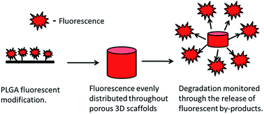
Degradable polymers such as poly(lactic-co-glycolic acid) (PLGA) are frequently chosen for tissue engineering, due to their ease of production, controllable degradation rates and Food and Drug Administration (FDA) approval. Within tissue engineering it is essential that the degradation profile of such biomaterials is understood and measured both in vitro and in vivo. The majority of techniques currently undertaken to study degradation are however destructive, leading to an over reliance on end point analysis. This study therefore defines a method of fluorescently tagging PLGA, via the addition of reactive amine groups and subsequent isothiocyanate reactions, with the purpose of monitoring degradation profiles through non-destructive techniques. The amine grafting and fluorescent labelling of the PLGA was confirmed using both X-ray photospectrometry and high performance liquid chromatography. The modification of the PLGA also had no significant effect on molecular weight or the hydrophilicity of the polymer. Both the release of fluorescent by-products and the changes in fluorescence retention within the modified PLGA were observed to be highly correlated to the changes in physical weight. This paper therefore demonstrates a novel method for the online and non-destructive monitoring of polymer degradation through the incorporation of a fluorescent marker, which can decrease the reliance on end point analysis and reduce the number of samples required both in vitro and in vivo.
Current methods utilised for the degradation profiling of biomaterials include weight loss, high performance liquid chromatography (HPLC), gel permeation chromatography (GPC) and micro-computed tomography (microCT).3–6 These techniques are however destructive end-point analyses which do not allow for real-time monitoring of degradation. Recent advances in the area have utilised non-invasive, fluorescent techniques to tag biomaterials and assess degradation through subsequent changes in fluorescence.7 These fluorescent tagging approaches have been utilised on several biomaterials to date, including chitosan,8 PEG–dextran,7 collagen,7 fibrin9 and thermoplastic polyurethane elastomers,10 with correlations being observed between changes in fluorescence intensity and weight loss during degradation, both in vitro and in vivo. Non-destructive assessment of degradation decreases the reliance on end-point analysis and allows for continued sample monitoring both in vitro and in vivo. This is especially important in vivo when adhering the policies to replace, reduce and refine (3Rs) the use of animals in medical research,11 as when combined with online techniques for tissue formation or cell migration it allows for a decrease in the number of animals required for experimentation.
Synthetic, biodegradable polymers with simple chemistry and low reactivity groups are often used in tissue engineering to provide the required support for tissue regeneration as they can be easily and reproducibly manufactured. Poly(lactic-co-glycolic acid) is one such polymer which has been Food and Drug Agency (FDA) approved12 and has been utilised in several tissue engineering applications including bone, cartilage, muscle and skin.13–16 One of the advantages of PLGA is that its degradation profile can be altered through the variation of the glycolic and lactic ratios and therefore it can be tailored to achieve maximum tissue regeneration. The relative inert reactivity of these polymers however means that there are no intrinsic molecular marks to detect or allow for the incorporation of active groups for degradation profile measurement. Hence degradation profiling of PLGA has relied heavily on destructive techniques.
In this paper we describe a novel method of fluorescently labelling PLGA using plasma treatment to produce an amine reactive polymer to which fluorescent molecule, rhodamine b isothiocyanate, was bound. It was shown that the degradation profile of the fluorescent material obtained through changes in fluorescence was correlated to the loss in physical mass. This gives rise to a novel new method of tagging inert polymers with various amine reactive fluorochromes.
To assess changes in the physical weight of the scaffold the PLGA was removed from the PBS solution, and placed on blotting paper for 30 seconds, to remove excess water from the surface, before weighing. To investigate the release of fluorescence into the PBS a 200 μL sample was taken and the fluorescence intensity (excitation: 530/emission: 590) was measured using a fluorescence plate reader (BioTek Synergy II). This was then compared to calibration curve of known concentrations of fluorescent PLGA.
Confocal laser scanning microscopy (CLSM) (Olympus, UK) was utilised to assess the fluorescence intensity retained within the PLGA throughout the degradation period. 3D visualisation of the PLGA was achieved by scanning through the scaffold at 10 μm intervals over a range of 200 μm from the base of the scaffold (z-axis) with a ×10 long working distance, air lens (Olympus, UK). The imaging parameters were maintained throughout the degradation period and changes in the average peak fluorescence values were calculated and normalized to a standard undegraded scaffold at each time point to account for internal variations within the system.
After PLGA surface modification through ammonia plasma treatment the rhodamine b isothiocyanate was shown to be taken up by the PLGA films and was retained throughout post reaction washing as shown by CLSM. It was observed that fluorescence was present on the modified surfaces of the PLGA films, with the highest peak being observed on the top surface of the film during treatment (Fig. 1a). Importantly once the modified films were recast into new films or 3D salt-leached scaffolds the fluorescence was observed to be evenly distributed throughout the PLGA (Fig. 1a), which is essential for investigating the degradation profile.
It was observed that that areas of PLGA covered during the plasma treatment were not fluorescently modified by the rhodamine b isothiocyanate (Fig. 1b), this suggests that the specificity of the reaction is limited to areas which are modified with reactive amine groups. Uptake rates, before washing (Fig. 1c), and retention rates, after washing (Fig. 1d), of rhodamine b isothiocyanate in PLGA films, however were shown to correlate to the hydrophilicity of the PLGA compositions. It was observed that the more hydrophilic films (50:
50 and 65
:
35), with a higher swelling degree, were shown to initially take up more rhodamine b isothiocyanate before washing (Fig. 1c). Therefore due to the innate swelling properties of the PLGA it was essential to ensure that the rhodamine was covalently bound to the modified PLGA and not merely entrapped during the natural material swelling. In order to fully represent degradation it is essential that the rhodamine b isothiocyanate is bound to the PLGA and that random leaching of the fluorescence out of the scaffold does not occur as this will affect the accuracy of the degradation calibration.
XPS survey scans of the plasma treated PLGA showed a new ammonia peak in the spectra (Fig. 2a) on the surface, this is confirmed when looking at the both high resolution ammonia and carbon scans (Fig. 2b and c respectively). High resolution ammonia scans showed peaks in both the plasma treated, and plasma treated, rhodamine b isothiocyanate grafted PLGA (Fig. 2b). High resolution carbon spectra showed a shift in the carbon–carbon/carbon–hydrogen (C–C/C–H) binding energy due to the formation of higher energy carbon-nitrogen (C–N) bonds as the PLGA surface was modified (Fig. 2c). The change in the surface chemistry of the PLGA films confirms the appearance of isothiocyanate reactive amine groups. Plasma treatment has been previously been used to modify PLGA surfaces in order to affect the cell–material interactions. For example oxygen and ammonia plasma treatment have both been shown to improve cell attachment and cell proliferation.18,19 The positive effect of amine modified surfaces suggests that any un-reacted amine groups left within the PLGA will not have a detrimental effect on cell attachment or proliferation and may increase cell–material interactions.
After rhodamine b isothiocyanate was reacted with the amine modified PLGA an increased peak in C–C/C–H bonds was observed due to the high number of these bonds within the rhodamine b isothiocyanate. This was confirmed through HPLC of degraded PLGA, which showed variation in the polarity of fluorescent products when compared to the rhodamine standard (Fig. 3a and b respectively). This increase in the number and the retention time of the peaks when compared to the rhodamine standard indicated the formation of highly polar fluorescent monomers and dimers of lactic/glycolic acid. However, a slight increase in the peak intensity for the C–C/C–H bonds was also observed in the non-ammonia plasma treated, rhodamine treated samples indicating that there was some non-specific uptake of the rhodamine. When investigating the XPS spectra no peak changes are observed in the hydroxyl (C–OH) or the carbonyl groups (CO) showing there was no non-specific binding of rhodamine b isothiocyanate in the absence of the ammonia plasma coating. This suggests that some of the rhodamine may remain unbound within the film during the modification of the PLGA, due to the swelling. Changes in the PLGA structure were also observed with FT-IR, where a new small peak can be observed around 2800 cm−1, which was methylamino stretch, showing the incorporation of amine groups within the structure and 1450 cm−1 where the aromatic ring stretch and methylene stretch was observed. This suggests the incorporation of both nitrogen and an increase in methylene bonds observed within the rhodamine b isothiocyanate (Fig. 4).
![]() | ||
Fig. 4 FT-IR spectra showing changes in the PLGA after labelling with rhodamine b isothiocyanate. Changes are marked with black arrows and can be observed around 3000 cm−1 and 1500 cm−1. |
It was essential to also ensure that the modification of the PLGA did not cause alterations in polymer chemistry, which may in turn affect the degradation rate. Molecular weight and hydrophobicity of the PLGA are two such factors which could have been affected by polymer modification. It was shown that the neither the molecular weight nor hydrophobicity were significantly altered through the fluorescent modification (Table 1). Both of these parameters have been shown to affect the degradation of PLGA, with decreased molecular weight and increased hydrophilicity increasing hydrolysis of the polymer.20 Therefore the fact that these parameters are not altered is essential for accurate monitoring of PLGA degradation.
PLGA composition | Contact angle | Mw | Mn | Pd |
---|---|---|---|---|
50/50 | 66 ± 3 | 94![]() |
52![]() |
1.78 |
Fluorescent 50/50 | 64 ± 2 | 89![]() |
48![]() |
1.84 |
65/35 | 70 ± 2 | 67![]() |
32![]() |
2.08 |
Fluorescent 65/35 | 68 ± 2 | 67![]() |
31![]() |
2.16 |
80/20 | 82 ± 3 | 204![]() |
118![]() |
1.72 |
Fluorescent 80/20 | 78 ± 4 | 206![]() |
118![]() |
1.75 |
After fluorescent labelling the PLGA was re-cast in order to disperse the fluorescence evenly across the depth of PLGA films (Fig. 1a) and across the transverse section of the three-dimensional scaffolds (Fig. 5) allowing for accurate determination of the degradation profile. It was however essential to determine the labelling efficiency and therefore the fluorescent signal per mg of PLGA (Table 2). It was shown that the labelling efficiency was varied dependent on the PLGA composition; however there was a high correlation between the weight and fluorescence, with Pearson's r values of 0.9842, 0.9549 and 0.9589 for 50/50, 65/35 and 80/20 PLGA composition respectively (ESI data 1†).
PLGA composition | Labelling efficiency (molar% TRITC/PLGA) |
---|---|
50/50 | 12.21 |
65/35 | 8.03 |
80/20 | 20.58 |
During degradation the fluorescence retained within the PLGA scaffolds was depleted, for all compositions, when monitored by CSLM (Fig. 5a and b). This depletion was shown to vary depending on the PLGA composition with higher glycolic acid content leading to increased fluorescence reduction when compared to the slower degrading PLGA compositions with higher lactic acid content (Fig. 5b). This change in fluorescence concurred with fluorescence release into the media, correlated to weight using calibration curves (ESI data 1†) and changes in the physical wet weight of the polymer for all three of the polymer compositions 50/50, 65/35 and 80/20 (Fig. 6a, b and c respectively). Overall, the measurements demonstrate an excellent correlation between weight loss, fluorescence retention and the release of soluble fluorescent by-products, with the all biomaterials exhibiting comparable weight losses independent of the parameters measured (Fig. 6).
This novel method for labelling inert PLGA with a fluorescent tag therefore enables accurate monitoring of the degradation of the scaffolds, in vitro. This is essential as it allows for online monitoring of biomaterial degradation of the same sample over a period of time. It has previously been reported that the degradation of fluorescent biomaterials can be monitored intra-vitally both in vitro and in vivo, decreasing the required number of samples and allowing for continual monitoring of the same specimen.7 PLGA mainly degrades through hydrolysis and therefore the degradation profiles in vitro and in vivo should correlate. However PLGA degradation has also been shown to vary with dimension and environmental conditions, such as pH and mechanical stresses and strains, which may be present in vivo that are not present in vitro and vice versa.21–23 It has also been suggested that, in vivo, enzymes may play a role in the degradation of PLGA, although there has been difficulty in identifying enzymes and their contributions.23
Degradation profiles of the various PLGA compositions were, therefore, also observed within the epiphyseal defect of the organotypic ex vivo chick femur and the percentage degradation quantified by the change in average photons emitted across the defect (Fig. 7). The degradation of the PLGA 50/50 was once again shown to be the fastest degrading polymer as assessed by changes in fluorescence. The degradation profile of the PLGA compositions was however altered from that seen in vitro (Fig. 6), with slower degradation observed within the ex vivo chick femur model (Fig. 7). These variations confirm that it is essential for biomaterial degradation to be investigated both in vitro and in vivo, in order to ensure that the profile is not altered to such an extent that will impact on tissue regeneration.
The change observed in the degradation profile from in vitro to ex vivo may also be due to the fact that the fluorescent degradation products may not be able to diffuse through the tissue and disperse. This method of fluorescent tagging therefore may also allow for the tracking of degradation by-products of the biomaterials used in tissue engineering. This is particularly important when investigating biomaterials where the accumulation of by-products may have a deleterious effect on the tissue repair. The degradation of PLGA in particular is known to produce an acidic local environment, because of the glycolic and lactic acid monomer by-products. In turn low pH environments are shown to have deleterious effects on proteins and nucleic acid.24 Therefore the ability to track fluorescent by-product diffusion and accumulation throughout tissues, may allow for better treatment design and maybe essential for the regulatory approval of these scaffold materials.
When investigating in vivo degradation it is also essential that the chosen fluorochromes or tag is able to penetrate the required tissue depth. Due to the high auto-fluorescence of both skin and fur, it is essential that the correct animal model and fluorochrome is selected. Fluorochromes that emit toward the red end of the spectra are of particular interest, as water and haemoglobin absorb less of the photons, therefore allowing for deeper tissue penetration.25 This novel method of tagging PLGA through the formation of a reactive amine surface has the advantage of being able to be used on a variety of polymers, which have no reactive groups intrinsically. There are several different fluorescent amine reactive moieties which could be used to bind a range of fluorochromes, such as fluorescein and rhodamine, these include isothiocyanate groups, used in this research, succinimidyl ester groups and dichlorotriazine groups.26 This ability to interchange between fluorochromes allows for the monitoring of multiple degradation profiles, which opens up the ability to investigate material interactions. For example polyglycolic acid and poly-L-lactic acid could be individually monitored to determine the individual degradation profiles of the two materials. This method could also be utilized for other methods of imaging, such as photoacustic imaging using other amine reactive compounds such as, monocarboxymethylene blue N-hydroxysuccinimide ester.27
Footnote |
† Electronic supplementary information (ESI) available. See DOI: 10.1039/c6ra04690h |
This journal is © The Royal Society of Chemistry 2016 |