Meng Wangab
aDepartment of Trace Examination, National Police University of China, Shenyang 110035, China. E-mail: mengwang@alum.imr.ac.cn
bKey Laboratory of Impression Evidence Examination and Identification Technology, Ministry of Public Security, China
First published on 7th April 2016
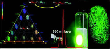
NaYF4:Yb,Er and NaYbF4:Er/Tm/Ho NIR-responsive upconversion fluorescent nanocrystals were used to develop latent fingermarks on the surfaces of various substrates. Development exhibited high developing contrast, high developing sensitivity, and high developing selectivity. In particular, one-year-old fingermarks, fingermarks on wet substrates, and fingermarks on surfaces with multicolored background and strong fluorescent properties could be clearly observed through our method.
Recently, the use of nanocrystals (NCs) in latent fingermark development, especially fluorescent NCs, has attracted much attention because of their unique physical and chemical properties, including small size, large surface area, strong fluorescent intensity, and good optical and chemical stability.3–13 Fluorescent NCs may have advantages in latent fingermark development, including enhancement of contrast, increase in sensitivity, and improvement of selectivity. The use of quantum dots (QDs) in latent fingermark development has also been of significant interest.14–21 QDs can emit strong visible fluorescence due to excitation by ultraviolet (UV) radiation, which plays a role in enhancing fingermark detection signals, resulting in enhanced developing contrast. However, the excitation of QDs and other traditional fluorescent powders usually requires UV radiation, which results in various drawbacks, such as UV-induced damage to the human skin, interference by background autofluorescence from the substrate, DNA lesions, and even DNA damage in fingermark residues caused by long-term irradiation.22–24 In addition, increasing concerns over the inherent toxicity of QDs have hindered the long-term use of QDs.25–27
Upconversion fluorescent nanocrystals (UCNCs), which can convert near-infrared (NIR) light to visible light by two/multi-photon mechanism,28–31 are an emerging class of fluorescent NCs for latent fingermark development.32–36 UCNCs can also emit strong visible fluorescence, ensuring high contrast in latent fingermark development. The excitation of UCNCs often requires NIR radiation, the absence of autofluorescence from the substrate thus prevents the background interference.32–36 Compared with UV light, NIR light has lower energy and is therefore harmless to human skin. Moreover, UCNCs have low toxicity, large Stokes shifts, narrow emission peak, as well as good physical and chemical stability.37–39 Therefore, UCNCs are promising candidates for use in latent fingermark development.
On the basis of the aforementioned concepts, we developed a novel suspension method for latent fingermark development using NaY0.78F4:Yb0.20,Er0.02 and NaYb0.98F4:RE0.02 (RE = Er, Tm, Ho) UCNCs. This work demonstrates that UCNCs can be suitable agents for latent fingermark development on a variety of substrates with high developing contrast, high developing sensitivity, and high developing selectivity. In particular, one-year-old fingermarks, as well as fingermarks on wet substrates and on surfaces with multicolored background and strong fluorescence could also be clearly developed by our method.
The general principle of this approach to the development method using NaY0.78F4:Yb0.20,Er0.02, and NaYb0.98F4:RE0.02 (RE = Er, Tm, Ho) UCNCs in this study is illustrated in Fig. 1. First, the whole fingermark is immersed in suspensions containing UCNCs and the surfactant sodium dodecyl sulfonate (SDS). During this process, the hydrophilic sulfonic groups on one end of SDS cap the surfaces of the UCNCs, and the hydrophobic alkyl chains on the other end of SDS adsorb onto the grease in fingermark residues. The adsorption involves hydrophobic interactions among the alkyl chains, thus staining fingermarks with high developing selectivity and high developing sensitivity. Then, NIR light is used as excitation light source to irradiate the fingermarks on various substrates (detailed developing procedures are described in the ESI†). The conversion of NIR to visible light provides high developing contrast in revealing fingermarks.
NaYF4:Yb,Er UCNCs with strong UC fluorescent intensity and uniform size were synthesized through a typical solvothermal approach (detailed synthetic procedures are described in the ESI†). The crystal structures and the phase purity of the as-prepared UCNCs were examined by X-ray powder diffraction (XRD). Typical XRD patterns of the UCNCs are presented in Fig. 2c. Diffraction peaks of the UCNCs are well-defined, and the peak positions and intensities agree well with the calculated values for hexagonal-phase NaYF4 (line pattern in Fig. 2c, JCPDS no. 028-1192), indicating that the as-prepared UCNCs have pure hexagonal phase with high crystallinity. The size and morphology of the as-prepared UCNCs were characterized by transmission electron microscopy (TEM), as shown in Fig. 2a. The TEM image shows that the UCNCs are spherical, well-dispersed, and uniform in size. A histogram of the particle diameter (Fig. 2b) reveals that the UCNPs are 60–85 nm in diameter, with an average value of 72.8 nm. Notably, UCNCs synthesized even under relatively harsh conditions (i.e., 175 °C for 36 h) present a regular spherical shape, which benefits their further application in latent fingermark development. The resultant UCNCs suspension exhibited bright green fluorescence under 980 nm light irradiation, as shown in Fig. 2e. Such strong fluorescence from UCNCs under NIR irradiation inspired us to use them for developing latent fingermarks. The fluorescence properties of the as-prepared UCNCs suspension were characterized by fluorescence spectroscopy (Fig. 2d). Strong green emissions at 520.5 and 540.5 nm could be assigned to 2H11/2 → 4I15/2 and 4S3/2 → 4I15/2 transitions, respectively; a weak red emission at 654.5 nm could be assigned to the 4F9/2 → 4I15/2 transition.40
The NaYF4:Yb,Er UCNCs suspension was used as a novel fluorescent label for latent fingermarks. In our UCNCs suspension, the surfactant SDS played a very important role in the conjugation between UCNCs and fingermarks. The SDS concentration and the staining time greatly affected the results of fingermark development. When no SDS was added, a poor fingermark image was observed (Fig. S1†) even when the staining time was long enough (10 min) because of nonspecific conjugation between UCNCs and fingermarks. Better fingermark images were observed when the SDS concentration was increased to 0.5% and when the staining time was extended to 1 min and even 3 min, in contrast to the hardly visible image when no SDS was used (Fig. S2†). As the SDS concentration was increased to 1.0%, clear fingermark images were observed even when the staining time was only 10 s (Fig. S3a†). This reveals fast development. With further extension of the staining time, more UCNCs adsorbed onto the fingermark surface (Fig. S3b–h†). With extension to more than 5 min, however, the UCNCs adsorbed not only on the papillary ridge of the fingermark but also on furrows, resulting in an indistinct fingermark image. When the SDS concentration was increased to 2.0%, all of the fingermark images observed were of poor quality (Fig. S4†), because part of the grease dissolved in higher concentrations of SDS. When the SDS concentration was further increased to 3.0%, more grease dissolved in SDS, resulting in an incomplete fingermark image (Fig. S5†). In consideration of the contrast and efficiency of developing, an SDS concentration of 1.0% and a staining time of 30 s were deemed as the optimal developing condition.
To determine the contrast in our fingermark development method, control experiments using different conventional powder suspensions were performed. A plastic plate with multiple background colors and fluorescence property was used as the substrate. Contrast in fingermark development refers to the contrast between the fingermarks and the substrate background. The fingermarks stained with zinc oxide powder suspension (Fig. 3a) suffered from serious interference by the background color, leading to low developing contrast. Although the fingermarks stained with ferric oxide powder suspension (Fig. 3b) showed a better result, the developing contrast still needed improvement. When green fluorescent powder suspension was used, the developed fingermarks gave strong green emissions under 254 nm UV radiation (Fig. 3c′). However, the developing contrast was still not high enough because of strong autofluorescence interference from the substrate under 254 nm UV radiation. When the fingermarks were stained with NaYF4:Yb,Er UCNCs under 980 nm NIR radiation, the developing contrast was enhanced remarkably (Fig. 3d′) because of the strong green emissions from the fingermarks and the absence of autofluorescence interference. Therefore, our UCNCs-based method could provide high contrast in latent fingermark development compared with conventional methods, especially on some substrates with multiple colors and/or strong fluorescent property.
To determine the sensitivity of our method, control experiments using different conventional powder suspensions were performed. Transparent glass was used as the substrate. Sensitivity in fingermark development refers to the visibility and clarity of the ridge details (e.g., arches, termination points) and tiny features (e.g., sweat pores). Some ridge details of fingermarks stained with zinc oxide powder suspension (Fig. 4a′) and ferric oxide powder suspension (Fig. 4b′) could be observed clearly. However, the tiny features of the fingermarks such as sweat pores could not be observed clearly, leading to a decreased developing sensitivity. When the green fluorescent powder suspension was used, the developing sensitivity was rather low because of the poor visibility, as well as low clarity of ridge details and tiny features (Fig. 4c′). Investigators have reported that powders with large size and unsuitable affinity can cover some tiny features such as sweat pores.38,39 When the fingermarks were stained with NaYF4:Yb,Er UCNCs, detailed features of the fingermarks, including sweat pores, could be observed clearly (Fig. 4d′), leading to an increased developing sensitivity. Therefore, our UCNCs-based method exhibited high sensitivity compared with conventional methods for latent fingermark development, especially for developing some tiny features such as sweat pores.
To determine the selectivity of fingermark development, control experiments using different conventional powder suspensions were performed. Transparent glass was used as the substrate. Selectivity in fingermark development refers to the specificity of developing materials (e.g., chemical reagents, fingermark powders) that adhere or react with only the ridges in fingermarks, but not with furrows on substrates. When the green fluorescent powder suspension was used, some regions that responded to furrows on substrates also bound the powders (Fig. 4c′), making them difficult to rinse and decreasing selectivity. When the fingermarks were stained with zinc oxide powder (Fig. 4a′), ferric oxide powder (Fig. 4b′), or NaYF4:Yb,Er UCNCs (Fig. 4d′), the powders adhered only to the fingermark ridges, without furrow staining, leading to improved developing selectivity. Therefore, our UCNCs-based method, could exhibit high selectivity in latent fingermark development compared with conventional methods.
To investigate the practicability of latent fingermark development on wet surfaces, various marbles with wet surfaces and complex patterns were used as the substrate. The results show that under 980 nm NIR irradiation, fingermark ridges with sharp edges could be clearly observed without background interference (Fig. S6a′–h′†). This indicates that our UCNCs-based method is suitable for developing latent fingermarks on wet surfaces.
To investigate the applicability of our method on various other substrates, experiments using UCNCs were also performed. The substrates included stainless steel sheets, aluminum alloy sheets, aluminum foil, ceramic tiles, marbles, plastic cards, painted wood, Chinese paper money, and glass. The developing method was versatile; it could be used to detect fingermarks on various substrates, as shown in Fig. 5a–g. All of our results show clear observation of well-defined ridges on various substrates without background interference. The development method could also be carried out on infiltrative substrates such as Chinese paper money. Chinese paper money emits strong background fluorescence under 254 nm UV light, which interferes with fingermark development. However, well-defined ridges on the substrate were clearly defined under 980 nm NIR light irradiation, without any background interference (Fig. 5h). Our method was also suitable for the development of old fingermarks. As shown in Fig. 5i, one-year-old latent fingermarks, which are hardly developed through traditional methods such as powder dusting and cyanoacrylate fuming, could also be developed through our method.
Development of latent fingermarks was carried out by using suspensions containing a novel fluorescent label, NaYb0.98F4:RE0.02 (RE = Er, Tm, Ho) UCNCs. NaYbF4:RE UCNCs emit multicolor light under 980 nm radiation, were used on latent fingermarks for the first time. Similarly, Fig. 6a–l shows clear fingermark images on glass substrates without background interference under 980 nm irradiation, resulting in high developing contrast, high developing sensitivity, and high developing selectivity.
In comparison with traditional powder methods, our method possesses two prominent advantages: (i) it does not involve dusting, thus making it safe; and (ii) it is a feasible, important supplement to the traditional powder methods. Our method has three main advantages over suspension methods using conventional powders such as zinc oxide powder, ferric oxide powder, and green fluorescent: (i) high developing contrast due to the strong multicolor emission and the absence of autofluorescence interference under 980 nm NIR excitation; (ii) high sensitivity due to the small particle size and the suitable affinity of UCNCs; and (iii) high selectivity due to the conjugation between the UCNCs and fingermark residues.
In conclusion, we have designed novel NaY0.78F4:Yb0.20,Er0.02 and NaYb0.98F4:RE0.02 (RE = Er, Tm, Ho) UCNCs for fingermark development on various surfaces, including metal, glass, marble with different textures, polymer surfaces, ceramic tile, wood, and special papers. In particular, one-year-old fingermarks, as well as fingermarks on wet substrates and on surfaces with multicolored and strongly fluorescent background, could also be visualized through our method. The procedure shows high performance in terms of high developing contrast, sensitivity, and selectivity. Our work introduces new research using UCNCs for developing latent fingermarks in forensics.
Footnote |
| † Electronic supplementary information (ESI) available: Experimental section and additional experimental data. See DOI: 10.1039/c6ra04573a |
| This journal is © The Royal Society of Chemistry 2016 |