DOI:
10.1039/C6RA04276G
(Paper)
RSC Adv., 2016,
6, 55257-55265
LPA1 extracellular loop residues 115 and 191 are not required for receptor activation but prevent Ki16425 super-antagonism†
Received
19th February 2016
, Accepted 18th May 2016
First published on 20th May 2016
Abstract
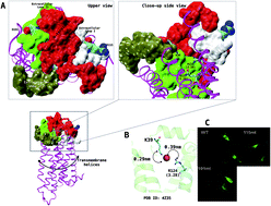
Lysophosphatidic acid receptor 1 (LPA1) is a clinically important target, which confers a biological response to lysophosphatidic acid-type agonists. Recently, the 3D structure of LPA1 resolved using X-ray crystallography and our site-directed mutagenesis has provided significant insight into the mechanism of LPA1 activation and antagonism. In the present study, we report that extracellular loop residues (R115 and D191) are not required for receptor activation but repress Ki16425-type super-antagonism but not LPA-analogue antagonists using a combination of site-directed mutagenesis and intracellular calcium assay procedures. The underlying mechanism was further queried using an all-atom molecular dynamics (MD) simulation. The result showed that Ki16425 when bound to LPA1 evolved characteristic conformational and optimal route communication signatures as a LPA (C18:1)-agonist which is reversed in R115A and D191A mutants. In conclusion, Ki16425 is a super-antagonist when bound to mutant (R115A and D191A) LPA1.
Introduction
The 3D structure of the second member of the endothelial differentiation gene (EDG) was recently determined,1 adding spectacular insight into our understanding of the structure and function of lipid receptors since the resolution of sphingosine-1 phosphate receptor 1 (S1PR1);2 the de facto first member of the family. Prior to this breakthrough, our research group investigated the involvement of lysophosphatidic acid receptor 1 (LPA1, EDG-2) in neuropathic pain3 with a view to developing LPA1 antagonists. One of the steps taken to improve our chances of successful drug design was modeling a full-length LPA1 receptor from the S1PR1 transmembrane (TM) region and extracellular loops (EL). N- and C-termini were modeled using an ab initio method.4 This approach provided a reliable starting structure for probing the involvement of the N-terminus and extracellular loops (EL) as activation partners with R3.28/Q3.29 (Zx.yy corresponds to Ballesteros–Weinstein nomenclature,5 where Z represents the amino acid residue, x represents the helix where the residue is located and yy is the position relative to the most conserved residue in that helix where the residue is located) during receptor activation; an activity signature reported in S1PR1.6
In a recent study, we observed that N-terminal lysine 39 was shown to partner with R3.28/Q3.29 during LPA1 activation7 forming a bridge-like scaffold where a carbonyl oxygen of LPA forms a hydrogen bond with lysine 39 while its phosphate head group hydrogen bonds with R3.28. The importance of this partnership is further reinforced by the recently crystallized LPA1; in these structures, co-crystallized ONO compounds or crystal water form this bridge.1 During our lysine 39/R3.28/Q3.29 study,7 being oblivious of the crystal structure of LPA1, two mutants (R115A, D191A) were also generated to further investigate the roles of charged extracellular residues in LPA1 in ligand binding, receptor activation and cell surface expression,8 drawing critical information from the vasopressin V2 receptor (V2R).9 With the benefit of the LPA1 crystal structure, it is clear that these residues are not direct participants in ligand binding, and neither is their mutation associated with gross changes in protein expression and membrane localization as established in this study. However, cells expressing R115A and D191A-LPA1 became super-sensitive to Ki16425 antagonism but not any of the lipid-based antagonists tested in this study. The underlying mechanism was also critically examined using molecular dynamics simulations and dynamical communication studies.
Materials and methods
Starting coordinates
Two wildtype LPA1 structures were built for this study; the first was modeled10 from the recently resolved X-ray structure of LPA1 co-crystallized with an antagonist (PDB ID 4Z35, inactive).1 The active conformation was built using molecular dynamics flexible fitting (MDFF,11 gscale = 0.2, simulation time = 100 ns, simulation software = NAMD12) of an inactive model to the density map of a nanobody-stabilized active state of beta2 adrenoceptor (PDB ID 3p0g) (active conformation).13 To ensure correct rotameric signatures of the amino acids during fitting, the target and input receptors were placed in POPC membrane models using the CHARMM-GUI server.14 POPC membrane coordinates were removed, followed by protonation of titratable residues at pH 7.2 using the GBSA solvation model to give the final LPA1 models (active and inactive). LPA (18:1, PubChem CID: 5311263) was docked (Autodock-vina)15 into the active conformation of LPA1 while Ki16425 (PubChem CID: 10367662) or BrP-LPA (PubChem CID: 16725999) was docked into the inactive conformation to generate the starting files. R115A and D191A mutants were generated using the mutagenesis suite in CHARMM-GUI. The schematic representation of the steps for the starting coordinate generation is presented in ESI Fig. 1A.†
Biosystems setup
Each of the oriented biosystems was re-inserted into a pre-equilibrated 1,2-dilauroyl-sn-glycero-3-phosphocholine (POPC, 68 lipids per leaflet) bilayer using the CHARMM-GUI webserver (http://www.charmm-gui.org). Ligand parameterization was performed using the ParamChem service (http://cgenff.paramchem.org) as implemented on the CHARMM-GUI webserver. The biosystems were solvated in the TIP3P explicit water model and neutralized with Na+/Cl− (0.15 M).
Molecular dynamics (MD) simulations
All molecular dynamics simulation runs were done using GROMACS (ver. 5.1) software using the CHARMM36 force field16 as we have previously reported.7 Briefly, during equilibration, the biosystems were subjected to constant pressure and temperature (NPT; 310 K, 1 bar) conditions using the Berendsen temperature and pressure coupling algorithms as implemented in GROMACS. Van der Waals interactions were estimated at 10 Å, long-range electrostatic interactions were computed using the particle mesh Ewald (PME) summation scheme while the equation of atomic motion was integrated using the leap-frog algorithm at a 2 fs time-step for a total time of 100 ns with positional restraints imposed on the heavy atoms (receptor/ligand) in all directions. To generate two starting coordinates for production-phase simulation (n = 2) for each complex, unrestrained molecular dynamics simulation was performed (20 ns) followed by 200 ns production simulations. In all the setups, lipid bilayer thickness was maintained between 3 and 4 nm throughout the simulation while the area per lipid was maintained at 80 Å. All calculations were performed on SuperMicro workstations (32-E2600 Intel Xeon CPUs, 4 Tesla K40 GPUs Accelerator PCI-E x16 Card/node).
Simulation data analysis
Dynamical networks (a set of nodes with connecting edges) for LPA1 TM helices were calculated as described7 using Carma (ver. 1.4), gncommunities and subopt scripts as implemented in VMD. Here, a pair of nodes was connected by an edge if the corresponding monomers resided within a 4.5 Å distance for at least 80% of the frames analyzed. The size of an edge corresponds to the weights of the monomers. LPA1 residues are represented using Ballesteros–Weinstein nomenclature.5
Site-directed mutagenesis experiments
PCXN2.1-LPA1, an expression vector for human LPA1, was kindly provided by Dr T. Shimizu (NCGM, Tokyo). The mutants of LPA1 (Arg115Ala, Asp191Ala) were generated by a two-step PCR. The cDNA of the 5′ portion of LPA1 was amplified by PCR using a forward primer containing a XhoI site and reverse primers containing the sequence encoding the mutated amino acids. The 3′ portion was also amplified with forward primers containing the mutated sequence and a reverse primer containing NotI and BglII sites. All cDNAs of the LPA1 variants were amplified using a mixture of the two PCR products together with the XhoI and NotI-containing primers. The resulting 1.0 KB products were cloned into a pCRII-Topo vector for sequence analysis. The cDNA of the LPA1 mutants was cut out from pCRII-Topo by XhoI and NotI, and ligated into the XhoI/NotI sites of pCAGGS-HA, to generate pCAGGS-HA-LPA1 mutants. Primer sequences for generating Arg115Ala and Asp191Ala and detailed protocols have been previously published.7
Cell culture and intracellular calcium ion (Ca2+) mobilization assay
B103 rat neuroblastoma cells that lack an LPA response were cultured in DMEM containing 10% fetal bovine serum at 37 °C in a 5% CO2 atmosphere. A transfection method for wildtype LPA1 (and mutants) into B103 cells, and the intracellular calcium ion mobilization assay protocol have been previously described.7 Briefly, B103 cells (1 × 10⁷) were transfected with plasmid encoding either wildtype or each mutant receptor using a NEPA21 Super Electroporator (Nepa Gene, Tokyo, Japan). After 24 h of transfection, the cells were harvested by centrifugation and suspended with 0.1% BSA supplied-DMEM. The cell suspension was plated in a 384-well plate with a density of 5 × 10³ cells per well. Following incubation for 18–24 h, cells were loaded with 10 μl Fluo-8 (8 μM) in 0.1% BSA supplied-DMEM containing 1 mg ml−1 amaranth. After 30 min, for agonist treatment, cells were stimulated with the LPA species (final concentration 0.001–100 μM); for antagonist treatment, cells were treated with specified antagonists (final concentration 0.001–10 μM except for Ki16425 (final concentration 0.001–100 μM)) for 30 min followed by 10 μM LPA 18:1 stimulation and immediate recording of the fluorescence using the Functional Drug Screening System/μCell (Hamamatsu Photonics K.K., Hamamatsu city, Japan). The fluorescence intensity was described as fura-2 ratio (tested value/basal value) or fold induction. Dose–response curves were plotted as mean ± S.E.M of at least two2 independent experiments using GraphPad Prism.
Results and discussion
The functional importance of ELs in GPCRs has been a kind of puzzle. More so, in most class A GPCRs, ELs appear tightly packed with the N-terminal region over and atop the transmembrane helices (TM) forming a cap-like structure.17 Curiously, each of these regions is now being studied in finer detail to delineate the individual contributions to ligand binding and receptor activation. Currently, it is now understood that the EL1 component of some class A GPCRs has an indirect role in ligand binding via interference with the space around the binding pocket.18 In peptidergic GPCRs for instance, perturbation of π–π interactions (WXFG motif) and charged residue interactions (DXXCR motif)19 within this region is associated with significant loss of receptor functions. Similarly, extensive studies of the β2 adrenergic receptor showed that disulfide-bond disrupting mutations within the EL2 component are associated with decreased ligand affinity.20 Additionally, unique secondary structures adopted by different class A GPCR types within this region are indicative of a strict requirement for conformational stability. 3D structures of sphingosine-1-phosphate receptor 1 (Edg-1)2 and lysophosphatidic acid receptor 1 (Edg-2, Fig. 1A upper plane, right)1 have further reinforced the notion that ELs partner with the N-terminus to form a compact structure over the TMs.
 |
| Fig. 1 LPA1 structure, orthosteric residues-N terminal cap and expression. (A) Ribbon representation of LPA1 showing the highly compact N-terminus (red surface), extracellular loop I (white surface), extracellular loop II (green surface) and extracellular loop III (green surface). The upper and close-up side views are shown for clarity. (B) Representation of LPA1 orthosteric site residues (green ribbon) showing the interaction between R124 (3.28) and lysine 39 bridged by a water molecule (red sphere). (C) Immunocytochemical demonstration of expressed wildtype LPA1 and the mutants (R115A and D191A) investigated. | |
Our recent work has established that to form a cap over LPA1, the N-terminus utilizes a hydrogen bond between lysine 39 and the carbonyl oxygen atom of lysophosphatidic acid.7 In its final pose, lysine 39 is now known to partner with previously reported orthosteric residues (R3.28/Q3.29, Fig. 1A upper plane, left).7 Indeed, in all of the three structures of LPA1 crystallized to date, the partnership is re-echoed; either by the ligand forming a bridge across the two or by water molecules (PDB ID 4Z35) forming an electrostatic bridge (Fig. 1B).1 In this study, to better understand the roles of EL1 and EL2, we took a cue from the previous work on the vasopressin V2 receptor (V2R)9 which depends on the net positive charge within EL1 for its biological function. Since superimposition of the V2R model on LPA1, LPA2, LPA3, and LPA5 shows that positive charged residues are conserved in similar or proximal residues in EL1 (V2R = R/R (104/106), LPA1 = R/R (115/116), LPA2 = R/R (95/98), LPA3 = K (95), and LPA5 = H/H (85/86), ESI Fig. 2A† (sequence alignment) & 2B (3D superposition)) of LPA receptors, we hypothesized that R115A may be associated with altered LPA1 expression, membrane localization, agonist binding and receptor activation. The LPA1-EL2 residue studied here (D191) is also related to D191 in V2R (V1aR (D204)) in terms of orientation and amino acid type. It is also noteworthy that a naturally occurring mutation (D191G) in human-V2R is linked with familial nephrogenic diabetes insipidus.9 The representative immunocytochemical snapshots (Fig. 1C) did not show any significant alteration in the signal, density or localization of LPA1 in B103 cells expressing the wildtype and mutant receptors. Western blot analysis in our previous report also established that R115A and D191A mutations did not result in a detectable difference in protein expression relative to the wildtype.7
Ki16425 is a super-antagonist when acting on R115A and D191A LPA1 mutants
To evaluate the biological consequence of R115A and D191A mutations, B103 cells transiently expressing the mutant and the wildtype receptors were treated with varying concentrations of different chain-length physiological agonists of LPA (C14:0, C16:0, C18:1 and C20:4). The dose–response curve showed that cells expressing the R115A LPA1 mutant treated with shorter-chain, saturated LPA agonists (C14:0 and C16:0) exhibited 10% higher efficacy (Emax 110%) in comparison with the wildtype. The D191A mutation did not result in any observable difference in the potency of the agonists in comparison with the wildtype. Shorter-chain, saturated LPA agonists (C14:0 and C16:0) did not alter the efficacy and potency parameters in the mutant receptors in comparison with the wildtype (Fig. 2Ai & ii). When B103 cells expressing the R115A-LPA1 receptors were exposed to longer-chain, unsaturated LPA agonists (C18:1 and C20:4), no observable difference was observed in comparison with the wildtype; rather the D191A mutation resulted in decreased efficacy (∼−15% vs. wildtype) of these agonists but not the potency (Fig. 2Bi & ii). Clearly, unlike the vasopressin V2 receptor (V2R),9 the R115A and D191A mutations may not significantly alter the receptor activation.
 |
| Fig. 2 Dose-dependent plots. (A, i and ii) Dose-dependent plot of LPA1 (wildtype and mutants) to saturated LPA-type agonists. (B, i and ii) Dose-dependent plot of LPA1 (wildtype and mutants) to unsaturated LPA-type agonists. (C, i–iv) Dose-dependent plot of LPA1 (wildtype and mutants) to LPA (18:1) in the presence of Ki16425 (i) and DGGP (ii), Br-LPA (iii) and 2CCPA (iv). The X-axes represent log10 of the μ-molar concentration of compounds. Plots are generated as mean of two2 or more independent assays using GraphPad Prism software. | |
To complete this study, we decided to test the effect of the mutations on the response of the receptors to antagonists; bearing in mind that LPA1 antagonists may play critical roles in the clinical treatment of diseases such as neuropathic pain,3 cancer21,22 and others.23 Firstly, we tested Ki16425; one of the most characterized LPA receptor antagonists (Ki values = LPA1 (0.34 μM), LPA2 (6.5 μM) and LPA3 (0.93 μM)) known to date which exhibits robust selectivity for the EDG-family.24 Surprisingly, both mutations equipotently converted Ki16425 into a super-antagonist, as the IC50 values of Ki16425 acting on the wildtype, R115A and D191A LPA1 were 0.11, 0.010, and 0.094 μM respectively following three independent determinations (Fig. 2Ci). To establish whether this finding is a more generalized event or specific for Ki16425, DGPP, Br-LPA and 2-CCPA (ESI Fig. 1B†) were also tested over the same concentration range with Ki16425 but in only two independent determinations. The results showed that none of the mutations have an effect on the potency of the latter antagonists (Fig. 2Cii–iv). These results further demonstrate that the mechanism of LPA1 antagonism in Ki16425 is distinct from those designed LPA analogues such as DGPP, Br-LPA and 2-CCPA, although, the detailed mechanism is not clear from the data. A few studies have hinted at the possibility that Ki16425 might represent an inverse agonist,24 or has the ability to bind different LPA1 conformations along its active and inactive conformational spectrum.25
Internal water path and TM3/TM6 network in wildtype and mutant LPA1
Next, we queried how the mutations caused a ligand-type dependent response in LPA1 using atomistic simulations. Previous studies have identified key activation signatures in GPCRs such as a continuous internal water path, and rupture of the TM3/TM6 ionic lock.7,26 In this study, two antagonists (Br-LPA and Ki16425) and a LPA agonist (C18:1) were studied for the evolution of (in)activation signatures in wildtype and mutants during a 500 ns simulation period. Our simulations show that R115A and D191A mutations did not significantly affect the continuous water tunneling through TM7 of the receptor bound to LPA-(C18:1) (Fig. 3Ai–iii). As expected,7,26 Br-LPA (antagonist) broke the water channel in both the wildtype and mutant receptors (Fig. 3Bi–iii). Ki16425-bound biosystems show a rather surprising pattern; in the wildtype LPA1, the internal water path was maintained but not in the mutant receptors (Fig. 3Ci–iii). Again, this observation lends credence to the hypothesis that Ki16425 is a representative inverse agonist of LPA1 24,25 and that the R115A/D191A mutation forces Ki16425 to behave like a classical antagonist; i.e. breaking the internal water tunnel through LPA1. When the inter-helical network was investigated, critical communication between TM3/TM6 was completely ruptured in the agonist bound wildtype and mutant receptors (Fig. 3Di–iii). In Ki16425-bound wildtype LPA1, no communication in this domain is observable. R115A and D191A LPA1 mutants show re-established communication within TM3/TM6 domains (Fig. 3Ei–iii), which indicates receptor inactivation26–28 as observed in Br-LPA bound biosystems (data not shown). From the foregoing, it seems R115A/D191A mutations preferentially reduce the energy barrier between two critical LPA1 conformations; one that exhibits high affinity for (inverse)-agonists and the other, which favours classical antagonist binding.
 |
| Fig. 3 Internal water path and TM3/TM6 salt-bridge formation in wildtype and mutant LPA1: (A, i–iii) internal water path in wildtype and mutant LPA1 in complex with LPA (18:1) agonist, (B, i and ii) internal water path in wildtype and mutant LPA1 in complex with Br-LPA (18:1) antagonist, and (C, i–iii) internal water path in wildtype and mutant LPA1 in complex with Ki16425 antagonist. Water is shown as the red surface, LPA1 is shown as the green cartoon, the phospholipid is removed for clarity, and where shown, ligands are shown as sticks. (D) TM3/TM6 interaction in LPA1-wildtype (i), R115A (ii) and D119A (iii) in complex with the agonist. (E) TM3/TM6 interaction in LPA1-wildtype (i), R115A (ii) or D119A (iii) in complex with the antagonist. | |
R115A and D191A alter optimal communication paths to TM3/TM6 intracellular hydrophobic clusters in LPA1
From the foregoing, R115A and D191A mutations may have the ability to convert Ki16425 from an inverse agonist into a classical antagonist but the basis for this conversion is rather a puzzle. Therefore, to gain further insight into the underlying mechanism(s), we traced the residues along the transmembrane helices (TM2 & TM5, purple cartoon, Fig. 4Ai) from R115 and D191 (extracellular) along the TMs (intracellular direction), and noticeably, the TMs (intracellular region) have a cluster of hydrophobic residues (M2.43,L2.48//M5.54,Y5.48) which may act as a possible reinforcement for the cluster of hydrophobic residues around the ionic lock zone (TM3 (L3.43,L3.46)/TM6 (V6.37,V6.40,L6.41), stick representation, Fig. 4Aii); dynamical changes around the TM3/TM6 ionic lock zone are closely linked with class A GPCR activation and inactivation.7,29 Relay of information from the orthosteric residues (R3.28/Q3.29, magenta stick, (Fig. 4Aiii)) towards the intracellular region along TM3 upon agonist binding is key to EDG-class GPCR activation via perturbation of the ionic lock.1,2,7,30 To investigate whether R115A and D191A mutations disrupt this communication, especially in the Ki16425 bound state, network analysis of the data was performed. In this analysis, optimal communication paths between the R115/M88(2.43) and D191/T225(5.58) pairs were computed using the last 50 ns of the trajectories. In the agonist bound biosystem (Fig. 4Bi), the optimal route for R115/M88(2.43) communication passed through TM3 along R124(3.28) as opposed to through TM2 as we had predicted before the simulation. The role of R124(3.28) in agonist binding and EDG-class GPCR activation provides a strong indication that R115 may play a subtle role in receptor activation. More interestingly, in agonist-bound LPA1, the D191/T225(5.58) pair tends to also communicate through TM6 (specifically engaging L275(6.52) and L278(6.55)) as opposed to through TM5. Whilst it is very interesting to note that the R115A and D191A optimal paths deviated in the agonist bound state in comparison to the wildtype (A115/M88 chose the TM2 path while A191/T255 chose the TM5 path (Fig. 4Bii and iii)), our experimental data however did not support this observation noting that the efficacy and potency of the receptors to the LPA-type agonist were not significantly altered by the mutations. Br-LPA bound to LPA1 (wildtype/mutant) showed a similar pattern to the agonist bound mutant LPA1 (Fig. 4Ci–iii). Ki16425 in complex with wildtype LPA1 showed a similar communication path with respect to the R115/M88(2.43) optimal route but preferentially engaged with TM5 from D191 en route to T225(5.58) (Fig. 4Di). In mutant LPA1, Ki16425 engaged with similar residues to the agonist (Fig. 4Dii and iii).
 |
| Fig. 4 Ligand-mediated communication in wildtype LPA1 is altered in R115A and D191A mutants. (A, i and ii) TM2/TM5/TM3 intracellular hydrophobic clusters (iii) relative position of the orthosteric site to the hydrophobic clusters. (B, i–iii) Communication path in wildtype and mutant LPA1 in complex with LPA (18:1) agonist. (C, i–iii) Communication path in wildtype and mutant LPA1 in complex with Br-LPA (18:1) antagonist. (D, i–iii) Communication path in wildtype and mutant LPA1 in complex with Ki16425. | |
Conclusion
LPA1 is an important clinical target, now newer insight into the 3D structure has been provided by the X-ray structures of S1PR1 and more recently LPA1. The contribution of the N-terminal region to receptor activation was also resolved by our group and later validated by the X-ray structures of LPA1. In this study, we provide further insight into the contribution of the extracellular loops focusing on R115 and D191 residues. These residues are well conserved in some GPCRs and have receptor activation roles in the vasopressin V2 receptor (V2R). Here, we report that although these residues do not affect receptor activation, they do, however, affect the receptor antagonism function of Ki16425. In the absence of clinical data, the importance of the R115A and D191A mutation to clinical response to Ki16425-type drug candidates cannot be fully established.
Acknowledgements
The authors show immense gratitude to Professor Hiroshi Ueda (Department of Molecular Pharmacology and Therapeutic Innovation, Nagasaki University, Nagasaki) for the gracious use of the Functional Drug Screening System, and for lending the hardware for GROMACS simulations for use at the Centre for Bio-Computing and Drug Development, Adekunle Ajasin University, Akungba-Akoko. The Advance Computing Centre, Nagasaki University also donated computer time for the NAMD-MDFF simulation. Dr Nagai, Jun is also appreciated for providing technical support.
References
- J. E. Chrencik, C. B. Roth, M. Terakado, H. Kurata, R. Omi and Y. Kihara, et al., Crystal Structure of Antagonist Bound Human Lysophosphatidic Acid Receptor 1, Cell, 2015, 161(7), 1633–1643 CrossRef CAS PubMed.
- M. A. Hanson, C. B. Roth, E. Jo, M. T. Griffith, F. L. Scott and G. Reinhart, et al., Crystal structure of a lipid G protein-coupled receptor, Science, 2012, 335(6070), 851–855 CrossRef CAS PubMed.
- H. Ueda, H. Matsunaga, O. I. Olaposi and J. Nagai, Lysophosphatidic acid: chemical signature of neuropathic pain, Biochim. Biophys. Acta, 2013, 1831(1), 61–73 CrossRef CAS PubMed.
- I. O. Omotuyi and H. Ueda, A Novel Unified Ab Initio and Template-Based Approach to GPCR Modeling: Case of EDG-LPA Receptors, Curr. Bioinf., 2013, 8(5), 603–610 CrossRef.
- J. A. Ballesteros and H. Weinstein, Integrated method for the construction of three dimensional models and computational probing of structure–function relations in G-protein coupled receptors, Methods Neurosci., 1995, 25, 366–428 CAS.
- C. O’Sullivan and K. K. Dev, The structure and function of the S1P1 receptor, Trends Pharmacol. Sci., 2013, 34(7), 401–412 CrossRef PubMed.
- O. I. Omotuyi, J. Nagai and H. Ueda, Lys39-Lysophosphatidate Carbonyl Oxygen Interaction Locks LPA1 N-terminal Cap to the Orthosteric Site and partners Arg124 During Receptor Activation, Sci. Rep., 2015, 5, 13343 CrossRef CAS PubMed.
- S. R. Hawtin, J. Simms, M. Conner, Z. Lawson, R. A. Parslow and J. Trim, et al., Charged extracellular residues, conserved throughout a G-protein-coupled receptor family, are required for ligand binding, receptor activation, and cell-surface expression, J. Biol. Chem., 2006, 281(50), 38478–38488 CrossRef CAS PubMed.
- M. F. Arthus, M. Lonergan, M. J. Crumley, A. K. Naumova, D. Morin and L. A. De Marco, et al., Report of 33 novel AVPR2 mutations and analysis of 117 families with X-linked nephrogenic diabetes insipidus, J. Am. Soc. Nephrol., 2000, 11(6), 1044–1054 CAS.
- M. Biasini, S. Bienert, A. Waterhouse, K. Arnold, G. Studer and T. Schmidt, et al., SWISS-MODEL: modelling protein tertiary and quaternary structure using evolutionary information, Nucleic Acids Res., 2014, 42, W252–W258 CrossRef CAS PubMed.
- R. McGreevy, A. Singharoy, Q. Li, J. Zhang, D. Xu and E. Perozo, et al., xMDFF: molecular dynamics flexible fitting of low-resolution X-ray structures, Acta Crystallogr., Sect. D: Biol. Crystallogr., 2014, 70(9), 2344–2355 CAS.
- J. C. Phillips, R. Braun, W. Wang, J. Gumbart, E. Tajkhorshid and E. Villa, et al., Scalable molecular dynamics with NAMD, J. Comput. Chem., 2005, 26(16), 1781–1802 CrossRef CAS PubMed.
- S. G. Rasmussen, H. J. Choi, J. J. Fung, E. Pardon, P. Casarosa and P. S. Chae, et al., Structure of a nanobody-stabilized active state of the beta(2) adrenoceptor, Nature, 2011, 469(7329), 175–180 CrossRef CAS PubMed.
- S. Jo, T. Kim, V. G. Iyer and W. Im, CHARMM-GUI: a web-based graphical user interface for CHARMM, J. Comput. Chem., 2008, 29(11), 1859–1865 CrossRef CAS PubMed.
- V. Y. Tanchuk, V. O. Tanin, A. I. Vovk and G. Poda, A New Scoring Function for Molecular Docking Based on AutoDock and AutoDock Vina, Curr. Drug Discovery Technol., 2015, 12(3), 170–178 CrossRef CAS.
- K. Vanommeslaeghe and A. D. MacKerell Jr, CHARMM additive and polarizable force fields for biophysics and computer-aided drug design, Biochim. Biophys. Acta, 2015, 1850(5), 861–871 CrossRef CAS PubMed.
- G. Shahane, C. Parsania, D. Sengupta and M. Joshi, Molecular insights into the dynamics of pharmacogenetically important N-terminal variants of the human beta2-adrenergic receptor, PLoS Comput. Biol., 2014, 10(12), e1004006 Search PubMed.
- S. Harterich, S. Koschatzky, J. Einsiedel and P. Gmeiner, Novel insights into GPCR-peptide interactions: mutations in extracellular loop 1, ligand backbone methylations and molecular modeling of neurotensin receptor 1, Bioorg. Med. Chem., 2008, 16(20), 9359–9368 CrossRef PubMed.
- M. C. Peeters, G. J. van Westen, Q. Li and A. P. IJzerman, Importance of the extracellular loops in G protein-coupled receptors for ligand recognition and receptor activation, Trends Pharmacol. Sci., 2011, 32(1), 35–42 CrossRef CAS PubMed.
- C. M. Fraser, Site-directed mutagenesis of beta-adrenergic receptors. Identification of conserved cysteine residues that independently affect ligand binding and receptor activation, J. Biol. Chem., 1989, 264(16), 9266–9270 CAS.
- Y. Liao, G. Mu, L. Zhang, W. Zhou, J. Zhang and H. Yu, Lysophosphatidic acid stimulates activation of focal adhesion kinase and paxillin and promotes cell motility, via LPA1-3, in human pancreatic cancer, Dig. Dis. Sci., 2013, 58(12), 3524–3533 CrossRef CAS PubMed.
- M. David, J. Ribeiro, F. Descotes, C. M. Serre, M. Barbier and M. Murone, et al., Targeting lysophosphatidic acid receptor type 1 with Debio 0719 inhibits spontaneous metastasis dissemination of breast cancer cells independently of cell proliferation and angiogenesis, Int. J. Oncol., 2012, 40(4), 1133–1141 CAS.
- Y. Kihara, H. Mizuno and J. Chun, Lysophospholipid receptors in drug discovery, Exp. Cell Res., 2015, 333(2), 171–177 CrossRef CAS PubMed.
- H. Ohta, K. Sato, N. Murata, A. Damirin, E. Malchinkhuu and J. Kon, et al., Ki16425, a subtype-selective antagonist for EDG-family lysophosphatidic acid receptors, Mol. Pharmacol., 2003, 64(4), 994–1005 CrossRef CAS PubMed.
- N. A. Moughal, C. M. Waters, W. J. Valentine, M. Connell, J. C. Richardson and G. Tigyi, et al., Protean agonism of the lysophosphatidic acid receptor-1 with Ki16425 reduces nerve growth factor-induced neurite outgrowth in pheochromocytoma 12 cells, J. Neurochem., 2006, 98(6), 1920–1929 CrossRef CAS PubMed.
- S. Yuan, S. Filipek, K. Palczewski and H. Vogel, Activation of G-protein-coupled receptors correlates with the formation of a continuous internal water pathway, Nat. Commun., 2014, 5, 4733 CrossRef CAS PubMed.
- R. Nygaard, Y. Zou, R. O. Dror, T. J. Mildorf, D. H. Arlow and A. Manglik, et al., The dynamic process of beta(2)-adrenergic receptor activation, Cell, 2013, 152(3), 532–542 CrossRef CAS PubMed.
- R. O. Dror, D. H. Arlow, P. Maragakis, T. J. Mildorf, A. C. Pan and H. Xu, et al., Activation mechanism of the beta2-adrenergic receptor, Proc. Natl. Acad. Sci. U. S. A., 2011, 108(46), 18684–18689 CrossRef CAS PubMed.
- E. Vardy and B. L. Roth, Conformational ensembles in GPCR activation, Cell, 2013, 152(3), 385–386 CrossRef CAS PubMed.
- S. Yuan, R. Wu, D. Latek, B. Trzaskowski and S. Filipek, Lipid receptor S1P(1) activation scheme concluded from microsecond all-atom molecular dynamics simulations, PLoS Comput. Biol., 2013, 9(10), e1003261 Search PubMed.
Footnote |
† Electronic supplementary information (ESI) available. See DOI: 10.1039/c6ra04276g |
|
This journal is © The Royal Society of Chemistry 2016 |
Click here to see how this site uses Cookies. View our privacy policy here.