DOI:
10.1039/C6RA03738K
(Paper)
RSC Adv., 2016,
6, 43563-43573
Photo-induced monomer/dimer kinetics in methylene blue degradation over doped and phase controlled nano-TiO2 films†
Received
10th February 2016
, Accepted 15th April 2016
First published on 18th April 2016
Abstract
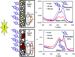
Transparent TiO2 thin films containing 1 to 6 wt% of Ni, Fe and Nb doping were synthesized via a hybrid non-aqua sol–gel dip-coating technique. Metal-doping is found to affect the phase formation, polymorphic transition, visible light absorbance and optical transparency of the TiO2 films. Physico-chemical characterization revealed that, in the given doping range, addition of Ni and Fe enhances the visible light absorbance, while Nb doping does not alter the electronic structure of TiO2. Higher doping concentrations (4 and 6 wt% of Ni and Fe, respectively) lead to the crystallization of a MTiO3 (M: Ni or Fe) phase along with anatase and rutile TiO2. On the other hand, higher Nb doping favors the formation of pure anatase TiO2. 6 wt% of Ni and Fe doping induces the reduction in band gap (3.1 and 2.9 eV), whereas Nb does not alter the TiO2 band gap (3.3 eV). In contrast to the low efficiency of undoped TiO2 films, Ni-doped films yields 71% (for monomer) and 55% (for dimer) photocatalytic degradation efficiency towards methylene blue (MB) under visible light irradiation (λ ∼ 420 nm). This demonstrates the selective and preferential photo-dynamics of the MB monomer during the photocatalytic process. The doped films show a significant Incident photon to current conversion efficiency for oxidation of water under wavelength λ ≥ 400 nm. Such superior improvement in the visible light activity of Ni doped TiO2 is attributed to the presence of nanosized NiTiO3 in the nanostructure of the TiO2 film.
1. Introduction
Anatase, rutile and brookite phases of titania (TiO2) are known for their self-cleaning properties based on the photocatalytic principle under ultraviolet radiation (UV, λ < 380 nm). They have high reactivity and chemical stability at various pH levels under UV light.1 However, in recent years, development of visible light active photocatalyst (λ > 400 nm) has received major attention in view of their potential applications for indoor self-cleaning and self-sterilizing surfaces such as glass and ceramic tiles.2 In order to achieve such visible light active surfaces, several new materials,3–5 as well as modifications of existing materials,6–15 are being explored. Such modifications includes transition metal ion doping,6–9 non-metal doping,10–13 sensitization14 and nanocomposites,15 Each type of doping has its relevant mechanism(s) that yields improved opto-electronic properties.
There are a number of reports describing dopant-dependent photocatalytic performance improvements, especially the self-cleaning activity of TiO2. Bahadur et al.6 have shown that Ni and Ag-doped TiO2 displays insignificant visible light activity. In yet another comparative study of various dopants, viz. W, V, Ce, Zr, Fe and Cu-doped TiO2, the systems showed lower activity than pure TiO2.9 Such observations were correlated to inadequate dopant concentrations that did not induce sufficient defect state densities in the band gap of TiO2. Jeong et al.16 on the other hand, demonstrated higher hydrogen evolution for Cr and Fe-doped TiO2 nanoparticles that were facilitated by efficient visible light photocatalytic water splitting. Similarly Li et al.17 reported an improved visible light activity for Fe-doped TiO2. In general, Fe-doped TiO2 has become a commonly accepted dopant to enhance visible light activity.18–20 Recent reports have revealed that Ni-doped TiO2 shows reasonably good visible light activity21,22 while dopants like Nb have been rarely considered for visible light activities in TiO2.19,20 In spite of several reports on transition metal-doped TiO2, there is a lack of systematic investigation with respect to the ionic radii and “d” orbital configuration of the dopant metal-ion. It is interesting to note that ions of Fe3+, Ni2+ and Nb5+ exhibit very similar ionic radii (0.64, 0.69 and 0.69 Å, respectively) to that of Ti4+ (0.68 Å) and this does not affect the TiO2 host lattice. In addition, these dopants show special roles of d-orbitals in the electronic structure of TiO2. Consequently Nb (4d4), Fe (3d6), and Ni (3d8) orbitals contribute dominantly towards the valence band of doped TiO2. However, the criterion for the selection of an effective dopant for visible light induced photocatalytic reaction has remained inconclusive with respect to the non-standard approach of the self-cleaning performance studies.
In the present work, we study the effect of Ni, Fe and Nb-doping in the TiO2 lattice with respect to (i) phase formation behaviour, and (ii) photocatalytic self cleaning properties using methylene blue (MB) stain in the solid state, as reported in our earlier work,23,24 as well as found in other related studies.25–28 In-line with our earlier work, the aggregation behaviour of the MB molecule yields inconclusive results. Thus, in this present study, we utilized our standard method to estimate the degradation efficiency of the doped TiO2 thin film. Furthermore, an important salient feature of this work is the possible effect of light scattering related to the grain size on the visible light activity that occurs due to the existence of a nanosized NiTiO3/or FeTiO3 phase.
2. Experimental procedure
2.1 Preparation of pure and doped TiO2 thin films
<STOP>Titanium(IV) isopropoxide (TTIP), ethanol and diethanolamine were procured from Sigma-Aldrich, India, and used without any further purification. A transparent precursor solution for titanium oxide was prepared by mixing the desired amount of TTIP in ethanol in the presence of diethanolamine (1
:
5
:
0.3 volume ratio of TTIP
:
ethanol
:
diethanolamine). The solution was stirred for 2 h at room temperature to enhance the reaction rate between diethanolamine and TTIP followed by ageing at room temperature for 24 h. Subsequently, Ni, Fe and Nb dopants (1 to 8 wt%) were introduced by mixing nickel nitrate, iron nitrate and niobium chloride, respectively, into the as-prepared and aged TiO2 sol. One part of the sol was used for titania powder preparation while the rest was used for the coating process. The powder samples were prepared by firing the same precursor solution that was used for the coating process at 600 °C for 30 min on a quartz plate.
Deposition of films: single side coated films of around 70 nm (ref. 24) thickness were deposited on several fused silica (FS) slides (75 × 25 × 1 mm) by a single-dip coating process at a constant withdrawal speed of 1 mm s−1. The FS slides were pretreated using dilute nitric acid solution, ultrasonically cleaned in distilled water and baked at 90 °C in a vacuum oven for 24 h prior to dip-coating. The single side coated films were prepared by the method explained by Sreemany et al.24,29 in which immediately after withdrawal from the solution, one side of the coated substrate was wiped off carefully using ethanol. The coatings were allowed to dry at room temperature for 60 min and fired at 600 °C for 10 to 60 min in a preheated furnace.
2.2 Structural and elemental characterization
The crystallization and phase transformation behaviour of titania with respect to the wt% of Ni, Fe and Nb was investigated by X-ray diffraction (Bruker AXS D8 Advance diffractometer) using Cu-Kα radiation. Powder samples were used for structural characterization. The data was collected in the step-scan mode with a step size of 0.01° and a dwelling time of 4 s per step using a highly sensitive LynxEye detector. The diffraction peaks of the pattern were indexed using ICDD PDF-4 database while quantitative estimates were obtained by the “Reference Intensity Ratio (RIR)” technique. The RIR method30 takes into account the intensities of all the peaks of the individual phases in the experimental profile as identified after background subtraction and Kα2 stripping, and is known to be an accurate method for quantitative analysis of a multiple phase mixture. The average crystallite size was estimated from the most intense diffraction peak of the identified phase using the Scherrer formula.31 Instrumental broadening at the appropriate Bragg angle was subtracted to obtain the exact full width at half maxima (FWHM) values.
The film thickness and surface morphology were studied on fractured surfaces of films using a high resolution scanning electron microscope (HR-SEM, Hitachi S-4300 SE/N SCM) equipped with energy dispersive spectroscopy (EDS).
The surface chemistry and oxidation states of the doped TiO2 thin films were analyzed using X-ray photoelectron spectroscopy (XPS; OMICRON instrument) equipped with aluminum anodes using Al-Kα radiation. The peak positions were calibrated with respect to the internal standard of the C 1s peak at 284.6 eV. In order to understand the various surface oxidation states of the dopant in TiO2 thin films, XPS analysis was done on 2 and 4 wt% metal-ion doped thin films. The XPS data was analyzed after normalizing the spectrum with reference to the C 1s peak resulting from the adventitious hydrocarbon present on the sample surface.
2.3 Optical properties
Optical absorbance studies were carried out using a double beam spectrophotometer (Perkin Elmer Lambda 650) at normal incidence in the 200–800 nm spectral range using air as a reference. The absorption coefficient (α) was determined from the region of strong absorption of the transmittance data by the method proposed by Manifacier et al.32 The fundamental absorption, which corresponds to electron excitation from the valance band to the conduction band, was used to determine the nature and value of the optical band gap. The optical energy band gap was calculated by extrapolating the linear portion of the (αhν)n vs. hν plot, where n depends on the type of transition (n = 2 for indirect allowed transitions).
2.4 Visible light active photocatalytic self-cleaning stain removal studies
The photocatalytic degradation of adsorbed methylene blue on the TiO2 thin film was studied by the method proposed in our earlier work.23 MB of more than 98% purity was used for preparing standard stock solutions of 0.01 mM concentration with water that was purified using a Millipore milli-Q lab purification system. The MB adsorption on TiO2 thin films was achieved with the stock solutions for 30 min adsorption time in the dark. The films were rinsed with pure water to remove any traces of un-adsorbed MB followed by air drying. The MB-adsorbed TiO2 thin films were subjected to simulated solar irradiation using an Oriel 300 W solar simulator with an air mass filter AM1.0 at a solar irradiation of 128.3 mW cm−2. The decomposition of MB adsorbed on the TiO2 thin film was calculated based on the reduction in the absorption of MB peak λmax at 666 nm (monomer) and λmax at 612 nm (dimer) using a Perkin Elmer Lambda 650 UV-visible spectrometer.
2.5 Photo-electrochemical studies on thin film
The photoelectrochemical (PEC) measurements were carried out using an electrochemical workstation (PARSTAT 2272). For the UV-vis light source, a xenon arc lamp (300 W Oriel) coupled with a monochromator (New port Oriel 74125) was used. The PEC measurements involved current–voltage (I–V) characteristics of the PEC cell consisting of three electrodes: the working electrode (semiconductor thin film), counter electrode (platinum electrode) and a reference electrode (Ag/AgCl, electrode, BASI, MF-2052) immersed in NaOH electrolyte maintained at pH 13. Samples of un-doped and doped TiO2 were used as the photoelectrode in the PEC cell and the I–V characteristics under darkness and illumination was recorded using a three-electrode system in a quartz cell. The capacitance at the semiconductor-electrolyte junction at an AC signal frequency of 1 kHz was measured using the same electrochemical workstation by varying the electrode potentials. The required electrodes were prepared using electrically conducting fluorine-doped tin oxide (FTO) glass.
3. Results
3.1 UV and visible light active self-cleaning studies on doped TiO2 thin films
UV and visible light active self-cleaning stain removal studies were done on the MB stained TiO2 surface. It has been observed from our previous studies24 that TiO2 films deposited under the optimized conditions, 0.3 vol% of diethanolamine (DEA) and 30 min of firing time at 600 °C, shows optimized photocatalytic degradation activity towards the monomer and dimer forms of MB when illuminated with simulated solar radiation. This was used as a reference for comparing the degradation efficiency of doped TiO2 in this study. In order to understand the effect of dopant on the UV activity, 1–6 wt% of Ni, Fe or Nb were doped in TiO2 thin films. These were subsequently utilized in the photocatalytic degradation studies. The activity plots for various wt% of dopants are given in the ESI (Fig. S1 to S3†). It can be easily deduced that under simulated solar radiation, 4 wt% of Ni doped film shows an efficient and rapid degradation of the monomer as well as the dimer of MB. This clearly demonstrates that there is preferential photo-kinetics of monomer during the photocatalytic process. A similar behavior was observed for Fe and Nb-doped films when compared to un-doped TiO2 film. Fig. 1a shows the UV-vis absorbance spectrum that indicates a change in the concentration of the bonded monomer and dimer of MB over TiO2 under 420 nm photons. It clearly demonstrates that under 420 nm photons, TiO2 is not capable of degrading MB. It is important to note that Ni, Fe and Nb-doped TiO2 films capably degrade the MB molecules (Fig. 1b) in a dopant concentration dependant manner (see Fig. S4 to S6†).
 |
| | Fig. 1 Change in concentration due to visible light (420 nm) photocatalytic degradation of bounded MB on (a) un-doped TiO2, (b) 4 wt% of Ni-doped TiO2 thin films. | |
The abovementioned discussion shows that 4 wt% Ni is the most efficient dopant to degrade MB under visible light (420 nm) photons. An in-depth analysis with respect to monomer and dimer degradation under visible light is shown in Fig. 2 for Ni (for other systems, see S7 to S9†). Fig. 2a shows how, with respect to illumination time at 420 nm, the monomers decrease rapidly within 20 min (>30%) and upto 70% within 120 min. In contrast, the dimer starts degrading at a slower rate only after 40 min, and shows 60% degradation after 180 min. Interestingly, TiO2 shows negligible degradation for monomer and dimer. The monomer degradation efficiency for the others can be ranked in order of increasing efficiency as Fe < Nb < Ni. Needless to say that Fe and Nb show no degradation efficiency for dimer MB (Fig. 2b). Extra details are also presented in Fig. S7 to S9† for the sake of completeness and clarity.
 |
| | Fig. 2 MB degradation efficiency (eff) on un-doped and 4 wt% doped TiO2 thin films. (a) Monomer and (b) dimmer. | |
3.2 Structural chemical properties of pure and doped TiO2 thin films
In order to understand the effect of dopant on the phase stability and crystallite size, doped TiO2 powders fired at 600 °C for 10 min were investigated by X-ray diffraction (XRD). The XRD profiles of the samples are shown in Fig. 3a–c for Ni, Fe and Nb-doped films. The analyzed results are shown in Table 1. It is evident from Table 1 that there is no change with addition up to 2 wt% for Ni and Fe. Further increases in the wt% of Ni and Fe shows reduction in the anatase fraction and an increase in the rutile fraction apart from NiTiO3 and FeTiO3 phase formation. (JCPDS no. for, NiTiO3: 00-017-0617, FeTiO3: 01-075-1211). No significant change in the crystallite size of anatase and rutile is observed with Ni addition. The crystallite size of the NiTiO3 phase is also comparable with the anatase and rutile crystallites. Whereas relatively larger crystallites are evident in the case of the FeTiO3 phase. Addition of Nb, on the other hand, strongly stabilizes the anatase phase and also reduces its crystallite size. Table 1 also presents the fraction of anatase/rutile and their crystallite sizes as estimated by XRD analyses of pure TiO2 powder synthesized using 1
:
5
:
0.3 vol% of titanium tetraisopropoxide, ethanol and diethanolamine. It may be noted that the role of diethanolamine was discussed in detail in our previous publication.24 Fig. 4a–d shows the SEM images of the respective film cross-sections of the TiO2 and doped TiO2. The SEM images reveal that the coating thickness is 70–80 nm.
 |
| | Fig. 3 XRD pattern of 0 to 8 wt% of dopants. (a) Ni-doped TiO2, (b) Fe-doped TiO2, (c) Nb-doped TiO2. (JCPDS no. for, NiTiO3: 00-017-0617, FeTiO3: 01-075-1211). | |
Table 1 XRD analysis of doped TiO2 powders after 10 min of firing time at 600 °C
| Dopant wt% |
Quantitative data vol% |
Crystallite size in nm |
| Anatase |
Rutile |
MTiO3a |
Anatase |
Rutile |
MTiO3a |
| MTiO3 (M = 1Ni and 2Fe). |
| Un-doped |
87 |
13 |
— |
27 |
34 |
— |
| 1 wt% of Ni |
87 |
13 |
— |
20 |
18 |
— |
| 2 wt% of Ni |
83 |
17 |
— |
20 |
18 |
— |
| 4 wt% of Ni |
53 |
34 |
114 |
18 |
17 |
117 |
| 6 wt% of Ni |
45 |
38 |
116 |
20 |
17 |
117 |
| 8 wt% of Ni |
42 |
38 |
119 |
20 |
16 |
116 |
| 1 wt% of Fe |
87 |
12 |
— |
16 |
28 |
— |
| 2 wt% of Fe |
88 |
21 |
— |
13 |
26 |
— |
| 4 wt% of Fe |
79 |
14 |
— |
13 |
37 |
— |
| 6 wt% of Fe |
83 |
31 |
25 |
11 |
29 |
256 |
| 8 wt% of Fe |
54 |
5 |
215 |
11 |
36 |
262 |
| 1 wt% of Fe |
95 |
— |
— |
14 |
16 |
— |
| 2 wt% of Nb |
100 |
— |
— |
11 |
— |
— |
| 4 wt% of Nb |
100 |
— |
— |
9 |
— |
— |
| 6 wt% of Nb |
100 |
— |
— |
8 |
— |
— |
| 8 wt% of Nb |
100 |
|
— |
8 |
— |
— |
 |
| | Fig. 4 HR-SEM image of (a) un-doped TiO2 thin film, (b) 4 wt% Ni-doped, (c) 4 wt% Fe-doped, and (d) 4 wt% Nb-doped TiO2 thin films. | |
3.3 Film surface state analysis: XPS
3.3.1 Un-doped TiO2 film. The oxidation state of the dopants in the TiO2 lattice was studied using X-ray photo electron spectroscopy (XPS). Fig. 5a and b shows the XPS regional spectrum for O 1s and Ti 2p levels. The Ti 2p peaks were observed at binding energies of about 457.68 eV (Ti 2p3/2) and 463.368 eV (Ti 2p1/2) (Fig. 5a). Deconvolution of the O 1s region (Fig. 5b) shows two contributions. The variation in binding energy values of Ti 2p and O 1s upon Ni, Fe and Nb addition are shown in Table 2. The spectra for 2 and 4 wt% of Ni, Fe and Nb-doped TiO2 thin films are shown in the ESI (Fig. S10†).
 |
| | Fig. 5 High resolution XPS core level spectra of TiO2 thin film on fused silica (a) Ti 2p and (b) O 1s. | |
Table 2 XPS analysis of doped TiO2 thin films
| |
Ti peak position (eV) |
O 1s peak position (eV) |
| |
Ti 2p3/2 |
Ti 2p1/2 |
| Undoped TiO2 |
|
457.68 |
463.37 |
528.9, 531.2 |
| Ni–TiO2 |
2 wt% |
457.94 |
463.65 |
529.17, 531.16 |
| 4 wt% |
457.84 |
463.53 |
529.0, 529.53, 530.85 |
| Fe–TiO2 |
2 wt% |
457.82 |
463.52 |
528.78, 530.76, 529.34 |
| 4 wt% |
457.84 |
463.54 |
529.1, 530.71 |
| Nb–TiO2 |
2 wt% |
458.15 |
463.85 |
529.64, 532.26, 530.81 |
| 4 wt% |
458.06 |
463.79 |
529.3, 530.64 |
The Ti 2p peaks were observed at binding energies of around 457.68 eV (Ti 2p3/2) and 463.368 eV (Ti 2p1/2) (Fig. 5a). Deconvolution of the O 1s region revealed the contribution from two peaks at 528.92 and 531.18 eV (Fig. 5b). Based on previously reported values,33,34 these peaks may be attributed to Ti–O and Ti–OH, respectively. Similarly, the XPS peak position for 2 and 4 wt% of Ni, Fe and Nb-doped TiO2 thin films are shown in Table 2. The XPS peak position shows changes in the Ti and O peak position with respect to the dopants. As evident from Table 2, there is an increase in the binding energy from 0.16 to 0.38 eV for the Ti 2p3/2, which confirms Ni substitution in the Ti lattice. Thus, an increase in the binding energy is expected as Ni has higher electronegativity when compared to Ti.
3.3.2 Ni-doped TiO2 film. The XPS spectrum of Ni in TiO2 (Fig. S10a†) shows peaks at 872.581 eV and 854.885 eV that correspond to NiO. This also confirms that nickel is in the Ni2+ state. Other than the Ni main peak, two satellite peaks at 860.978 eV and 879.232 eV are also observed. The peak at 860.978 eV also corresponds to Ni 2p3/2, as mentioned by Oswald et al.35 The satellite peak at 879.23 eV, on the other hand, may be due to the formation of a new phase such as NiTiO3 at 4 wt% Ni. The binding energy component of the Ni (2p3/2 and 2p1/2) is in good agreement with previous work.36–38 It may be noted that the NiTiO3 phase can form even at 2 wt%. However, it may be in trace amounts and thus difficult to be detected by XRD. Furthermore, no significant XRD peak shift for the anatase phase could be observed with Ni addition, possibly due to the very close ionic radii of Ti and Ni. (rTi4+ = 0.68 Å, rNi2+ = 0.69 Å). These observations suggests that the incorporation of Ni in TiO2 is substitutional and possible defect formation is given by eqn (1).| |
 | (1) |
Considering the XRD and XPS data, addition of surplus nickel above 4 wt% confirms the formation TiO2–NiTiO3 nanocomposites.
3.3.3 Fe-doped TiO2 film. XPS analysis of Fe-doped TiO2 shows the binding energy peak positions at 710.84 eV for Fe 2p3/2 and 724.13 eV for Fe 2p1/2 (Fig. S10b†). These can be assigned to the tetravalent state of iron.39–42 The peak at 715.65 eV and the satellite peaks between 714 and 720 eV may be attributed to Fe3O4 as reported earlier by Fujii et al.42 The broad intensity at 716 eV can be mainly assigned to the octahedral Fe2+ structure. In our experiment, the 4 wt% Fe-doped sample showed the satellite peak at 715.66 eV, while the peak is at 718.35 eV in the 2 wt% Fe (Fig. S10b†). Thus, it appears that with the increase in the Fe wt%, the Fe2+ state increases for fixed film synthesis parameters. However, XRD analysis shows no trace of Fe2O3 or Fe3O4 peaks even at 6 wt% of Fe. As in the Ni-doped samples, a new phase viz. FeTiO3 was observed in the Fe-doped samples (Fig. 3b). This indicates that Fe in TiO2 may substitutionally diffuse and show existence of both Fe2+ and Fe3+. This explains the possible defect formation mechanism, as shown in eqn (2) and (3).| |
 | (2) |
| |
 | (3) |
3.3.4 Nb-doped TiO2 film. In the Nb-doped TiO2 thin films, the binding energies of Nb 3d5/2 and Nb 3d3/2 were 206.54 and 209.31 eV, respectively, which are in good agreement with standard XPS data.43,44 The XPS results of these films indicate that Nb exists mainly as Nb5+ and there is no new phase formation unlike in Ni and Fe-doped TiO2. The possible equation for the aliovalent substitution is given below.| |
 | (4) |
The eqn (1)–(4) indicate that the oxygen vacancies are responsible for the extra rutile in Fig. 3a, which was also reported earlier by Riyas et al.45 These observations are in good agreement with previously reported results.46–48 Similar to Ni doping in TiO2, Fe doping also creates oxygen vacancies, which in turn supports rutile phase formation in the case of low doping concentrations. For higher doping concentrations, formation of additional phases as NiTiO3 or FeTiO3 is expected along with the host lattice of TiO2. In the case of 1 wt% Nb doping, rutile formation is suppressed whereas reduction in the TiO2 crystallite size is observed for higher Nb concentrations. Such an observation may be attributed to the formation of titanium vacancies in the presence of Nb, which is unlike the oxygen vacancies formed with Ni and Fe addition.
3.4 Optical properties of pure and doped TiO2 thin films
The variation in optical absorbance was studied using UV-visible absorption spectroscopy. Fig. 6a–c shows the optical absorption spectra for different dopants with respect to their concentration variation (1–8 wt%). Result of Tauc analysis from the respective spectra are shown in the inset of the figures. Accordingly, the estimated band gaps are shown in Table 3. The un-doped TiO2 band gap value was found to be 3.3 eV. In all the cases, 1 wt% of dopant shows undetectable changes in the band gap value. With increasing dopant concentrations of Ni and Fe, there are measurable band gap reductions and no change for Nb doping. In depth analysis clearly indicates that in comparison to TiO2, Ni and Fe-doped films show enhanced absorbance in the visible light region (350 to 600 nm) as shown in Fig. S11.† This shows that, specifically in the wavelength range 350–600 nm, TiO2 shows a notable hump at 420 nm, which shows a red shift with respect to Ni and Fe-doped films (λmax = 500 nm). This can be attributed to the existence of NiTiO3 and FeTiO3 phases that are in-line with the XRD results shown in Fig. 4 and Table 1. Unexpectedly, there is no interesting change for the Nb-doped film.
 |
| | Fig. 6 UV-visible absorbance spectra of 0 to 8 wt% doped TiO2 thin film (insets shows the linear portion of the Tauc relation): (a) Ni, (b) Fe, and (c) Nb-doped films. | |
Table 3 Optical energy band gap variation and dye degradation efficiency with respect to various wt% of dopant
| Wt% of dopent |
Effect of dopant |
| In energy band gap (eV) |
In dye degradation efficiency in 180 min (%) |
| Ni |
Fe |
Nb |
Ni |
Fe |
Nb |
| Mo |
Di |
Mo |
Di |
Mo |
Di |
| Un-doped |
3.3 |
3.3 |
3.3 |
2.9 |
0 |
2.9 |
0 |
2.9 |
0 |
| 1 |
3.3 |
3.3 |
3.3 |
64.7 |
34.9 |
46.1 |
22.1 |
23 |
22 |
| 2 |
3.2 |
3.2 |
3.3 |
58.0 |
34.2 |
40 |
0 |
63 |
0 |
| 4 |
3.2 |
3.0 |
3.3 |
71.0 |
54.6 |
33 |
0 |
54 |
0 |
| 6 |
3.1 |
2.9 |
3.3 |
60.8 |
18.7 |
32.2 |
3.2 |
72.3 |
13.3 |
| 8 |
— |
2.9 |
— |
— |
— |
— |
— |
|
|
4. Discussion
4.1 Visible light (420 nm) photocatalytic activity of doped TiO2 films
The abovementioned results lead to important findings with respect to monomer and dimer degradation over doped TiO2 films. It is thus important to discuss this observation in detail. Fig. 1 and 2 shows the change in the concentration of bonded MB on un-doped and Ni-doped TiO2 thin films, respectively. Fig. 1a shows that there is no reduction in MB concentration over un-doped TiO2; however, there is a little reduction in the monomer concentration during attainment of the monomer–dimer equilibrium between the adsorbate–substrate (MB–TiO2) interaction. In contrast, Fig. 1b shows a tremendous reduction in monomer/dimer concentration in the case of Ni-doped TiO2 films. Fig. 2a and b shows the visible light photocatalytic degradation activity plot for monomer and dimer with various dopants. It is clear from the figure that 4 wt% of Ni-doped TiO2 shows 70% and 60% reduction of monomer and dimer, respectively. It can be noted that for lower Ni concentrations (1 & 2 wt%) there is only 40% monomer MB and 30% dimer MB degradation, as seen in Fig. S7.† Similarly, for Ni concentration of 6 wt%, there is a reduction in the degradation activity. Fig. 2a clearly indicates that, as compared to the Ni-doped film, Fe and Nb-doped films shows lower degradation activity towards monomer MB. Further Fig. 2b shows that Ni-doped TiO2 displays significant degradation activity, whereas other films (Fe/Nb-doped) exhibit no activity. Surprisingly, within the first 20 min of illumination, exclusive monomer degradation is seen whereas no degradation is observed for the dimer MB. This can be attributed to the occurrence of a virtual equilibrium phase of dimer formation in the presence of photons. It can be noticed that Fe and Nb-doped films show a similar behavior towards monomer and dimer MB degradation (Fig. S8 and S9†), except that their activity is too low as compared to Ni-doped films. In contrast to the present observation, earlier reports20 considered the degradation of MB monomer species alone; accordingly, their study led to a higher estimate of MB degradation efficiency for the Nb–TiO2 system as compared to the other (Ni, Fe) doped TiO2 systems. Thus, the present study clearly demonstrates that it is important to consider mainly the concentration of monomer and dimer species of the model pollutant during calculation of degradation efficiency. The percentage of degradation efficiency of monomer and dimer MB are summarized in the Table 3.
As an outdoor application of the present study demands a better understanding of the response of doped films under solar light, accordingly, we investigated the variation in the photocatalytic degradation activity of monomer and dimer forms of the MB under simulated solar irradiation on un-doped, as well as 1 and 2 wt% of Ni-doped TiO2 films. Higher amounts of Ni dopant led to a drop in the activity (Fig. S1†). Similarly, for the Fe-doped TiO2 up to 4 wt%, there is no detectable variation in the degradation kinetics while the activity comes down with further increases in the Fe content (Fig. S2†). In the case of Nb-doped TiO2, there is no significant variation in the monomer decomposition activity (Fig. S3†). However, the dimer degradation activity declines with an increase in Nb content. It may be noted that for all these three dopants, the UV photocatalytic degradation of monomer and dimer up to 2 wt% shows no change. If we compare these results, it appears that such a difference in the behavior may be attributed to the formation of new phases such as NiTiO3 for Ni doping and FeTiO3 for Fe doping (see Fig. 3a and b) and may act as recombination centers. This is in good agreement with most of the reported results on enhanced UV photocatalytic degradation.49–51 It may be noted that in the case of visible light photocatalytic degradation, 4 wt% of Ni shows better degradation kinetics when compared to the others. To understand the results of visible light activity of Ni-doped TiO2, variations in the phase fraction, grain size, surface morphology and cross-section analysis were evaluated by SEM, surface chemical states of dopant, variation in optical energy band gap and visible light absorbance.
High resolution SEM (image not presented in this paper) revealed that there is no difference in the surface morphology of the doped and un-doped TiO2 surface. However, there exists a small variation in the film thickness from 70 to 80 nm for 4 wt% of dopant (Fig. 4a–d). This may be caused by chemically bound water present in the Ni precursor (Ni(NO3)2·6H2O), which also enhances the dehydration rate due to hydrolysis. It was earlier reported that un-controlled hydrolysis yields to a relatively thicker coating.24 The presence of the hydroxyl group on the coating may enhance the UV activity.52,53 This is in very good agreement with 2 wt% of Ni-doped TiO2, which initially shows faster degradation kinetics under simulated solar irradiation (Fig. S1†).
In present study, the substitution of metal ions (Ni2+, Fe3+ and Nb5+) were achieved by a non-aqua sol–gel method. The incorporation of the metal ion and validation of the oxidation state of the respective dopants was confirmed by XPS analysis (Table 2). As discussed earlier, 4 wt% of Ni-doped TiO2 thin films showed better visible light activity against monomer and dimer forms of MB, though the band gap of 4 wt% of Fe is less (3.0 eV) than that for 4 wt% of Ni (3.2 eV). However, simulated solar irradiation activity shows no significant variation for the initial degradation kinetics. The results indicate that there is an energy band gap reduction in the Ni and Fe-doped TiO2 thin films possibly due to the population of different surface states that absorb the visible light as reported by Kisch et al.54 The oxygen vacancies, probably produced during Ni and Fe-doped TiO2 film deposition (ref. eqn (1)–(3)), are expected to play a role in rendering visible light activity. It is known that formation of oxygen vacancies can be depicted by the defect chemistry eqn (1)–(3).
In the case of Nb doping, there is no change in the band gap, but surprisingly, the grain size estimation indicates that nanosized grains are retained. This observation of un-altered band gap and retention of nano-grain dimension can be attributed to the role of Nb that arrests the change in the anatase grain size in Nb-doped films as found by XRD studies (shown in Table 1). It important to note that Lin et al.55 reported an increase in the energy band gap mainly due to the reduction in the anatase grain size.
4.2 Effect of doping on visible light activity
In order to understand the origin of the visible light photo-degradation property of doped TiO2 films, it is important to understand their optical properties. Fig. 6a–c shows the optical absorption of Ni, Fe and Nb-doped TiO2 films. In-depth analyses of the results using the Tauc formalism is shown in the inset of the respective figures. Accordingly, estimation of the optical band gap is presented and compared in Table 3. Irrespective of the dopant, doping is known to affect (1) the band gap and (2) increase the overall film absorption with respect to dopant concentration. It is clearly seen from Table 3 that the band gap of the film 4 wt% Ni-doped TiO2 is reduced to 3.2 eV, and to 3.0 eV for the Fe-doped film, but does not alter the band gap for the Nb-doped film. Secondly, the variation in the film absorption, with change in the dopant concentration, can be understood in the following manner based on the analysis of the films' optical behavior in the visible light wavelength region of 350–600 nm (see Fig. S11†). One can note that the λmax at 420 nm shifts (Fig. 6) to 500 nm in the case of Ni-doped TiO2 (for magnified view, see Fig. S11†), whereas it shifts to 450 nm in Fe-doped film and displays no shift in the case of the Nb-doped film. Consequently, the reduction in the film absorbance is possibly caused by nano-grains that lead to a dominant scattering rather than absorption of light. This can be correlated to the analogouss Tyndall scattering, where the particles smaller than the wavelength of incident light affect transmitted intensity to a negligible extent. This is clearly depicted in Fig. 7 that shows that in the case of the Ni-doped film (4 wt%) the NiTiO3 grains (17 nm) affect the film absorption to a lesser extent, whereas in the case of the Fe-doped film (≥4 wt%) the larger sized FeTiO3 grains (56 nm) dominantly scatter the incident intensity, thereby increasing the absorbed intensity as seen in optical absorption in the visible range (Fig. S11†). Needless to say that in case of very heavy doping (i.e. >8 wt%), the absorption increases to a maximum value. The above discussion clearly validates that such an increase in the absorption and a synchronous shift in the λmax at the 420 nm peak is the result of a competitive optical processes of scattering due to nano-grains. This is in line with the XRD and cross-sectional SEM analysis, which indicates that the grains of the secondary phase does exist and thus, are bound to significantly affect the absorbance properties of the film.
 |
| | Fig. 7 Schematic of the possible light scattering mechanism in Ni- and Fe-doped TiO2 thin films in the visible light region. | |
The above mentioned conclusion is additionally investigated with respect to particle size estimation that is obtained from the XRD analysis. Accordingly, the analysis show that the crystallite size gradually reduces with an increase in the dopant concentration in all cases (Fig. S11c†). In line with the above discussion, the reduction in size induces the formation of surface states, in addition defect states, also induced in the band gap of TiO2. Such states facilitate an absorption in the higher wavelength region. In the case of the Fe-doped films, rutile and FeTiO3 crystallite sizes showed an increase with Fe concentration. In order to verify the scattering phenomena, 8 wt% Fe-doped film was prepared and analyzed by absorption spectroscopy. It was observed that the formation of rutile and FeTiO3 with large crystallite sizes led to an abrupt increase in the apparent absorbance due to the scattering (Fig. S11b†). In the case of the Nb-doped films, the anatase grain size gradually reduces to 8 nm with increasing Nb content. The reduction in the grain size also reduces the scattering contribution and no significant peak shift is observed (Fig. S11b†), which is possibly responsible for the null activity from the visible light. From this study it is quite clear that the visible light absorbance has several contributions such as intrinsic absorbance (actual absorbance due to electronic excitation), scattering (apparent absorption due to scattering with respect to particle or grain size) and the dopant effect. Thus, even if there is low intrinsic absorption in the visible region, a negligible number of photons reach the detector indicating an apparent drop in the absorbance intensity. In previous a report on metal-doped TiO2, with secondary nano particles dispersed in the matrix, the visible light activity was explained by the formation of a metal-induced energy state in the band gap of TiO2.56 In our studies, the sample with a uniform dispersion of NiTiO3 compared to the anatase and rutile crystals showed better visible light degradation of the monomer and dimer of MB. On the other hand, the Fe-doped TiO2 films, those consisting of larger (56 nm) FeTiO3 crystallites yielding larger film thickness, showed relatively lower degradation kinetics. From these observations it is clear that the new phase, viz. NiTiO3 and FeTiO3, may act as a sensitizer and their size distribution in the TiO2 (anatase/rutile mixtures) influences the visible light activity. Based on the results presented in Table 3 from the structural and optical characterization, a schematic is presented to explain the effect of NiTiO3 and FeTiO3 grain size on the photo-degradation properties (Fig. 7).
4.3 PEC characterization of doped TiO2 films
In order to understand the reason behind the visible light activity of metal-doped TiO2 films (Ni and Fe), PEC studies were undertaken. The PEC behaviour of un-doped and doped TiO2 thin films are shown in Fig. 8a–d. Fig. 8a shows the Mott–Schottky plot for 4 wt% of Ni and Fe-doped TiO2 thin films in the dark. The flat band potential of the thin film at the interface of the electrolyte/film junction was obtained by extrapolating the linear portion of the Mott–Schottky plot.40,41 These values were used to depict and draw the band diagram. The possible band edge position for the doped and un-doped TiO2 films estimated from the Mott–Schottky and Tauc plots are shown in Fig. 8b. Fig. 8c shows the incident photo current conversion efficiency (IPCE) of the un-doped and doped films. The onset of IPCE was observed at 380 nm for un-doped and Fe-doped TiO2 thin films, whereas the onset of IPCE for Ni-doped TiO2 was around 420 nm. In order to compare the action spectrum with the absorbance spectrum, the region of interest is shown in Fig. 8d. The conduction band edge position −0.55 eV on the standard hydrogen electrode potential for un-doped TiO2 is in good agreement with the already reported values.57,58 The conduction band edge position of the doped TiO2 thin films are more negative when compared to that of the un-doped TiO2. The non-ideality (linear behavior) in the Mott–Schottky plot for Ni-doped TiO2 indicates the presence of energy states associated with the surface that are different from those in the bulk of the semiconductor. The overall PEC analysis confirms that the Ni and Fe-doped TiO2 films show band energetics that are favourable for yielding significant photocatalytic activity under visible light irradiation.
 |
| | Fig. 8 PEC characteristics of un-doped and doped TiO2 thin films. (a) Mott–Schottky plot for un-doped and 4 wt% of Ni and Fe-doped TiO2 thin films. (b) Proposed energy band position. (c) Incident photocurrent efficiency (action spectrum). (d) Visible light absorbance spectrum. (NiTiO3 and FeTiO3 band edge position are estimated and displayed for reference). | |
5. Conclusions
A simple hybrid chemical methodology can be used to deposit metal doped TiO2 films to fabricate visible light active photocatalysts. Fe and Ni 4 wt% doping reduces the band gap of TiO2 (3.3 eV) to 3.0 eV and 3.2 eV, respectively. The 4 wt% Ni-doped TiO2 shows significantly higher visible light activity than the Fe-doped TiO2 towards photo-degradation of MB. Dopant dependant visible light photoactivity is observed. Activity is also contributed by the existence of NiTiO3 and FeTiO3 crystallites for dopant concentrations above 2 wt%. Scattering due to the large grains of rutile and FeTiO3 is responsible for the inferior visible light activity of 4 wt% Fe-doped TiO2 thin films. Nb doping shows no effect on the band gap, but shows an increase in the band gap due to dopant-induced quantum confinement as Nb inhibits the grain growth of TiO2 phases. Degradation of MB over doped TiO2 films mainly occurs by the monomer degradation rather than degradation of dimers or MB aggregates adsorbed over the film surface. It is demonstrated that, in order to design an ideal photocatalyst, the adsorbed monomer population should be monitored during the degradation kinetics. The dimer population affects the MB degradation to a lesser extent. It is shown that MB degradation under visible light photons over doped TiO2 films can be controlled by the dopant and its concentration as well as the grain size. These results indicate that visible light activity is not only determined by the quantity of visible light absorbance of the catalyst, but is also strongly affected by the scattering of light in the visible region. Thus, in order to identify a better dopant for visible light active catalysis, the aggregation behavior of MB species on the catalyst surface needs to be taken into account.
Acknowledgements
The authors gratefully acknowledge Dr Neha Hebalkar (of ARCI) for support in the XPS data collection in this work.
References
- A. Fujishima, T. N. Rao and D. A. Tryk, J. Photochem. Photobiol., C, 2000, 1, 1 CrossRef CAS
. - A. Fujishima, K. Hashimoto and T. Watanabe, TiO2 Photocatalysis: Fundamentals and Applications, BKC, Inc., Tokyo, 1999 Search PubMed
. - D. F. Li, N. Xu, Y. F. Chen and Z. G. Zou, Res. Chem. Intermed., 2005, 31, 521 CrossRef CAS
. - X. Lin, F. Huang, W. Wanga and J. Shi, Scr. Mater., 2007, 56, 189 CrossRef CAS
. - S. Zhang, J. Shen, H. Fu, W. Dong, Z. Zheng and L. Shi, J. Solid State Chem., 2007, 180, 1456 CrossRef CAS
. - N. Bahadur, K. Jain, A. K. Srivastava, G. Gupta, R. Gakhar, D. Haranath and M. S. Dulat, Mater. Chem. Phys., 2010, 124, 600 CrossRef CAS
. - M. Zhou, J. Yu, B. Cheng and H. Yu, Mater. Chem. Phys., 2005, 93, 159 CrossRef CAS
. - J. C. S. Wu and C.-H. Chen, J. Photochem. Photobiol., A, 2004, 163, 509 CrossRef CAS
. - K. Nagaveni, M. S. Hegde and G. Madras, J. Phys. Chem. B, 2004, 108, 20204 CrossRef CAS
. - S. Sakthivel and H. Kisch, Angew. Chem., Int. Ed., 2003, 42, 4908 CrossRef CAS PubMed
. - T. Umebabayashi, T. Yamaki and H. Asai, Appl. Phys. Lett., 2002, 81, 454 CrossRef
. - T. Ohmo, T. Mitsui and M. Matsumura, Photocatalytic Chem. Lett., 2003, 32, 364 CrossRef
. - A. Zaleska, J. W. Sobczak, E. Grabowska and J. Hupka, Appl. Catal., B, 2007, 78, 92 CrossRef
. - D. Chatterjee and A. Mahata, Appl. Catal., B, 2001, 33, 119 CrossRef CAS
. - T. Hirai, K. Suzuki and I. Komasawa, J. Colloid Interface Sci., 2001, 244, 262 CrossRef CAS
. - E. D. Jeong, P. H. Borse, J. S. Jang, J. S. Lee, O. S. Jung, H. Chang, J. S. Jin, M. S. Won and H. G. Kim, J. Ceram. Process Res., 2008, 9, 250 Search PubMed
. - J. Li, J. Xu, W. L. Dai, H. Li and K. Fan, Appl. Catal., B, 2009, 85, 162 CrossRef CAS
. - Y. Zhang, J. Gu, M. Murugananthan and Y. Zhang, J. Alloys Compd., 2015, 630, 110 CrossRef CAS
. - R. Rajendran, Z. Yaakob, M. Pudukudy, M. S. A. Rahaman and K. Sopian, J. Alloys Compd., 2014, 608, 207 CrossRef CAS
. - R. Subasri, M. Tripathi, K. Murugan, J. Revathi, G. V. N. Rao and T. N. Rao, Mater. Chem. Phys., 2010, 124, 63 CrossRef CAS
. - X. Zhang and Q. Liu, Appl. Surf. Sci., 2008, 254, 4780 CrossRef CAS
. - N. Murakami, T. Chiyoya, T. Tsubota and T. Ohno, Appl. Catal., A, 2008, 348, 148 CrossRef CAS
. - K. Murugan, T. N. Rao, A. S. Gandhi and B. S. Murty, Catal. Commun., 2010, 11, 518 CrossRef CAS
. - K. Murugan, T. N. Rao, G. V. N. Rao, A. S. Gandhi and B. S. Murty, Mater. Chem. Phys., 2011, 129, 810 CrossRef CAS
. - V. S. Shelkovsky, M. V. Kosevich, O. A. Boryak, V. V. Chagovets, I. V. Shmigol and V. A. Pokrovskiy, RSC Adv., 2014, 4, 60260 RSC
. - H. C. Junqueira, D. Severino, L. G. Dias, M. S. Gugliotti and M. S. Baptista, Phys. Chem. Chem. Phys., 2002, 4, 2320 RSC
. - D. Gabrielli, E. Belisle, D. Severino, A. J. Kowaltowski and M. S. Baptista, Photochem. Photobiol., 2004, 79, 227 CrossRef CAS PubMed
. - B. Liu, L. Wen, K. Nakata, X. Zhao, S. Liu, T. Ochiai, T. Murakami and A. Fujishima, Chem.–Eur. J., 2012, 18, 12711 Search PubMed
. - M. Sreemany and S. Sen, Mater. Res. Bull., 2007, 42, 177 CrossRef CAS
. - J. W. Visser and P. M. de Wolff, Absolute Intensities, Report 641.109, Technisch Physische Dienst, Delft University, Netherlands, 1964 Search PubMed
. - B. D. Cullity, Elements of x-ray diffraction, Addison-Wesley, USA, 2nd edn, 1978, p. 102 Search PubMed
. - J. C. Manifacier, M. de Murcia, J. P. Fillard and E. Vicario, Thin Solid Films, 1977, 41, 127 CrossRef CAS
. - L. Ge, M. Xu and H. Fang, Thin Solid Films, 2007, 515, 3414 CrossRef CAS
. - J. Yu, X. Zhao and Q. Zhao, Thin Solid Films, 2000, 379, 7 CrossRef CAS
. - S. Oswald and W. Bruckner, Surf. Interface Anal., 2004, 36, 17 CrossRef CAS
. - M. Chigane and M. Ishikawa, J. Chem. Soc., Faraday Trans., 1998, 94, 3665 RSC
. - Z. Yao, F. Jia, S. Tian, C. X. Li, Z. Jiang and X. Bai, ACS Appl. Mater. Interfaces, 2010, 2, 2617 CAS
. - H.-Y. Lin, Y.-W. Chen and W.-J. Wang, J. Nanopart. Res., 2005, 7, 249 CrossRef CAS
. - N. R. Mathews, M. A. C. Jacome, E. R. Morales and J. A. T. Antonio, Phys. Status Solidi C, 2009, 6, 219 CrossRef
. - L. Pan, G. Zhang, C. Fan, H. Qiu, P. Wu, F. Wang and Y. Zhang, Thin Solid Films, 2005, 473, 63 CrossRef CAS
. - M. Aronniemi, J. Lahtinen and P. Hautojärvi, Surf. Interface Anal., 2004, 36, 1004 CrossRef CAS
. - T. Fujii, F. M. F. de Groot and G. A. Sawatzky, Phys. Rev. B: Condens. Matter Mater. Phys., 1999, 59, 3195 CrossRef CAS
. - Y. Sato, H. Akizuki, T. Kamiyama and Y. Shigesato, Thin Solid Films, 2008, 516, 5758 CrossRef CAS
. - A. Ahmad, S. Buzby, C. Ni and S. I. Shah, J. Nanosci. Nanotechnol., 2008, 8, 2410 CrossRef CAS PubMed
. - S. Riyas, V. A. Yasar and P. N. M. Das, Bull. Mater. Sci., 2002, 25(4), 267 CrossRef CAS
. - Y. M. Lim, J.-H. Jeong, J.-H. An, Y.-S. Jeon, K.-O. Jeon, K.-S. Hwang and B.-H. Kim, J. Ceram. Process Res., 2005, 6, 302 Search PubMed
. - S. Riyas, G. Krishnan and P. N. Mohan Das, Ceram. Int., 2006, 32, 593 CrossRef CAS
. - K.-S. Hwang, J.-H. Jeong, J.-H. Ahn and B.-H. Kim, Ceram. Int., 2006, 32, 935 CrossRef CAS
. - Z. Ambrus, N. Balázs, T. Alapi, G. Wittmann, P. Sipos, A. Dombi and K. Mogyorósi, Appl. Catal., B, 2008, 81, 27 CrossRef CAS
. - S. Rengaraj, S. Venkataraj, J.-W. Yeon, Y. Kim, X. Z. Li and G. K. H. Pang, Appl. Catal., B, 2007, 77, 157 CrossRef CAS
. - Y. Huo, J. Zhu, J. Li, G. Li and H. Li, J. Mol. Catal. A: Chem., 2007, 278, 237 CrossRef CAS
. - R. S. Sonawane, B. B. Kale and M. K. Dongare, Mater. Chem. Phys., 2004, 85, 52 CrossRef CAS
. - J. Ryu and W. Choi, Environ. Sci. Technol., 2008, 42, 294 CrossRef CAS PubMed
. - H. Kisch, S. Sakthivel, M. Janczarek and D. Mitoraj, J. Phys. Chem. C, 2007, 111, 11445 CAS
. - H. Lin, C. P. Huand, W. Li, C. Ni, S. Ismat Sha and Y.-H. Tseng, Appl. Catal., B, 2006, 61, 1 CrossRef
. - H. H. Tsenga, M.-C. Wei, S.-F. Hsiunga and C.-W. Chioua, Chem. Eng. J., 2009, 150, 160 CrossRef
. - A. P. Sing, S. Kumari, R. Shrivastav, S. Dass and V. R. Satsangi, Int. J. Hydrogen Energy, 2008, 33, 5363 CrossRef
. - S. J. Hong, H. Jun, P. H. Borse and J. S. Lee, Int. J. Hydrogen Energy, 2009, 34, 3234 CrossRef CAS
.
Footnote |
| † Electronic supplementary information (ESI) available: Details on photocatalytic degradation activity and XPS spectra. See DOI: 10.1039/c6ra03738k |
|
| This journal is © The Royal Society of Chemistry 2016 |
Click here to see how this site uses Cookies. View our privacy policy here.