Bacterial protease-triggered clove oil release from proteoliposomes against S. aureus biofilms on dried soybean curd

Haiying Cui, Wei Li and Lin Lin*
School of Food and Biological Engineering, Jiangsu University, Zhenjiang 212013, China. E-mail: linl@ujs.edu.cn

Received 5th February 2016 , Accepted 23rd March 2016

First published on 24th March 2016


Abstract

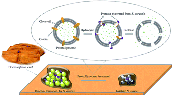
The purpose of this study is to develop a new approach to deliver antimicrobials against bacterial infections by taking advantage of the hydrolysis of protease by casein. Clove oil was encapsulated into proteoliposomes (liposomes inlaid with casein) to enhance their stability and prolong action time. The controlled release of clove oil from proteoliposomes was triggered via bacterial proteases secreted from S. aureus. The average particle size of proteoliposomes containing clove oil was 701.5 nm with a polydispersity index (PDI) of 0.019, zeta potential of −38.6 mV and encapsulation efficiency of clove oil of 40.01%. The antibiofilm activities of proteoliposomes containing clove oil against S. aureus on dried soybean curd were assessed by a colony forming units (CFU) counting method. Compared to traditional liposomes, the application of the proteoliposomes could further enhance the antibiofilm effect of an essential oil against food-borne pathogens.


1 Introduction

In recent years, one of the main public health problems has been food-related diseases, and in particular food-borne diseases, which are the cause of numerous complications and many deaths all over the world.1 Soybean curd, also known as tofu, is an important component in East Asian cuisines. There are many different varieties of soybean curd, including fresh soybean curd and soybean curd, which are processed in some way. Dried soybean curd is one of the most important and widely accepted forms of traditional oriental products derived from soybeans. Due to its rough surface texture and rich nutrient composition, dried soybean curd can be easily contaminated by the adhesion of S. aureus biofilm.2 Since bacteria in biofilms can render their inhabitants more resistant to antibiotics, they have become problematic in a wide range of food industries.3

To minimize the potential risk of contamination by S. aureus biofilm, various antimicrobial additives are employed for soybean curd processing. Among them, essential oils (EOs) of natural plants have gained special interest because of the increasing public concern about the safety of food and the potential impact of synthetic additives on health. Unfortunately, the present applications of essential oils are restricted due to their volatility and chemical instability in the presence of air, light, moisture and high temperatures.4

One popular approach to overcome these limitations is the use of liposomal technologies. However, the antibacterial activity of liposome-encapsulated essential oils has been limited due to uncontrollable release rate. Currently, a new generation of liposomes has been developed, such as a stimuli-sensitive liposome, which is a type of liposome that generally depends on different environmental factors (including pH, light, magnetism, temperature, and ultrasonic waves) in order to improve release efficiency.5 In consideration of the possible safety problems of these stimulating agents, many research studies have focused on new stimulating agents with favorable bio-compatibility.

Herein, inspired by the hydrolysis of protease to protein, a new stimulating agent with favorable biological safety and high sensitivity was introduced. The selective strategy is depicted in Fig. 1. In this study, liposomes inlaid with proteins were prepared, which were defined as proteoliposomes.6,7 The proteoliposomes could release antibiotics (clove oil) when they were activated by proteases secreted from S. aureus.8 The released clove oil would then exert its antimicrobial activity rapidly and locally. As a proof of concept, the antibiofilm activity of proteoliposomes containing clove oil against S. aureus on dried soybean curd was evaluated.


image file: c6ra03363f-f1.tif
Fig. 1 Schematic of S. aureus protease-triggered clove oil release from proteoliposomes.

2 Experiments

2.1 Materials and culture

The clove oil was obtained from J.E International (Caussols plateau, France). The Staphylococcus aureus ATCC 25923 strain was shake cultured in nutrient broth (NB) medium at 37 °C for 48 h and stored at −80 °C in NB.

2.2 The antibiofilm activity of clove oil

2.2.1 The determination of minimum biofilm inhibitory concentration (MBIC) and minimum biofilm eradication concentration (MBEC). The antibiofilm activity of clove oil was measured by MBIC and MBEC assay.9,10 The MBIC was defined as the lowest concentration of clove oil without visible growth of biofilm in 96-well platforms. The wells were filled with Tryptone Soya Broth (TSB) containing S. aureus (105–6 CFU mL−1) and clove oil (from 0.125 mg mL−1 to 4.0 mg mL−1). After incubation at 37 °C for 48 h, the plate was washed and 0.05% (v/v) 3-(4,5-dimethyl-2-thiazolyl)-2,5-diphenyl-2-H-tetrazoliumbromide (MTT) was added (MTT Cell Proliferation and Cytotoxicity Assay Kit, Beyotime Biotechnology, Shanghai, China). After incubation at 37 °C for 4 h, MTT was replaced by formazan dissolving solution and the absorbance was measured at 550 nm. MBEC indicates the lowest concentration leading to a clear well containing biofilm. Clove oil (from 0.5 mg mL−1 to 8.0 mg mL−1) in PBS was added to the formed biofilms. A biofilm-containing tube without clove oil was used as the control. The plate was incubated at 37 °C for 24 h, and then the absorbance at 550 nm was measured.
2.2.2 Antibiofilm effect of clove oil on S. aureus. Biofilm viability was assessed with the MTT staining11 and biofilm colony forming unit (CFU) counting12 method. Inhibitory and eradicative effects of clove oil against S. aureus biofilms were studied as follows. The concentrations of clove oil were 0.125 mg mL−1, 0.25 mg mL−1, 0.5 mg mL−1, 1.0 mg mL−1, 2.0 mg mL−1, and 4.0 mg mL−1. The absorbance was determined at 550 nm. Biofilm CFU counting method was utilized for viable cell counting of biofilm formed on stainless steel (1 cm × 1 cm). The biofilm-containing tubes were sonicated moderately using a water table sonicator for 5 min for viable cell counting.

2.3 Preparation and characterization of proteoliposome-encapsulated clove oil

2.3.1 Preparation of proteoliposome-encapsulated clove oil. Proteoliposomes were prepared following a novel method described as follows. Soy lecithin and cholesterol (5[thin space (1/6-em)]:[thin space (1/6-em)]1) were dissolved in trichloromethane and mixed with clove oil (5.0 mg mL−1). The mixture was evaporated in a rotary evaporator until a thin film was formed and dried in a vacuum oven at 30 °C for 24 h. Then, the lipid film was suspended in phosphate buffer solution (PBS, pH 7.2) and homogenized in a cell ultrafine grinding instrument (Ymnl-1000Y, NanJing Immanuel Instrument Equipment Co., Ltd., Nanjing, China) for 30 min at 360 W. The mixture was centrifuged at 6000 rpm for 15 min. Finally, the liposome was filtered through a 0.22 μm membrane.5

Casein (20 mg mL−1) was dissolved in PBS (pH 7.2) containing 50 mg mL−1 surfactant Brij-35 and incubated for 3 h at room temperature. Then, the resulting liposomes and casein at the desired concentration (0.2 mg mL−1) were mixed together, followed by 10 min of bath sonication. The mixture was freezed at −18 °C for 2 h with consequent rupture of vesicle membranes, and then slowly thawed at 0 °C for 2 h, causing insertion of the protein solubilized in surfactant into the membrane, followed by mild sonication (low energy in pulse mode) to facilitate the sealing of the proteoliposomes.13

2.3.2 The particle size, PDI and zeta potential. The particle size and PDI were determined by a dynamic light scattering zetasizer (Nano ZS90, Malvern Instruments, Worcester, UK).14 The zeta potential was measured in folded capillary cells at 25 °C.
2.3.3 Encapsulation efficiency of essential oil. Clove oil (0.1 mg mL−1, 0.2 mg mL−1, 0.4 mg mL−1, 0.6 mg mL−1, and 0.8 mg mL−1) was analyzed by a gas chromatography-mass spectrometer (GC-MS, Agilent 6890N/5973N, Agilent, Santa Clara, California, USA). From this, the standard curve was obtained. Subsequently, proteoliposomes were centrifuged at 20[thin space (1/6-em)]000 rpm for 1 h. The vesicles were dissolved in ethanol and then treated with ultrasonic waves for 3 h. Finally, the mixture was centrifuged at 6000 rpm for 15 min. The supernatant was kept and analyzed through GC-MS.15
2.3.4 Determination of embedding efficiency of protein. The embedding efficiency of casein, i.e., the amount of total protein incorporated into proteoliposomes, was quantified indirectly using the BCA (bicinchoninic acid) assay (Jiancheng Bioengineering Institute, Jiangsu, China) by determining the amount of protein remaining in the supernatant after the ultracentrifugation step.7
2.3.5 Stability study of proteoliposomes containing clove oil. Proteoliposomes containing clove oil (approximately 8 mL) were stored in bottles at 4 °C for 90 days. The particle size, PDI, zeta potential, and encapsulation efficiency of the essential oil and embedding efficiency of the protein were determined at 0 d, 10 d, 30 d, 60 d, and 90 d.

2.4 Determination of release rate of proteoliposomes

To estimate the release rate of proteoliposomes in vitro, the leakage of calcein sodium salt from the liposomal interior was monitored by a fluorescence method. After removing the free calcein sodium salt, the suspensions of liposomes or proteoliposomes containing calcein sodium salt (20% v/v) were added to NB containing S. aureus (105–6 CFU mL−1) and incubated at 37 °C with shaking. At 0 d, 1 d, 2 d, 3 d, and 4 d, the suspension was sampled and detected by a fluorescence spectrophotometer (F-4500, Hitachi Limited, Tokyo, Japan). In order to make a comparison, the liposomes or proteoliposomes suspension mixed with NB without S. aureus treatment was subjected to the same procedure.16

2.5 The antibiofilm activity of proteoliposome-encapsulated clove oil and its application in soy products

2.5.1 The antibiofilm activity of proteoliposome-encapsulated clove oil on stainless steel. The biofilm CFU counting method was used to assess the antibiofilm effect of proteoliposomes on S. aureus. Inhibitory and eradicative effects of clove oil against biofilms were studied as follows. The sterile stainless steel plates (1 cm × 1 cm) were added to TSB medium containing S. aureus (105 CFU mL−1) and proteoliposomes (10%, 20%, and 40% (v/v)). After 48 h incubation at 37 °C, the samples were sonicated moderately using a water table sonicator for 5 min for viable cell counting. After 48 h biofilm formation on sterile stainless steels (1 cm × 1 cm), the TSB was gently aspirated and the tubes were rinsed. Proteoliposomes in PBS were inserted into each tube. A sample with no antibacterial agent was used as the control. The tubes were incubated at 37 °C for 24 h. Then, the biofilm-containing tubes were sonicated for viable cell counting.17
2.5.2 The antibiofilm activity of proteoliposome-encapsulated clove oil on dried soybean curd. The dried soybean curds were sterilized at 121 °C for 30 min and cooled to room temperature. One sterile dried soybean curd (1 cm × 1 cm) was vertically placed in each tube. Then, the inhibitory and eradicative effects of the proteoliposomes were evaluated as previously described.
2.5.3 Biofilm formation by S. aureus on dried soybean curd during storage. With the purpose of comparing the antibiofilm activity of clove oil (0.4 mg mL−1), liposome-encapsulated clove oil (20% v/v) and proteoliposome-encapsulated clove oil (20% v/v) on dried soybean curd at 25 °C and 37 °C during 4 d storage, the population of S. aureus biofilm was measured by the CFU counting method every day.
2.5.4 Laser scanning confocal microscopy (LSCM) and environmental scanning electron microscopy (ESEM) analyses. The biofilms were detected using the 4′,6-diamidino-2-phenylindole (DAPI) staining method with modification according to Carvalho, Puppin-Rontani, & Fúcio.18 The biofilm of S. aureus was treated with 20% (v/v) proteoliposome-encapsulated clove oil for 24 h. Equal volume of DAPI (10 μg mL−1, Roche Diagnostics GmbH, Mannheim, Germany) was dropped onto the biofilms, which were then kept in the dark for 10 min. The sample was observed by LSCM (Leica TCS SP5 II, Leica, Wetzlar, Germany). The biofilm in sterile PBS without proteoliposome treatment was observed as the control. After being treated with 20% (v/v) proteoliposomes for 24 h at 37 °C, the dried soybean curds were taken out and observed by ESEM (XL-30, Philips, Eindhoven, Netherlands). As control, the biofilm without proteoliposome treatment was also observed.19,20

2.6 Color and texture evaluation

The same size and type of dried soybean curd pieces were used for evaluation after exposure to clove oil, liposomes or proteoliposomes. The changes of dried soybean curd surface color were evaluated with a Chromatic meter (Color Quest XE, Hunter Lab Co., Reston, Virginia, USA). Hunter color values, L* (lightness), a* (redness), and b* (yellowness) were determined. Surface texture measurement of the dried soybean curd was performed using a TA.XT. Plus (Stable Micro Systems Ltd, Godalming, Surrey, UK). Hardness, springiness and chewiness were used to evaluate the texture quality of the dried soybean curd.

2.7 Statistical analysis

All experiments were conducted in triplicate, and the results were analyzed with the SPSS software (SPSS16.0 for Windows). The one-way ANOVA was used to determine the level of significance and P < 0.05 was considered to be significant.

3 Results and discussion

3.1 Antibiofilm activity of clove oil

The antibiofilm activity of clove oil was assessed using the MTT staining and biofilm CFU counting method.11,12 The inhibitory effect of different concentrations of clove oil on the ability of biofilm formation of S. aureus was evaluated after 48 h incubation time. The optical density (OD) values of S. aureus biofilms containing 1.0 mg mL−1 clove oil were reduced from 1.73 to 0.21 (Fig. 2a). However, the antibiofilm effects had no evident improvement when the concentration of clove oil was higher than 1.0 mg mL−1. This indicated that 1.0 mg mL−1 of clove oil was enough to inhibit the formation of S. aureus biofilm. Similar results were observed when S. aureus biofilms formed on stainless steels were dropped into tubes containing TSB with different concentrations of clove oil. After 48 h, the population of S. aureus in the biofilm containing 1.0 mg mL−1 clove oil was decreased by 5.20 log units from that of the control group (Fig. 2b).
image file: c6ra03363f-f2.tif
Fig. 2 Inhibitory effect of clove oil on S. aureus biofilm. Viability was determined by MTT staining (a) or plate colony-counting (b) methods. Each symbol indicates the mean ± standard error of the mean for three independent experiments. *P < 0.05 versus the control groups.

The eradicative effect of different concentrations of clove oil on the viability of the S. aureus biofilm was evaluated after 24 h of treatment. The OD values of S. aureus biofilms treated with 1.0 mg mL−1 clove oil were reduced from 1.71 to 0.24 (Fig. 3a). However, the antibiofilm effects had no evident improvement when the concentration of clove oil was higher than 1.0 mg mL−1. This indicated that 1.0 mg mL−1 of clove oil was enough to eradicate the S. aureus biofilm. Similar results were observed when S. aureus biofilms formed on stainless steels were dropped into tubes containing PBS with different concentrations of clove oil. After 24 h, populations of S. aureus decreased 4.07 log units using 1.0 mg mL−1 clove oil treatment (Fig. 3b).


image file: c6ra03363f-f3.tif
Fig. 3 Eradicative effect of clove oil on S. aureus biofilm. Viability was determined by MTT staining (a) or plate colony-counting (b). Each symbol indicates the mean ± standard error of the mean for three independent experiments. *P < 0.05 versus the control groups.

Therefore, the MBIC and MBEC values of clove oil against S. aureus biofilm were both 1.0 mg mL−1. As such, clove oil showed prominent antibiofilm activity against S. aureus and could be an effective inhibitor and bactericide of S. aureus biofilm.

3.2 Characterization and stability evaluation of proteoliposome-encapsulated clove oil

After preparing the proteoliposomes containing clove oil by the thin film-dispersion and freeze-thawing methods, the physicochemical properties of proteoliposome-encapsulated clove oil were evaluated. As shown in Fig. 4c, the average particle size of the proteoliposomes was 701.5 ± 14.03 nm with a PDI of 0.019 ± 0.004. Compared to the liposome-encapsulated clove oil,5 significant increase of proteoliposome-encapsulated clove oil size was obtained at the clove oil concentration of 5.0 mg mL−1. The result might confirm the existence of casein on the surface of the liposomes. The PDI value was used to characterize the particle size distribution. The distribution of PDI values of the proteoliposomes in this study is narrow. From the data obtained, the proteoliposomes containing clove oil had a high negative zeta potential of −38.6 ± 0.77 mV (Fig. 4c). In general, <−30 mV or >+30 mV would be considered high zeta potentials.21 Therefore, the proteoliposomes prepared in the study with uniform particle size and high zeta potential could prevent the occurrence of aggregation or precipitation. The linear regression equation between peak areas of eugenol and concentration of clove oil was y = 44[thin space (1/6-em)]668[thin space (1/6-em)]381.8571x − 15[thin space (1/6-em)]928.4000, R2 = 0.9892. In the case of 5.0 mg mL−1 clove oil encapsulation, the value of entrapment efficiency was 40.01 ± 0.80% (Fig. 4c). The embedding efficiency of casein was also investigated by BCA protein assay. The value of embedding efficiency of casein was 21.8 ± 0.44% (Fig. 4c).
image file: c6ra03363f-f4.tif
Fig. 4 Characterization and stability evaluation of proteoliposomes with 5.0 mg mL−1 clove oil encapsulation. (a) Diagram of the proteoliposome-encapsulated clove oil, (b) physical figures of proteoliposome samples stored for different numbers of days, and (c) property parameters of proteoliposomes stored for different numbers of days.

The stability of the proteoliposomes containing clove oil was evaluated by examining the physicochemical properties after 10 d, 30 d, 60 d, and 90 d of storage at 4 °C. As shown in Fig. 4b, the turbidity of proteoliposomes did not significantly change during the storage. Compared to 0 d of storage, the proteoliposomes were generally stable. The size of proteoliposomes was increased from 701.5 ± 14.03 nm to 725.8 ± 14.52 nm over time. The PDI value was altered from 0.019 ± 0.004 to 0.274 ± 0.005 (Fig. 4c). However, the encapsulation efficiency of clove oil and embedding efficiency of casein values were identical to those obtained at 0 d. The results suggested that the proteoliposomes retained the clove oil and casein constituents during the storage. Based on the abovementioned results, the proteoliposomes containing clove oil possessed good stability.

3.3 Release rate of proteoliposomes

In order to calculate the release rate of an internal substance in vitro, the leakage of fluorescent agent (calcein sodium salt) into the supernatant was monitored by a fluorescence method. During a 4 day assay period, the release of fluorescent agent was continuously detected when the liposomes and proteoliposomes were incubated with S. aureus in NB.

The release rate of calcein sodium salt was calculated using the following equation:

image file: c6ra03363f-t1.tif
where R is the release rate (%), I2 is the fluorescence intensity of calcein sodium salt released from the S. aureus treated liposomes or proteoliposomes, I1 was the fluorescence intensity of calcein sodium salt from Triton x-100 treated liposomes or proteoliposomes, and I0 is the fluorescence intensity of calcein sodium salt released from liposomes or proteoliposomes without treatment.

As shown in Fig. 5, after 1 d of S. aureus treatment, the fluorescence intensity of calcein sodium salt from proteoliposomes was 23.50 (Fig. 5a) and the release rate was 28.66% (Fig. 5b). In contrast, the fluorescence intensity of calcein sodium salt was 4.72 when the proteoliposome was incubated without S. aureus. From 1 d to 4 d, the release rate of calcein sodium salt from liposomes and proteoliposomes increased gradually by the stimulation of S. aureus. The release rate of proteoliposomes at 2 d, 3 d, and 4 d was 60.45%, 75.21%, and 82.07%, respectively (Fig. 5b). The values were higher than those for the liposomes. This might be contributed by the proteolysis of S. aureus protease. However, the fluorescence intensity of proteoliposomes without S. aureus treatment did not significantly change after 1 d of incubation. This indicated that liposomes or proteoliposomes containing calcein sodium salt remained stable during the storage.


image file: c6ra03363f-f5.tif
Fig. 5 (a) The fluorescence intensity of calcein sodium salt released from liposomes and proteoliposomes, and (b) the release rate of liposomes and proteoliposomes.

3.4 The antibiofilm activity of proteoliposome-encapsulated clove oil and its application in dried soybean curd

The antibiofilm effects of different concentrations of liposomes and proteoliposomes containing clove oil against S. aureus on stainless steels were measured by a plate colony-counting method, and the results are shown in Fig. 6e and f. After 24 h treatment, compared to the control groups, the amount of viable S. aureus in biofilms treated with 10% (v/v) proteoliposomes was reduced by 1.84 log units (Fig. 6e). Under the same conditions, the number of S. aureus cells reduced by 0.99 log units when treated with liposomes (Fig. 6e). The number of viable S. aureus treated with liposomes or proteoliposomes showed a gradual and consecutive decrease with the increase of concentration. However, compared to liposomes, the inhibitory (Fig. 6e) and eradicative (Fig. 6f) activities of proteoliposomes against S. aureus biofilm were significantly improved. The improvement was attributed to the increase of essential oil release rate by embedded casein.22 In addition, LSCM analysis was employed to illuminate the quantitative changes of S. aureus biofilms treated by proteoliposomes. As reflected by the LSCM images in Fig. 6a–d, the untreated biofilms (Fig. 6a and c) were crowded spheres. However, in contrast, there were obvious decreases in the fluorescence intensities of biofilms treated with proteoliposomes (Fig. 6b and d).
image file: c6ra03363f-f6.tif
Fig. 6 The inhibitory and eradicative effects of different concentrations of proteoliposomes against S. aureus biofilms on stainless steels. The LSCM images of the inhibitory (a and b) and eradicative (c and d) effect of proteoliposomes on S. aureus biofilm. The inhibitory (e) and eradicative (f) effects were determined by a plate colony-counting method. Each symbol indicates the mean ± standard error of the mean for three independent experiments. *P < 0.05 versus the control groups.

The antibiofilm properties of proteoliposomes against S. aureus on the surface of dried soybean curds were compared with those of liposomes. Similar to biofilm formation on stainless steel, for S. aureus biofilms formed on dried soybean curds (Fig. 7e), the antibiofilm activity of proteoliposomes was higher than that of liposomes. In particular, the counts of viable S. aureus in biofilms formed on dried bean curd treated with 10% (v/v) liposomes and proteoliposomes were reduced by 0.95 and 2.09 log units, respectively, (Fig. 7f) after 24 h incubation. Correspondingly, the counts of viable S. aureus treated with increased concentrations of liposomes and proteoliposomes were reduced more. The antibiofilm activities of proteoliposomes were significantly improved from that of liposomes. The ESEM analysis was utilized to observe the morphological and quantitative changes of S. aureus biofilms on the surface of dried soybean curds treated with proteoliposomes containing clove oil. In the case of ESEM images, it could be found that the untreated biofilms (Fig. 7a and c) adhered were crowded sphere connected by extracellular compounds. However, there was an obvious decrease in the quantity of biofilms treated with proteoliposomes (Fig. 7b and d). The results underlined the inhibitory and eliminative effects of proteoliposomes on S. aureus biofilms.


image file: c6ra03363f-f7.tif
Fig. 7 The inhibitory and eradicative effects of different concentrations of proteoliposomes against S. aureus biofilms on dried soybean curds. The ESEM images of the inhibitory (a and b) and eradicative (c and d) effect of proteoliposome on S. aureus biofilm. The inhibitory (e) and eradicative (f) effects were determined by a plate colony-counting method. Each symbol indicates the mean ± standard error of the mean for three independent experiments. *P < 0.05 versus the control groups.

3.5 Biofilm formation by S. aureus on dried soybean curd during storage

The ability of biofilm formation by S. aureus on dried soybean curds at 25 °C and 37 °C was measured by a CFU counting method. As Fig. 8 showed, the population of S. aureus biofilm without treatment was increased constantly from 1 d to 4 d, whereas the population of the liposome and proteoliposomes treated S. aureus biofilm steadily declined over time. Compared with the control group, the population of S. aureus biofilm treated with clove oil was increased more slowly over time. However, the antibiofilm activity of clove oil was weaker than that of liposome and proteoliposome. When incubated at 25 °C (Fig. 8a), almost 0.51 log units and 1.08 log units reduction was observed after 1 d of liposome and proteoliposome treatment. Moreover, the numbers of S. aureus biofilms after 2 d, 3 d, and 4 d treatment of proteoliposomes were reduced by 3.13, 4.83, and 5.93 log units, respectively. When incubated at 37 °C (Fig. 8b), almost 0.22 log units and 0.37 log units reduction was observed after 1 d of liposomes and proteoliposomes treatment, respectively. Furthermore, the numbers of S. aureus biofilms after 2 d, 3 d, and 4 d treatment of proteoliposomes were reduced by 3.39, 4.80, and 5.93 log units. Whether the environmental temperature was 25 °C or 37 °C, the antibiofilm activity of the proteoliposomes was higher than that of the liposomes and clove oil (Fig. 8a and b). Moreover, most of the S. aureus strains had a higher biofilm production at 37 °C than at 25 °C.23 The formation of S. aureus biofilm at 25 °C was also slower than that at 37 °C in this study. Thus, the antibiofilm activity of liposomes or proteoliposomes was better at 25 °C than at 37 °C.
image file: c6ra03363f-f8.tif
Fig. 8 Biofilm formation by S. aureus on dried soybean curds at 25 °C (a) and 37 °C (b). Each symbol indicates the mean ± standard error of the mean for three independent experiments. *P < 0.05 versus the 1st d.

3.6 Color and texture evaluation

The qualities of dried soybean curd treated with clove oil, liposomes, and proteoliposomes were evaluated by color and texture. As shown in Table 1, the L*, a*, and b* values were obtained from surface color measurements. The values did not significantly alter after different treatments. Furthermore, the hardness, springiness, and chewiness were tested in this study, and no significant differences were observed between controls and treated samples. Therefore, the treatment of liposomes or proteoliposomes containing clove oil could not only inhibit and eliminate the S. aureus biofilms, but also maintain the quality of dried soybean curd.
Table 1 Effects of different treatments on the color and texture qualities of dried soybean curda
Parameter Different treatments
Control Clove oil Liposome Proteoliposome
a Values are expressed as mean ± standard deviation. All the values were considered to be insignificant (P > 0.05 versus the control groups).
L* 82.80 ± 0.33 82.86 ± 0.06 82.70 ± 0.28 82.30 ± 0.08
a* 2.26 ± 0.21 1.99 ± 0.17 2.21 ± 0.08 2.07 ± 0.40
b* 23.93 ± 0.65 23.28 ± 0.87 23.82 ± 0.58 22.67 ± 0.99
Hardness (N) 348.391 ± 2.591 325.336 ± 3.578 320.408 ± 2.025 342.176 ± 1.140
Springiness (%) 97.628 ± 0.244 98.195 ± 2.291 99.598 ± 0.955 99.521 ± 0.906
Chewiness (N) 325.907 ± 25.597 287.361 ± 43.706 271.500 ± 20.948 311.928 ± 28.277


4 Conclusions

As a novel and safe antimicrobial agent, the proteoliposomes containing clove oil can not only increase the antimicrobial effect of clove oil on S. aureus biofilms by improving release efficiency, but also preserve the original quality of soybean products. In addition, the proteoliposomes were physically stable after 90 days of storage at 4 °C. Thus, the proteoliposomes possess a wide array of potential applications in the preservation and storage of food products.

Abbreviations

PDIPolydispersity index
CFUColony forming units
PFTsPore-forming toxins
NBNutrient broth
S. aureusStaphylococcus aureus
MBICMinimum biofilm inhibitory concentration
MBECMinimum biofilm eradication concentration
TSBTryptone soya broth
MTT3-(4,5-Dimethyl-2-thiazolyl)-2,5-diphenyl-2-H-tetrazolium bromide
PBSPhosphate buffer solution
GC-MSGas chromatography-mass spectrometry
BCABicinchoninic acid
DAPI4′,6-Diamidino-2-phenylindole
LSCMLaser scanning confocal microscopy
ESEMEnvironmental scanning electron microscopy

Acknowledgements

The authors acknowledge the financial support from the National Natural Science Foundation of China (grant no. 31301573), the Natural Science Foundation of Jiangsu Province (grant no. BK20130493), the Jiangsu University Research Fund (grant no. 11JDG050), the Innovation Fund Designated for Graduate Students of Jiangsu Province (grant no. KYLX15-1092) and the Priority Academic Program Development of Jiangsu Higher Education.

References

  1. G. Pesavento, C. Calonico, A. R. Bilia, M. Barnabei, F. Calesini, R. Addona, L. Mencarellia, L. Carmagninia, M. C. Di Martinoc and A. Lo Nostroa, Food Control, 2015, 54, 188–199 CrossRef CAS.
  2. K. Jakub, E. Sigrun and W. Kinga, Int. J. Antimicrob. Agents, 2009, 33, 343–347 CrossRef PubMed.
  3. S. Srey, I. K. Jahid and S. D. Ha, Food Control, 2013, 31, 572–585 CrossRef.
  4. E. Zehra Karagöz, Y. Gökçe Polat, C. Betül Kodal and C. Kezban, Meat Sci., 2010, 86, 283–288 CrossRef PubMed.
  5. H. Y. Cui, C. T. Zhao and L. Lin, Food Control, 2015, 56, 128–134 CrossRef CAS.
  6. P. Pingitore, L. Pochini, M. Scalise, M. Galluccio, K. Hedfalk and C. Indiveri, Biochim. Biophys. Acta, Biomembr., 2013, 1828, 2238–2246 CrossRef CAS PubMed.
  7. P. Freixeiro, E. Diéguez-Casal, L. Costoya, B. Seijo, C. M. Ferreirós, M. T. Criado and S. Sánchez, Int. J. Pharm., 2013, 443, 1–8 CrossRef CAS PubMed.
  8. S. B. Sørensen, T. L. Sørensen and K. Breddam, FEBS Lett., 1991, 294, 195–197 CrossRef.
  9. W. S. Wu, C. C. Chen and Y. C. Chuang, J. Microbiol., Immunol. Infect., 2013, 46, 89–95 CrossRef CAS PubMed.
  10. P. Sabaeifard, A. Abdi-Ali and M. R. Soudi, J. Microbiol. Methods, 2014, 105, 134–140 CrossRef CAS PubMed.
  11. H. J. Tang, C. C. Chen, W. C. Ko, W. L. Yu, S. R. Chiang and Y. C. Chuang, Int. J. Antimicrob. Agents, 2011, 38, 46–51 CrossRef CAS PubMed.
  12. T. Bodur and A. Cagri-Mehmetoglu, Food Control, 2012, 25, 1–9 CrossRef.
  13. M. Scalise, L. Pochini, N. Giangregorio, A. Tonazzi and C. Indiveri, Pharmaceutics, 2013, 5, 472–497 CrossRef CAS PubMed.
  14. H. H. Song, X. Gong, G. R. Williams, J. Quan, H. L. Nie, L. M. Zhu, E. L. Nan and M. Shao, Mater. Res. Bull., 2014, 53, 280–289 CrossRef CAS.
  15. Z. Wen, B. Liu, Z. K. Zheng, X. K. You, Y. T. Pu and Q. Li, Chem. Eng. Res. Des., 2010, 88, 1102–1107 CrossRef CAS.
  16. D. Pornpattananangkul, L. Zhang, S. Olson, S. Aryal, M. Obonyo, K. Vecchio, C. M. Huang and L. F. Zhang, J. Am. Chem. Soc., 2011, 133, 4132–4139 CrossRef CAS PubMed.
  17. L. Lin, H. F. Zhang, H. Y. Cui, M. Q. Xu, S. S. Cao, G. H. Zheng and M. D. Dong, Colloids Surf., B, 2013, 101, 97–100 CrossRef CAS PubMed.
  18. F. G. Carvalho, R. M. Puppin-Rontani and S. B. Fúcio, J. Appl. Oral Sci., 2012, 20, 568–575 CrossRef PubMed.
  19. J. H. Priester, A. M. Horst, L. C. Van De Werfhorst, J. L. Saleta, L. A. Mertes and P. A. Holden, J. Microbiol. Methods, 2007, 68, 577–587 CrossRef CAS PubMed.
  20. L. Lin, H. Y. Cui, G. H. Zeng, M. L. Chen, H. F. Zhang, M. Q. Xu, X. Q. Shen, C. Bortolini and M. D. Dong, J. Mater. Chem. B., 2013, 21, 2719–2723 RSC.
  21. Q. Lu, P. M. Lu, J. H. Piao, X. L. Xu, J. Chen, L. Zhu and J. G. Jiang, LWT--Food Sci. Technol., 2014, 57, 686–695 CrossRef CAS.
  22. M. Craig, M. Amiri and K. Holmberg, Colloids Surf., B, 2015, 127, 200–205 CrossRef CAS PubMed.
  23. P. D. Ciccio, A. Vergara, A. R. Festino, D. Paludi, E. Zanardi, S. Ghidini and A. Ianieri, Food Control, 2015, 50, 930–936 CrossRef.

This journal is © The Royal Society of Chemistry 2016
Click here to see how this site uses Cookies. View our privacy policy here.