Systems metabolic engineering of Escherichia coli to enhance the production of flavonoid glucuronides

Yan Yangab, Hui-Min Wangb, Yuan-Feng Tongb, Min-Zhi Liub, Ke-Di Chengb, Song Wu*ab and Wei Wang*ab
aState Key Laboratory of Bioactive Substance and Function of Natural Medicines, Institute of Materia Medica, Peking Union Medical College & Chinese Academy of Medical Sciences, 1 Xian Nong Tan St., 100050 Beijing, China. E-mail: wwang@imm.ac.cn; ws@imm.ac.cn
bKey Laboratory of Biosynthesis of Natural Products of National Health and Family Planning Commission, Institute of Materia Medica, Peking Union Medical College & Chinese Academy of Medical Sciences, 1 Xian Nong Tan St., 100050 Beijing, China

Received 4th February 2016 , Accepted 24th March 2016

First published on 29th March 2016


Abstract

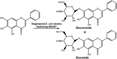
Large-scale application of whole-cell glycosylation is still hampered by inefficient UDP-sugar formation. Using a module-based approach, an engineered Escherichia coli strain was constructed to enhance the production of flavonoid glucuronides. The engineered metabolic pathway was partitioned into two modules: an endogenous upstream biosynthetic pathway to produce the sugar donor UDP-glucuronic acid (UDPGA) and a heterologous downstream UDP-dependent glycosyltransferase (UGT) to catalyse the glucuronidation of flavonoids. First, two UGTs (SbUGT-W76 and SbUGT-W112) were isolated from Scutellaria baicalensis Georgi. Enzymatic assays showed that the recombinant SbUGT exhibited regiospecificity towards the C7-OH position of flavonoid substrates, and conflicting results on the specificity of sugar donor and acceptor compared with that of reported SbUBGAT were obtained. Then, the native upstream module of the UDPGA synthetic pathway was strengthened to increase the endogenous level of UDPGA. Using these strategies, up to 797 mg L−1 baicalein-7-O-glucuronide was biosynthesized. Furthermore, systemic engineering of upstream and downstream genes in the glucuronide biosynthetic pathway was conducted for characterization and the capability to biotransform baicalein. The results showed that UDP-glucose 6-dehydrogenase (Ugd) catalysed the rate-determining step for glucuronide production. SbUGT displayed the same catalytic properties both in vivo and in vitro.
Introduction

Flavonoids, a large family of polyphenol compounds, exhibit a variety of important biological activities, such as anti-inflammation,1 anti-tumour,2 and anti-viral activities3 and cardiovascular system effects.4,5 The glucuronidation and deglucuronidation cycles are key steps for in vivo activation of flavonoid glucuronides.6,7 Thus, glucuronosylation is an important approach for improving the pharmacological properties of flavonoids, such as toxicity, pharmacokinetics, organ distribution, or bioavailability.8

The development of preparative phytochemistry enables clinical use of flavonoid glucuronides from plant extraction. For example, breviscapine injection, mainly containing scutellarein-7-O-glucuronide with minor amounts of apigenin-7-O-glucuronide, is approved by the China Food and Drug Administration (CFDA) and used to treat cardio-cerebral vascular diseases.9 Additionally, baicalein-7-O-glucuronide (i.e., baicalin), a compound isolated from the root of S. baicalensis Georgi, also attracts interest from the pharmaceutical industry due to its excellent biological effects.10 However, the current limited plant resource insufficiently meets clinical demand. The low regioselectivity of polyhydroxy glycosylation could be a hurdle for synthetic chemists seeking to synthetically prepare various flavonoid glucuronides. Even for a simple flavonoid glucuronide, chemical synthesis is typically a low-yield process that requires multiple protection and deprotection steps.11 Although enzymatic synthesis of glucuronides under mild reaction conditions enables regio- and stereo-specificity in a single step, this approach is also impractical to scale-up due to the high cost of the activated donor UDPGA, or the potential for substrate and product inhibition under the high concentrations.11–13

Synthetic biology and metabolic engineering have allowed the heterologous biosynthesis of core flavonoid structures (e.g., naringenin and pinocembrin)14,15 and subsequently modified glycosides, such as glucosides, rhamnosides, and xylosides, using an E. coli system in which the native pathway of UDP-glucose (UDPG) was shunted to other UDP-sugars combined with the expression of a glycosyltransferase.16–18 To the best of our knowledge, there have only been three research reports on the metabolic biosynthesis of glucuronides. Heterologous production of metabolic glucuronides was first described in fission yeast, where a human UDP-glucose 6-dehydrogenase (Ugd) together with several human or rat UGT isoforms were expressed to catalyse glucuronidation of 4-methylumbelliferone (space time yield of 52.8 mg L−1 day−1) and testosterone (space time yield of 15.1 mg L−1 day−1).19 An E. coli strain was constructed to synthesize naringenin and quercetin glucuronides (yield not mentioned) by combining expression of glucose-1-phosphate uridyltransferase (galU), UDP-glucose dehydrogenase (calS8), and glycosyltransferase (atGt-5).20 In recent reports, an E. coli strain was modified to express UGT (AmUGT from Antirrhinum majus or VvUGT from Vitis vinifera) and ugd genes from E. coli, Arabidopsis thaliana, or Glycine max to produce luteolin-7-O-glucuronide (yield of 300 mg L−1) and quercetin-3-O-glucuronide (yield of 687 mg L−1).21 All of the above studies inspired us to hypothesize that combinatorial approaches could provide a solution for the biosynthesis of flavonoid glucuronides, but we identified that an insufficient supply of UDP-sugars might limit the yield of glucuronides. Therefore, in this work, we construct an engineered E. coli with an endogenous upstream biosynthetic pathway to efficiently produce an increased supply of the sugar donor UDPGA and a heterologous downstream SbUGT to catalyse the glucuronidation of flavonoids. Furthermore, we studied how a change in the UDPGA biosynthetic pathway affected production of flavonoid glucuronides in our engineered E. coli strain.

Experimental

Materials and media

All chemicals and reagents were obtained from Sigma-Aldrich (St Louis, MO, USA) and BioBioPha (Kunming, China) unless otherwise stated. The pTWIN1 vector, together with all restriction enzymes and DNA ligase, were purchased from New England Biolabs (Ipswich, MA, USA). Analyses of substrate specificity and determinations of conversion rates were performed on a Thermo U3000 series HPLC system (Thermo Electron Corp., USA).

Transformed E. coli cells were grown on Luria–Bertani (LB) medium supplemented with antibiotics (100 mg L−1 ampicillin, 50 mg L−1 kanamycin and 25 mg L−1 chloramphenicol). The standard medium used for all shaker-flask production studies consisted of a defined M9 salts medium (pH 7.0, Na2HPO4 6.8 g L−1; KH2PO4 3 g L−1; NaCl 0.5 g L−1; NH4Cl 1 g L−1; MgSO4 0.24 g L−1; CaCl2 0.01 g L−1) supplemented with glucose (2 g L−1) as the main carbon source.

Plasmid construction

Construction of plasmids harboring pgm, galU, and ugd fusion genes. The DNA fragments encoding phosphoglucomutase (pgm), galU, and ugd were amplified by polymerase chain reaction (PCR) from E. coli BL21 (DE3) genomic DNA. All primers used for plasmid construction are listed in Table S1 in the ESI. Details of plasmid construction are supplied in the ESI.
Construction of a plasmid with the SbUGT fusion fragment. Total RNA was extracted from aseptic seedlings of S. baicalensis Georgi using an RNeasy Plant Mini Kit (TianGen Biotech, China), and cDNA was synthesized by reverse transcription using a GeneRacer™ RACE Kit (Invitrogen, USA) with a sequence-specific primer, UGT-5, designed according to the sequence of S. baicalensis Georgi SbUBGAT (GenBank: AB479151). The full-length flavonoid glycosyltransferase DNA homologs were generated by nested-PCR with the primers UGT-1 to UGT-4, verified by DNA sequencing, and then subcloned into a derivative (designated as pTWINB) of the plasmid pTWIN1 constructed by replacing the C-intein region of the DNA fragment with a multiple cloning site (MCS) from the plasmid pET-28a (Novagen, Germany), which was amplified by PCR using the primers Twin1-B1 and Twin1-B2. To co-express the SbUGT enzyme along with the other three enzymes of the upper module in host cells, the SbUGT DNA fragment obtained by Nco I and Xho I double digestion of pTWINB-SbUGT was inserted into the plasmid pAI22 derived from the vector pACYC184 (Novagen, Germany), resulting in the plasmid pACYC184-SbUGT.

Site-directed mutation

Site-directed mutation was performed by the overlap-extension PCR method23 with mutant-specific primers (W76-1 to W76-6, Table S1) containing appropriate base substitution(s) using the clone (SbUGT-W76) as the template. The mutagenized ORF fragments (SbUGT-W112G76) were cloned into Pst I and Bgl II sites of the plasmid pTWINB-SbUGT-W112 to generate the plasmid pTWINB-SbUGT-W112G76. To characterize SbUGT-W112G76 in vivo, the plasmid pACYC184-SbUGT–W112G76 was constructed using the same enzyme digestion and ligation described above. The presence of each mutation was verified by DNA sequencing. The mutagenized ORF fragment of SbUGT-W112G76 was identical to previously reported SbUBGAT.24

Expression and purification of recombinant proteins

Transformants harboring the plasmids pTWINB-SbUGT-W76, pTWINB-SbUGT-W112, and pTWINB-SbUGT-W112G76 were cultured according to the previously published method.25,26 The recombinant SbUGT was induced with 0.5 mM isopropyl-β-D-thiogalactopyranoside (IPTG) at 16 °C for 18 h. All subsequent purification procedures were conducted at 4 °C. The cells were pelleted by centrifugation and resuspended in buffer I (20 mM HEPES, 500 mM NaCl, pH 8.5) and then sonicated for 15 min in 5 s bursts at 6 s intervals. After centrifugation at 17[thin space (1/6-em)]000 rpm for 16 min, the supernatant containing recombinant proteins was removed, subjected to affinity chromatography on a chitin column, and eluted with 100 ml of buffer II (20 mM HEPES, 500 mM NaCl, pH 6.5). The target protein SbUGT was incubated at 25 °C overnight and released from the beads by cleaving off the chitin binding domain (CBD) with buffer II. The eluted fraction was balanced to 10 mM citrate buffer (pH 6.5) using ultrafiltration membranes (Millipore, USA). Protein purity was confirmed by SDS-PAGE to be >90%, and the protein concentration for all studies was determined using a Protein Quantitative Kit (Bradford) (Bio-Rad, USA).

Functional analysis of recombinant SbUGTs

The sugar donor specificity of purified enzymes SbUGT-W76 and SbUGT-W112 was characterized with two sugar donors, UDPG and UDPGA, respectively. To assay for substrate specificity of SbUGT-W76 and SbUGT-W112, 22 flavonoids (Fig. S1) were used as sugar acceptors.

Enzymatic reactions (500 μL total volume) were conducted in a 1.5 mL tube containing reaction buffer (10 mM citrate, pH 6.5), UDP-sugar (0.6 mM UDPGA-Na or UDPG-Na), substrates (0.2 mM of diverse flavonoids dissolved in DMSO), and purified enzyme (50 μg SbUGT-W76 or SbUGT-W112). The reaction mixture was incubated at 30 °C for 30 min. The reactions were initiated by addition of glucuronosyltransferase and terminated by adding 500 μL of methanol. Subsequently, the samples were centrifuged at 13[thin space (1/6-em)]500 rpm for 10 min, and aliquots were analysed by HPLC.

Determination of kinetic parameters of SbUGTs

To determine the initial velocity of SbUGTs, the assays were performed under steady state conditions using the standard reaction system (details in the ESI). The reaction mixtures contained certain concentrations of enzyme and saturating UDP-sugar (800 μM), with varying concentration (0–400 μM) of baicalein, narigenin, apigenin, or 4′-demethycalycosin or with constant concentrations of enzyme and excess baicalein (200 μM) with varying UDPGA (0–800 μM) or UDPG (0–10[thin space (1/6-em)]000 μM) in a final volume of 200 μL of 50 mM PBS–Na (pH 7.0). After incubation at 30 °C for 10 min, the reaction was terminated by addition of 400 μL of ice cold MeOH, and the samples were centrifuged at 13[thin space (1/6-em)]500 rpm for 10 min. Supernatants were analysed by analytical reverse-phase HPLC, peak areas were integrated with Thermo Workstation Software, and the product conversion was calculated using the external standard curve method. All experiments were performed in triplicate. The Km, Vmax, and kcat values were plotted using Lineweaver–Burk plots.

Optimization of culture medium for whole-cell biocatalysis

The engineered strain BPGUT (B, refers to the host strain BL21 (DE3); P, G, U, and T refer to genes encoding Pgm, GalU, Ugd, and SbUGT, respectively) was pre-cultured in LB medium. The recombinant proteins were induced by addition of 0.5 mM IPTG and then incubated for an additional 15 h at 16 °C. After induction, cells were harvested by centrifugation and resuspended in eight media: LB, LB plus 2% glucose, M9, M9 plus 2% glucose, M9 plus 5% glucose, M9 plus 10% glucose, M9 plus 15% glucose, and M9 plus 20% glucose. At this step, the cell density was adjusted to an OD600 of 3.26,27 Baicalein (1.0 mM) was added to the reaction mixture, and the bioconversion reaction was kept shaking at 30 °C for 24 h. The reaction mixture was collected and lyophilized. The dried sample was dissolved in methanol and analysed by HPLC.

The glucuronidation reaction procedure for whole cells

The engineered E. coli strains with the upstream and downstream modules were pre-cultured in LB broth. The overnight cultured cells were harvested and resuspended in M9 containing 2% glucose. At this step, cell density was kept at an OD600 of 3.26,27 The aromatic substrates (0.6 mM) were added to the reaction system, which was then incubated at 30 °C for 24 h. The reaction mixture was processed using a standard post-processing method and then subjected to HPLC for analysis.

To study the maximum transformation of the engineered E. coli strain BPGUT, baicalein (0.6, 1.0, 1.5, or 2.0 mM) was added to the reaction mixture in M9 containing 10% glucose, and the reaction was kept shaking at 30 °C for 24 h. The final product conversion was calculated according to a standard curve of baicalin.

HPLC-based separation method

An aliquot of 20 or 40 μL of the sample supernatant was used for analysis on a Thermo HPLC system equipped with a Hibar® C18 reverse-phase column (Hibar, 4.60 × 250 mm, 5 μm particle size). The mobile phase was water with 0.05% TFA (A) and acetonitrile with 0.05% TFA (B) with a gradient program. Analysis of flavonoids and their corresponding glucuronides was conducted as described in the ESI.

Results and discussion

Identification of UDP-glucuronosyltransferase from S. baicalensis Georgi

Access to a suitable glucuronosyltransferase (GAT) is a prerequisite for flavonoid glucuronidation. With primers designed according to the nucleotide sequence of the SbUBGAT (UGT88D1) gene, we were able to identify two cDNA clones, SbUGT-W76 and SbUGT-W112 (GenBank: KP183911 and KP183914), from S. baicalensis Georgi by RT-PCR.24,28 The deduced amino acid sequences of SbUGT-W76 and SbUGT-W112 shared high identity (≥99%) with that of SbUBGAT.24,28

To characterize the sugar acceptor specificity of SbUGT-W76 and SbUGT-W112, 22 flavonoids (Fig. S1) were tested using purified SbUGT enzymes. Based on the high sequence identity between SbUGTs and SbUBGAT, the two enzymes SbUGT-W76 and SbUGT-W112 were most likely to be counterparts of SbUBGAT. However, purified SbUGT-W76 and SbUGT-W112 possessed different substrate specificity than SbUBGAT,24 glucuronosylating the 7-OH group of flavones with or without the hydroxyl or methoxyl substituent at the ortho position. Additionally, the recombinant enzymes also catalysed reactions of some isoflavones and two flavanones (naringenin and robtin) (Fig. S2).

Further research was conducted on the sugar donor specificity of SbUGTs. Enzymatic assays indicated that SbUGT-W76 and SbUGT-W112 could also accept UDPG as a sugar donor and catalyse glucosylation of baicalein, wogonin, oroxylin A, scutellarein, chrysin, apigenin, kaempferol, genistein, and naringenin (Fig. S3), and the catalytic abilities were also different from that of SbUBGAT. The glucosylated products were identified by high performance liquid chromatography electrospray ionization mass spectrometry (HPLC-ESI-MSn) analyses (Fig. S4). The mass spectra of all of the products presented a [M + H]+ ion peak in the positive mode and [M + H − 162]+ fragment ions in the MS/MS detection, suggesting that a glucose group had been introduced into the skeleton of flavonoid aglycone.

To confirm the conflicting results from the characterization of UGTs from S. baicalensis Georgi, site-directed mutant of the SbUBGAT gene (designated as SbUGT-W112G76) was performed using recombinant PCR. Enzymatic assays showed that SbUGT-W112G76 could also accept UDPGA and UDPG as sugar donors and catalyse glucuronosylation of baicalein, apigenin (targeting the 7-OH group of flavones without the hydroxyl or methoxyl substituent at the ortho-position), naringenin (flavanone), and 4′-demethycalycosin (isoflavone). Kinetic analyses of SbUGT-W112G76 revealed that both the kcat of glucuronosyl transfer and the affinity for UDPGA and UDPG were similar to those of SbUGT-W76, while the kcat values of SbUGT-W112 for UDPGA and UDPG were 37% and 39% of the values of SbUGT-W112G76 for UDPGA and UDPG, respectively (Table 1). The kcat value for baicalein was somewhat larger than those for apigenin, narigenin, and 4′-demethycalycosin. Overall, the specificity constants (i.e., the kcat/Km value) for baicalein in the glucosyl transfer activity of SbUGT-W76, SbUGT-W112, and SbUGT-W112G76 were 1.6%, 5.3%, and 23.6%, respectively, of the values for baicalein in the glucuronosyl transfer activity. The specificity constants for apigenin, narigenin, and 4′-demethycalycosin in the glucuronosyl transfer activities of SbUGT-W76, SbUGT-W112, and SbUGT-W112G76 were 2–55%, 5–11%, and 4–25%, respectively, of that for baicalein in the glucuronosyl transfer activity. The specificity constants (with baicalein) for UDPG of SbUGT-W76, SbUGT-W112 and SbUGT-W112G76 were 0.5%, 0.5%, and 0.3%, respectively, of the values for UDPGA. It is noteworthy that although SbUGT-W76, SbUGT-W112, and SbUGT-W112G76 showed the highest activity toward baicalein and UDPGA, they exhibited glucosyl transfer activity toward baicalein and glucuronosyl transfer activity toward apigenin, narigenin, and 4′-demethycalycosin.

Table 1 Kinetic parameters of SbUGT-W76, SbUGT-W112, and point mutant SbUGT-W112G76
Substrate kcat (min−1) Km (μM) kcat/Km (min−1 μM−1)
a Kinetic parameters were determined at pH 7.0 with the appropriate UDP-sugar, as described in materials and methods.b 200 μM baicalein was used as the sugar acceptor.
Wild-type SbUGT-W76 (GAT activity)
Baicaleina 0.053 ± 0.003 34.9 ± 1.1 1.5 × 10⁻³
Apigenina 0.0138 ± 0.0002 410.6 ± 14.8 3.4 × 10⁻⁵
Narigenina 0.024 ± 0.001 305.1 ± 17.8 7.9 × 10⁻⁵
4′-Demethycalycosina 0.0074 ± 0.0002 121.0 ± 3.3 6.1 × 10⁻⁵
UDP-glucuronic acidb 0.099 ± 0.003 84.0 ± 8.4 1.2 × 10⁻³
[thin space (1/6-em)]
Wild-type SbUGT-W76 (GlcT activity)
Baicaleina 0.0020 ± 0.0001 84.9 ± 10.5 2.4 × 10⁻⁵
UDP-glucoseb 0.0015 ± 0.00001 248.0 ± 13.5 6.0 × 10⁻⁶
[thin space (1/6-em)]
Wild-type SbUGT-W112 (GAT activity)
Baicaleina 0.048 ± 0.008 260.1 ± 37.4 1.8 × 10⁻⁴
Apigenina 0.0180 ± 0.00006 175.5 ± 4.6 1.0 × 10⁻⁴
Narigenina 0.010 ± 0.0008 500.6 ± 56.9 2.0 × 10⁻⁵
4′-Demethycalycosina 0.0017 ± 0.0002 380.2 ± 49.9 4.5 × 10⁻⁵
UDP-glucuronic acidb 0.0276 ± 0.0006 121.1 ± 6.0 2.3 × 10⁻⁴
[thin space (1/6-em)]
Wild-type SbUGT-W112 (GlcT activity)
Baicaleina 0.00038 ± 0.00002 39.8 ± 1.4 9.5 × 10⁻⁶
UDP-glucoseb 0.00066 ± 0.00005 566.7 ± 36.2 1.2 × 10⁻⁶
[thin space (1/6-em)]
Point mutant SbUGT-W112G76 (SbUBGAT, GAT activity)
Baicaleina 0.104 ± 0.004 96.2 ± 5.4 1.1 × 10⁻³
Apigenina 0.0073 ± 0.0007 124.2 ± 12.1 5.9 × 10⁻⁵
Narigenina 0.041 ± 0.003 368.8 ± 40.5 1.1 × 10⁻⁴
4′-Demethycalycosina 0.0076 ± 0.0003 83.0 ± 2.1 9.2 × 10⁻⁵
UDP-glucuronic acidb 0.074 ± 0.006 54.2 ± 3.7 1.4 × 10⁻³
[thin space (1/6-em)]
Point mutant SbUGT-W112G76 (SbUBGAT, GlcT activity)
Baicaleina 0.0018 ± 0.0001 68.2 ± 3.0 2.6 × 10⁻⁴
UDP-glucoseb 0.0017 ± 0.0001 376.4 ± 37.3 4.5 × 10⁻⁶


Module manipulation of pgm, galU, ugd and SbUGT in E. coli strains

To increase the endogenous level of UDPGA of E. coli BL21 (DE3), the native upstream module of the UDPGA synthetic pathway (three genes: pgm, galU and ugd) was modified. Initially, the synthesis of UDPG, which interacts with glycolysis, the pentose phosphate pathway, nucleotide synthesis, and energy production, was strengthened by overexpressing the pgm and galU genes.29 To broaden the metabolic branch point and enhance production of UDPGA, the recombinant pgm and galU genes with the T7 promoter and T7 terminator were cloned into a low-copy plasmid, pSLB208/EG12. To maintain uniform expression, the ugd gene was subjected to the same T7 promoter and induced to express using a high-copy plasmid, pEG12.30 Given that the pACYC184 vector contains the p15A replicon, which is compatible with the replicons ColE1 of pEG12 and pSC101 of pSLB208/EG12, SbUGT was expressed under the control of the T7 promoter on a medium-copy plasmid, pAI, a derivative of pACYC184.30 As indicated from SDS-PAGE analysis of recombinant Pgm, GalU, Ugd, and SbUGT, SbUGT could be induced to co-express with the other three enzymes in host cells, and the non-codon-optimized ORF of heterologous SbUGT might result in lower expression compared to the other three enzymes (Fig. S5).

Engineering the strain increases glucuronide production in shaker-flask cultures

To assay for enhanced flavonoid glucuronide production in engineered strains, multivariate modules with different combinations of upstream and downstream genes for the glucuronide biosynthetic pathway were constructed (Table 2), and engineered strains were assayed for whole-cell biotransformation of baicalein. As shown in Fig. 1A–C and S6, baicalin was not detected in strain BPGU, while strain BT with overexpression of SbUGT-W76 produced 62 mg L−1 baicalin. The biotransformation yield of strain BT was similar to the yields achieved by strains BPT, BGT, and BPGT. Most remarkably, production of baicalin by strains BUT, BPUT, BGUT, and BPGUT increased 3−fold compared to strains without overexpression of the ugd gene (Fig. 1C). These results indicated that Ugd might be the key enzyme required to boost the production of UDPGA and glucuronides.
Table 2 Plasmids and strains used in this study
Plasmids or strains Relevant properties or genetic markera Source or reference
a AmpR, KanR, and CmR represent ampicillin, kanamycin, and chloramphenicol resistance, respectively.
Plasmids
pEG12 ColE1 ori, AmpR 22
pSLB208/EG12 pSC101 ori, KanR 22
pAI A derivative of pACYC184 with P15A ori, CmR 22
pSLB208-Pgm pSLB208/EG12 plus pgm from E. coli This study
pSLB208-GalU pSLB208/EG12 plus galU from E. coli This study
pSLB208-Pgm-T7-GalU pSLB208/EG12 plus pgm-T7-galU from E. coli This study
pEG-Ugd pEG12 plus ugd from E. coli This study
pACYC184-SbUGT pACYC184 plus SbUGT from S. baicalensis This study
[thin space (1/6-em)]
E. coli strains
BL21 (DE3) F ompT hsdSB (rB mB) gal dcm lon (DE3) Novagen
BPT BL21 (DE3) KanR CmR harboring pSLB208-Pgm and pACYC184-SbUGT-W76 This study
BGT BL21 (DE3) KanR CmR harboring pSLB208-GalU and pACYC184-SbUGT-W76 This study
BUT BL21 (DE3) AmpR CmR harboring pEG-Ugd and pACYC184-SbUGT-W76 This study
BPGT BL21 (DE3) KanR CmR harboring pSLB208-Pgm-T7-GalU and pACYC184-SbUGT-W76 This study
BPUT BL21 (DE3) AmpR KanR CmR harboring pSLB208-Pgm, pEG-Ugd and pACYC184-SbUGT-W76 This study
BGUT BL21 (DE3) AmpR KanR CmR harboring pSLB208-GalU, pEG-Ugd and pACYC184-SbUGT-W76 This study
BPGUT BL21 (DE3) AmpR KanR CmR harboring pSLB208-Pgm-T7-GalU, pEG-Ugd and pACYC184-SbUGT-W76 This study
BPGU BL21 (DE3) AmpR KanR harboring pSLB208-Pgm-T7-GalU and pEG-Ugd This study
BT BL21 (DE3) CmR harboring pACYC184-SbUGT-W76 This study


image file: c6ra03304k-f1.tif
Fig. 1 Characterization of SbUGT-W76 in engineered E. coli strains. (A) Schematic of the glucuronidation and glucosidation of baicalein by E. coli strains; (B) HPLC analysis of baicalein products generated by strains BPGUT (i), BT (ii), and BPGU (iii); (C) comparison of baicalin production in different engineered E. coli strains. Error bars represent the standard deviation from three replicates.

In an attempt to maximize production of baicalin in the engineered strains, glucose supplementation in the medium was investigated. As shown in Fig. 2, no baicalin was produced in M9 medium without the addition of glucose. Among the tested cultures, M9 plus 10% glucose was the most productive medium for baicalin synthesis. When the glucose supplementation increased to 15% and 20%, glucuronide production decreased and the biomass was slightly increased. Baicalein-7-O-glucoside production in M9 medium was elevated with increase of glucose, and the glucoside production in M9 medium plus 20% glucose increased 10 fold compared to M9 medium plus 2% glucose. Although the initial cell concentration was identical in all eight different media, the final cell density in each medium varied. However, further cell growth was not a key factor for enhancing baicalin production, as M9 plus glucose is the optimal medium for production, not cell growth. Finally, the strain BPGUT produced approximately 797 mg L−1 baicalin in M9 plus 10% glucose.


image file: c6ra03304k-f2.tif
Fig. 2 Effects of glucose supplementation on baicalin production in strain BPGUT. Error bars represent the standard deviation from three replicates.

The time course for baicalin production from baicalein by strain BPGUT was monitored (Fig. 3). The initial cell density was set to an OD600 value of 3, and baicalein (0.6 mM) was added at 0 h. Conversion of baicalein to baicalin dramatically increased within 6 h, and then the production incrementally slowed, with approximately 244 mg L−1 baicalin produced after 72 h and a 91% conversion yield.


image file: c6ra03304k-f3.tif
Fig. 3 Time course for the production of baicalin in strain BPGUT. Error bars represent the standard deviation from three replicates. Error bars represent the standard deviation from three replicates.

The substrate selectivities of 22 flavonoids in strain BPGUT were investigated (Fig. 4). Eighteen of 22 flavonoids, including isoflavonoids and flavanone, were accepted as substrates, with a glucuronide group regioselectively introduced into the C7-OH position of flavonoid aglycones (Structure elucidation in ESI). Further analysis of the substrate structures revealed that C-glucosides at the ortho-position of the 7-OH prevented the glucuronidation reaction, replicating the results from the purified enzyme assay.


image file: c6ra03304k-f4.tif
Fig. 4 The relative activities of strain BPGUT on flavones and flavanones detected at 280 nm (A), and isoflavones detected at 254 nm (B). The specific activity on baicalein and genistein are set as 100%. Error bars represent the standard deviation from three replicates. N.D. indicates “not detected”.

Characterization of SbUGT enzymatic activity in engineered E. coli strains

Specific enzymatic assays showed that SbUGT could accept UDPGA and UDPG as sugar donors. As shown in Fig. 1B-(ii), strain BT with overexpression of only SbUGT-W76 produced both glucoside and glucuronide under an insufficient supply of UDPGA, as did the E. coli strains with overexpression of only SbUGT-W112 and SbUGT-W112G76 (Fig. 5). By combining these results with the results showing that the strain BPGUT produced glucoside and glucuronide in M9 medium plus glucose, it could be concluded that SbUGT-W76, SbUGT-W112, and SbUGT-W112G76 still displayed glucuronosyl and glucosyl transfer activities in whole-cell-based biotransformation assays.
image file: c6ra03304k-f5.tif
Fig. 5 Production of baicalein-7-O-glucoside and baicalein-7-O-glucuronide by biotransformation of strain BT harboring SbUGT-W76, SbUGT-W112, and SbUGT-W112G76. Error bars represent the standard deviation from three replicates.

Conflicting results in the characterization of UDP-glucuronosyltransferase sugar donors and acceptors

Flavonoid-specific UDP-dependent glycosyltransferases (UGTs) from plants have been widely investigated, and they are considered to be the final or terminal steps in flavonoid biosynthesis. Generally, glucosyltransferase is the most common UGT in naturally occurring flavonoid glycosides. The crystal structures of several plant UGTs have revealed three distinct domains: N-terminal acceptor-binding domains, middle envelope helices, and C-terminal sugar-donor-binding domains.31–35 A small number of key amino acid residues, especially an arginine residue positioned in the substrate-binding pocket, were identified as crucial for sugar donor specificity. The corresponding Trp mutants of Arg-350 of PfF7GAT (UGT88D7)28 and Arg-140 of VvGT5 (ref. 36) displayed strong glucosyl transfer activities. The mutants R25S, R25G, and R25K located at the N-terminal domain of BpUGT (UGT94B1)37 all exhibited a 3 fold increase in activity with UDP-glucose. However, in this study, Arg-194, which is located in the middle region, and Arg-378, which is positioned in the C-terminal region, do not correspond to Arg-350 of PfF7GAT, Arg-140 of VvGT5, or Arg-25 of BpUGT in the primary structure. Examination of the kinetic parameters of UDPGA and UDPG does not show obvious differences among wild-type SbUGT-W76 and SbUGT-W112 and the mutant SbUGT-W112G76. All of the purified enzymes show a strong preference for UDPGA.

On average, a 2- to 3 fold increase in production of glucuronide over glucoside was achieved by strain BT (Fig. 5), which was different from that of in vitro enzymatic assays. One previous study reported that poly-His-containing peptides on the C-terminus of human UGT cause a minor increase in Km values.38 The SbUGTs expressed in the engineered E. coli strains carry an N-terminal fusion tag that is composed of an intein-tag that can be removed after chitin-binding-domain system purification. Strain BT with overexpression of only the intein-tagged SbUGT for 24 hours leads to accumulation of glucuronosyltransferase, and combined with the small amount of UDP-glucose and UDP-glucuronic acid naturally synthesized by E. coli, that accumulation might decrease the preference for UDP-sugar donors.

To our best knowledge, there are only eight plant UDP-dependent glucuronosyltransferase that have been functionally characterized.28,36,37 Among them, six GATs belong to the Lamiales order. Some previous reports concluded that the sugar-acceptor preference of flavonoid 7-O-glucuronosyltransferases (F7GATs) can be divided into two classes: non-Scutellarein F7GATs (e.g., S. indicum UGT88D6) and Scutellarein F7GATs (e.g., S. baicalensis SbUBGAT). The latter class specifically catalyses flavones with ortho-substituents at the 7-position, such as baicalein.24,28,39 Based on the highest sequence similarity to SbUBGAT, the two enzymes were most likely counterparts of SbUBGAT. However, the results indicated that the two purified SbUGT enzymes glucuronosylated the 7-OH group of flavones with or without the hydroxyl or methoxyl substituent at the ortho-position. Additionally, reactions of isoflavonoids were also catalysed by the recombinant enzymes with the consistency of narigenin and robtin (Fig. S2). The same sugar-acceptor preference of SbUGT was present in the engineered E. coli strains (Fig. 4). A previous paper reported that a few specific amino acid residues as well as the overall size and shape of the acceptor pocket define substrate specificity.37 These results prompted us to examine the possible effects of Arg-194 and Arg-378 on substrate specificity. The sugar-acceptor preference of R194G, R194K, and R378K mutants (data not shown) and the point mutant SbUGT-W112G76 were similar to those of non-Scutellarein F7GATs.

Ugd in the upstream module is the rate-determining enzyme to boost UDPGA and glucuronide production

Although wild type E. coli cells do synthesize UDPG and UDPGA, their intracellular concentrations are usually low.40 To increase the endogenous level of UDPGA in E. coli and then increase the conversion of flavonoid aglycones into corresponding glucuronides, it is necessary to modulate the biosynthetic pathway of UDPGA. The sugar donor UDPG, which is a precursor for synthesis of UDPGA, is synthetized from G6P by two different enzymes: Pgm and GalU. Previous studies reported that increasing intracellular levels of UDPG is more effective for production of polysaccharides than enhancing the availability of UDPGA.40,41 The sugar donor UDPGA, a critical precursor for the biosynthesis of lipopolysaccharides in the host strain, is synthesized from UDP-glucose by Ugd.42 The productivity of the ugd gene in engineered E. coli strain exhibited that native E. coli ugd was more effective than those from plants, such as Arabidopsis thaliana and Glycine max.21 Thus, systemic investigation of different combinations of upstream genes involved in the biosynthesis of UDPGA combined with SbUGT was performed in shaker-flask cultures to evaluate the production of glucuronides. As shown in Fig. 1C, the production of baicalin in strains BUT, BPUT, BGUT, and BPGUT with the overexpression of Ugd increased 3- to 4 fold compared to production in strain BT. Simultaneously, the co-overexpression of Pgm or GalU with Ugd enhanced production of baicalin. Thus, we conclude that Ugd is the rate-determining enzyme for glucuronide production. However, the availability of Ugd in engineered E. coli strains was limited, as UDP-glucose continued to accumulate, and the baicalein-7-O-glucoside production in M9 medium increased with the addition of glucose (Fig. 2).

Conclusions

In summary, a heterologous biosynthetic system with a strengthened UDPGA metabolic pathway was constructed using a module-based approach to efficiently enhance the production of flavonoid glucuronides. Using these strategies, up to 797 mg L−1 of baicalein-7-O-glucuronide (baicalin) was biosynthesized. The current results, together with related studies, strongly suggest that the availability of Ugd seems to be a bottleneck for increased glucuronide production in engineered E. coli strains. Based on experimental results, it is still a challenge to scale up production of flavonoid glucuronides, as ugd gene activity needs to be further upregulated. Most important, by diversifying sugar donors in the upstream module or replacing the SbUGT in the downstream module, it is possible in our further investigations to extend the regiospecific glycosylation to more target compounds.

Abbreviations

GalUGlucose-1-phosphate uridyltransferase
GATGlucuronosyltransferase
IPTGIsopropyl-β-D-thiogalactopyranoside
PgmPhosphoglucomutase
UDPGUDP-glucose
UDPGAUDP-glucuronic acid
UgdUDP-glucose 6-dehydrogenase
UGTUDP-dependent glycosyltransferase

Acknowledgements

This work was financially supported by the National Science Foundation of China (grant No. 30772677, 81072562). We are also grateful to Mr Li Changkun (Analytical Applications Center, Shimadzu, China) for technical assistance with HPLC-MS/MS analyses.

References

  1. K. Suk, H. Lee, S. S. Kang, G. J. Cho and W. S. Choi, J. Pharmacol. Exp. Ther., 2003, 305, 638–645 CrossRef CAS PubMed.
  2. S. Ikemoto, K. Sugimura, N. Yoshida, R. Yasumoto, S. Wada, K. Yamamoto and T. Kishimoto, Urology, 2000, 55, 951–955 CrossRef CAS PubMed.
  3. C. Li, G. Lin and Z. Zuo, Biopharm. Drug Dispos., 2011, 32, 427–445 CrossRef CAS PubMed.
  4. M. Yamamoto, H. Jokura, K. Hashizume, H. Ominami, Y. Shibuya, A. Suzuki, T. Hase and A. Shimotoyodome, Food Funct., 2013, 4, 1346–1351 CAS.
  5. J. Terao, K. Murota and Y. Kawai, Food Funct., 2011, 2, 11–17 CAS.
  6. F. Perez-Vizcaino, J. Duarte and C. Santos-Buelga, J. Sci. Food Agric., 2012, 92, 1822–1825 CrossRef CAS PubMed.
  7. R. Bartholomé, G. Haenen, P. C. H. Hollman, A. Bast, P. C. Dagnelle, D. Roos, J. Keijer, P. A. Kroon, P. W. Needs and I. C. W. Arts, Drug Metab. Pharmacokinet., 2010, 25, 379–387 CrossRef.
  8. I. Tranoy-Opalinski, T. Legigan, R. Barat, J. Clarhaut, M. Thomas, B. Renoux and S. Papot, Eur. J. Med. Chem., 2014, 74, 302–313 CrossRef CAS PubMed.
  9. C. Guo, Y. Zhu, Y. Weng, S. Wang, Y. Guan, G. Wei, Y. Yin, M. Xi and A. Wen, J. Ethnopharmacol., 2014, 151, 660–666 CrossRef CAS PubMed.
  10. M. R. Deoliveira, S. F. Nabavi, S. Habtemariam, E. I. Orhan, M. Daglia and S. M. Nabavi, Pharmacol. Res., 2015, 100, 296–308 CrossRef PubMed.
  11. A. V. Stachulski and X. Meng, Nat. Prod. Rep., 2013, 30, 806–848 RSC.
  12. M. Yang, G. J. Davies and B. G. Davis, Angew. Chem., Int. Ed., 2007, 46, 3885–3888 CrossRef CAS PubMed.
  13. S. M. Wilkinson, M. A. Watson, A. C. Willis and M. D. McLeod, J. Org. Chem., 2011, 76, 1992–2000 CrossRef CAS PubMed.
  14. J. Wu, T. Zhou, G. Du, J. Zhou and J. Chen, PLoS One, 2014, 9, e101492 Search PubMed.
  15. A. Luque, S. C. Sebai, B. Santiago-Schübel, Y. Lecoz, D. Jenot, O. Ramaen, V. Sauveplane and R. Pandjaitan, Metab. Eng., 2014, 23, 123–135 CrossRef CAS PubMed.
  16. J. Roepke and G. G. Bozzo, ChemBioChem, 2013, 14, 2418–2422 CrossRef CAS PubMed.
  17. S. H. Han, B. G. Kim, J. A. Yoon, Y. Chong and J. Ahn, Appl. Environ. Microbiol., 2014, 80, 2754–2762 CrossRef CAS PubMed.
  18. R. P. Pandey, S. Malla, D. Simkhada, B. G. Kim and J. K. Sohng, Appl. Environ. Microbiol., 2013, 97, 1889–1901 CAS.
  19. C. A. Drăgan, D. Buchheit, D. Bischoff, T. Ebner and M. Bureik, Drug Metab. Dispos., 2010, 38, 509–515 CrossRef PubMed.
  20. D. Simkhada, N. P. Kurumbang, H. C. Lee and J. K. Sohng, Biotechnol. Bioprocess Eng., 2010, 15, 754–760 CrossRef CAS.
  21. S. Y. Kim, H. R. Lee, K. Park, B. G. Kim and J. H. Ahn, Appl. Microbiol. Biotechnol., 2015, 99, 2233–2242 CrossRef CAS PubMed.
  22. W. Wang, C. Meng, P. Zhu and K. D. Cheng, China Biotechnol., 2005, 25, 35–39 CAS.
  23. K. L. Heckman and L. R. Pease, Nat. Protoc., 2007, 2, 924–932 CrossRef CAS PubMed.
  24. S. Nagashima, M. Hirotani and T. Yoshikawa, Phytochemistry, 2000, 53, 533–538 CrossRef CAS PubMed.
  25. M. Hirotani, R. Kuroda, H. Suzuki and T. Yoshikawa, Planta, 2000, 210, 1006–1013 CAS.
  26. J. A. Yoon, B. G. Kim, W. J. Lee, Y. Lim, Y. Chong and J. H. Ahn, Appl. Environ. Microbiol., 2012, 78, 4252–4262 CrossRef PubMed.
  27. D. H. Kim, B. G. Kim, N. R. Jung and J. H. Ahn, J. Microbiol. Biotechnol., 2009, 19, 1612–1616 CrossRef CAS PubMed.
  28. A. Noguchi, M. Horikawa, Y. Fukui, M. Fukuchi-Minzutani, A. Iuchi-Okada, M. Ishiguro, Y. Kiso, T. Nakayama and E. Ono, Plant Cell, 2009, 21, 1556–1572 CrossRef CAS PubMed.
  29. Z. Mao, H. D. Shin and R. R. Chen, Biotechnol. Prog., 2006, 22, 369–374 CrossRef CAS PubMed.
  30. W. Wang, J. Q. Kong, C. Meng, P. Zhu and K. D. Cheng, Chin. Pharm. J., 2005, 40, 1428–1431 CAS.
  31. M. Brazier-Hicks, W. A. Offen, M. C. Gershater, T. J. Revett, E. K. Lim, D. J. Bowles, G. J. Davies and R. Edwards, Proc. Natl. Acad. Sci. U. S. A., 2007, 104, 20238–20243 CrossRef CAS PubMed.
  32. H. Shao, X. He, L. Achnine, J. W. Blount, R. A. Dixon and X. Wang, Plant Cell, 2005, 17, 3141–3154 CrossRef CAS PubMed.
  33. W. Offen, C. Martinez-Fleites, M. Yang, E. Kiat-Lim, B. G. Davis, C. A. Tarling, C. M. Ford, D. J. Bowles and G. J. Davies, EMBO J., 2006, 25, 1396–1405 CrossRef CAS PubMed.
  34. L. Li, L. V. Modolo, L. L. Escamilla-Trevino, L. Achnine, R. A. Dixon and X. Wang, J. Mol. Biol., 2007, 370, 951–963 CrossRef CAS PubMed.
  35. L. V. Modolo, L. Li, H. Pan, J. W. Blount, R. A. Dixon and X. Wang, J. Mol. Biol., 2009, 392, 1292–1302 CrossRef CAS PubMed.
  36. E. Ono, Y. Homma, M. Horikawa, S. Kunikane-Doi, Y. Kawai, M. Ishiguro, Y. Fukui and T. Nakayama, Plant Cell, 2010, 22, 2856–2871 CrossRef CAS PubMed.
  37. S. A. Osmani, S. Bak, A. Imberty, C. E. Olsen and B. L. Møller, Plant Physiol., 2008, 148, 1295–1308 CrossRef CAS PubMed.
  38. H. Zhang, A. Patana, P. I. Mackenzie, S. Ikushiro, A. Goldman and M. Finel, Drug Metab. Dispos., 2012, 40, 1935–1944 CrossRef CAS PubMed.
  39. E. Ono, M. Ruike, T. Iwashita, K. Nomoto and Y. Fukuki, Phytochemistry, 2010, 71, 726–735 CrossRef CAS PubMed.
  40. D. Cimini, E. Carlino, A. Giovane, O. Argenzio, I. D. Iacono, M. Derosa and C. Schiraldi, Biotechnol. J., 2015, 10, 1307–1315 CrossRef CAS PubMed.
  41. E. Roman, I. Roberts, K. Lidholt and M. Kusche-Gullberg, Biochem. J., 2003, 374, 767–772 CrossRef CAS PubMed.
  42. S. D. Breazeale, A. A. Ribeiro, A. L. Mcclerren and C. R. H. Raetz, J. Biol. Chem., 2005, 280, 14154–14167 CrossRef CAS PubMed.

Footnote

Electronic supplementary information (ESI) available. See DOI: 10.1039/c6ra03304k

This journal is © The Royal Society of Chemistry 2016
Click here to see how this site uses Cookies. View our privacy policy here.